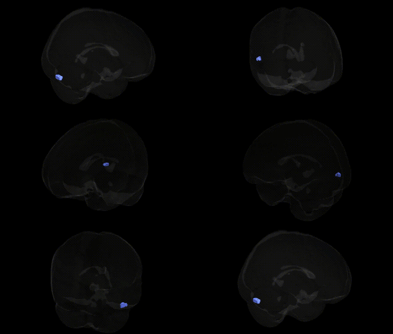
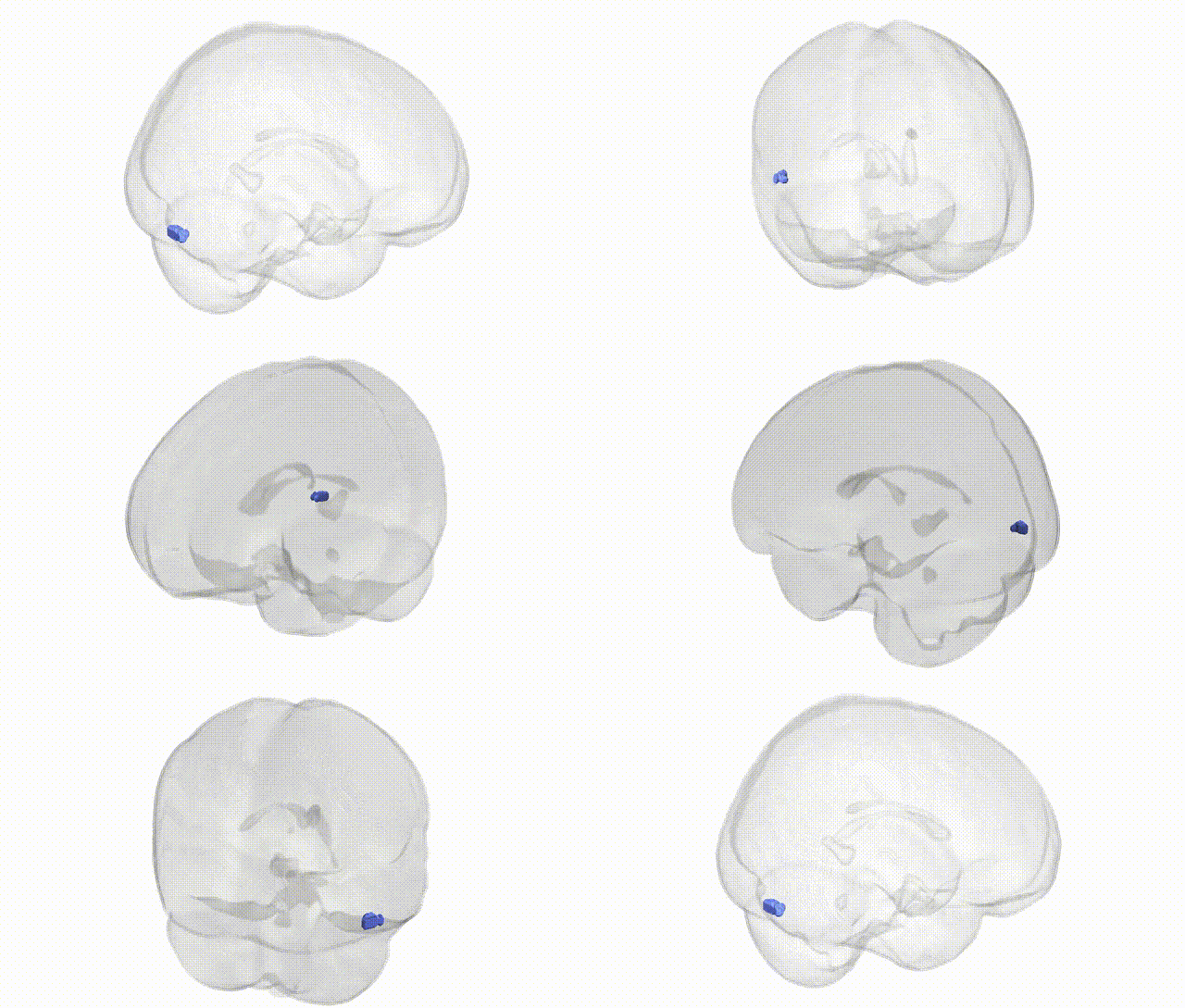
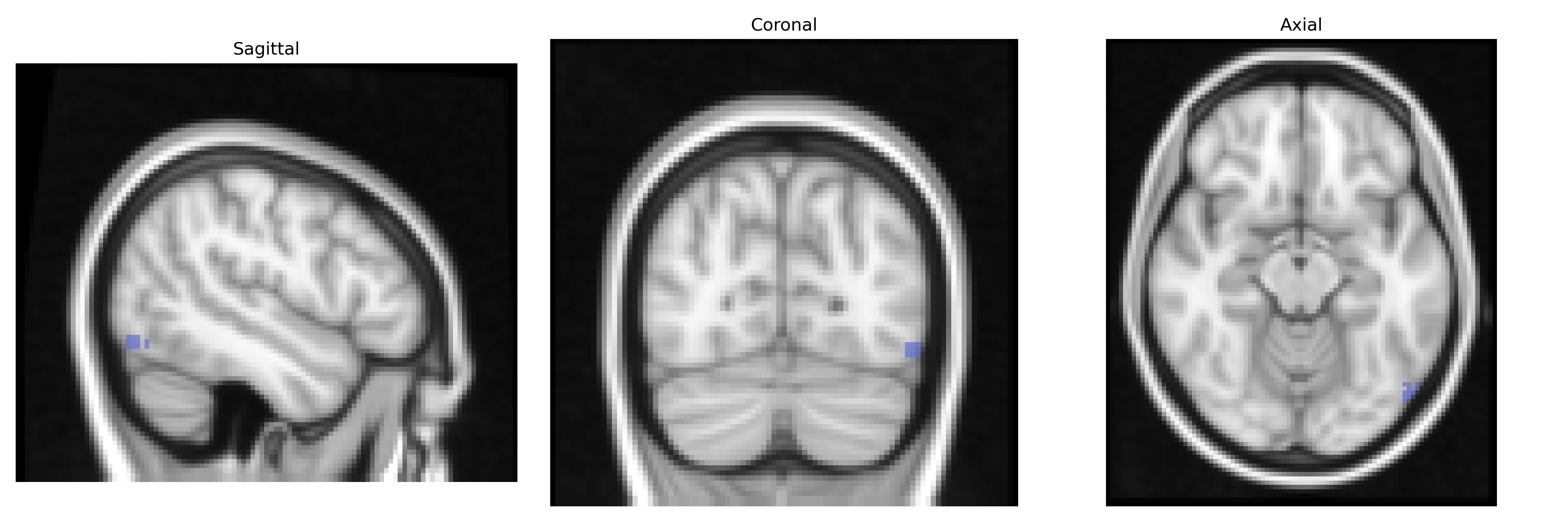
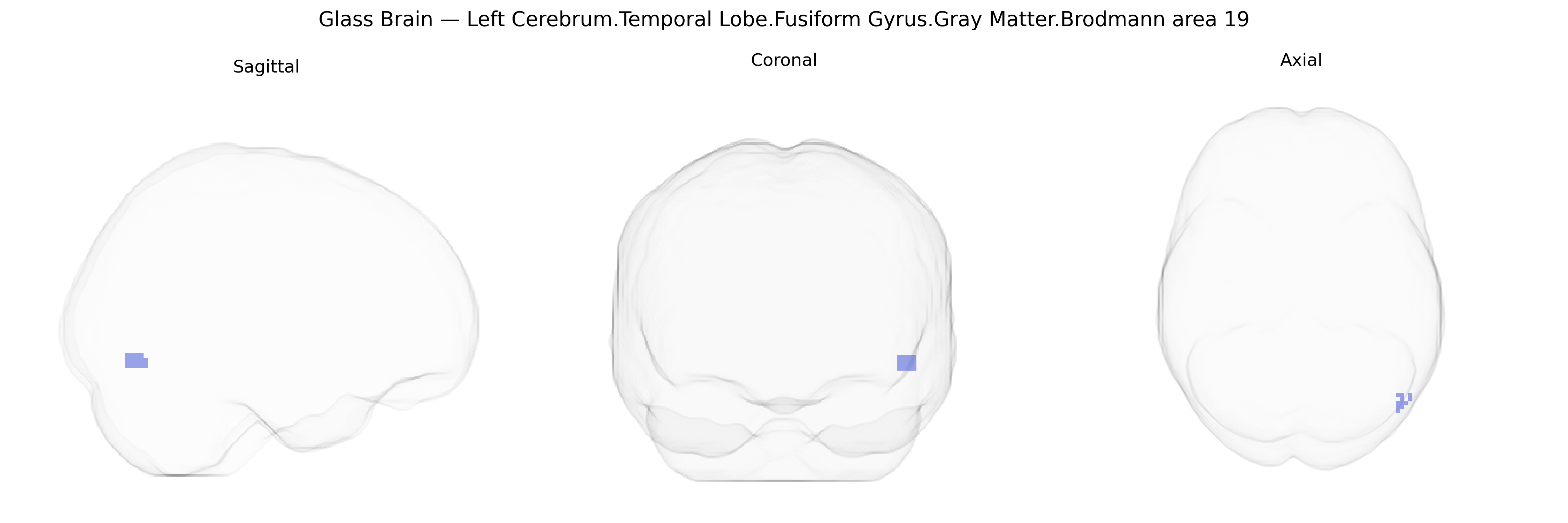
The bilateral Left Cerebrum.Temporal Lobe.Fusiform Gyrus.Gray Matter.Brodmann area 19 region in the Talairach 2 mm Atlas corresponds to cortical tissue within the fusiform gyrus that falls functionally into the territory of Brodmann area 19, a higher-order visual association area traditionally localized to the occipital lobe. In this temporal-lobe extension, BA19 participates in complex visual processing, including integration of form, color, and motion information and the recognition of visually presented objects and patterns, contributing to ventral stream (“what pathway”) functions. Gray matter in this zone contains pyramidal neurons and interneurons organized into six cortical layers, forming part of a distributed network for visual perception and object recognition that interfaces with neighboring regions such as BA37 and the lateral occipital complex. There is no direct Wikipedia article for this exact composite region; a closely related structure is the fusiform gyrus: Fusiform gyrus.
The bilateral left temporal fusiform gyrus gray matter in Brodmann area 19, corresponding to a high-level visual association region, has been implicated in multiple genetic and GWAS findings, largely through imaging genetics and neuropsychiatric studies rather than region-specific Talairach-based analyses. Variants in genes involved in synaptic plasticity, neurodevelopment, and glutamatergic signaling (such as BDNF, NRGN, CACNA1C, and GRIN2B) have been associated with structural and functional differences in adjacent fusiform and occipitotemporal cortices, influencing face and object recognition, visual word processing, and social cognition. Large-scale GWAS of brain cortical thickness and surface area have linked common variants near genes like DACT1, TBL1XR1, and others to occipitotemporal and fusiform measures, suggesting polygenic influences on this region’s morphology. In psychiatric genetics, schizophrenia, autism spectrum disorder, and major depressive disorder risk variants (e.g., in CACNA1C, ZNF804A, and certain 16p11.2 CNVs) have been associated with altered fusiform and temporo-occipital gray matter volume or activation patterns, potentially contributing to social perception and language-related deficits. Additionally, GWAS of reading ability and dyslexia implicating genes such as DCDC2 and KIAA0319 converge with imaging findings that involve left fusiform/BA19 territory in orthographic and phonological processing, indicating that common and rare genetic variation affects this region’s structure and function in relation to visual cognition, language, and neuropsychiatric vulnerability.
Overview generated by GPT-4o (2026).
Region ID: 265
Hemisphere: bilateral
Atlas: Talairach labels 2mm
Full Quality Version: Download MP4
Full Quality Version: Download MP4
Wali Sidiqyar*, Gaurav Rudravaram*, Elyssa M. McMaster, Trent M. Schwartz, Adam M. Saunders, Kurt G. Schilling, Bennett A. Landman "Introducing SPINS: A Shared Public Visualization Library of Neuroanatomical Structures." Medical Imaging with Deep Learning- short paper
This resource is licensed under CC0 1.0 Universal (Public Domain).