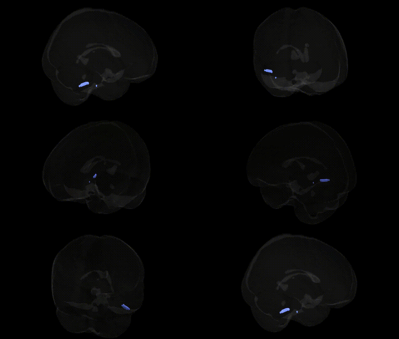
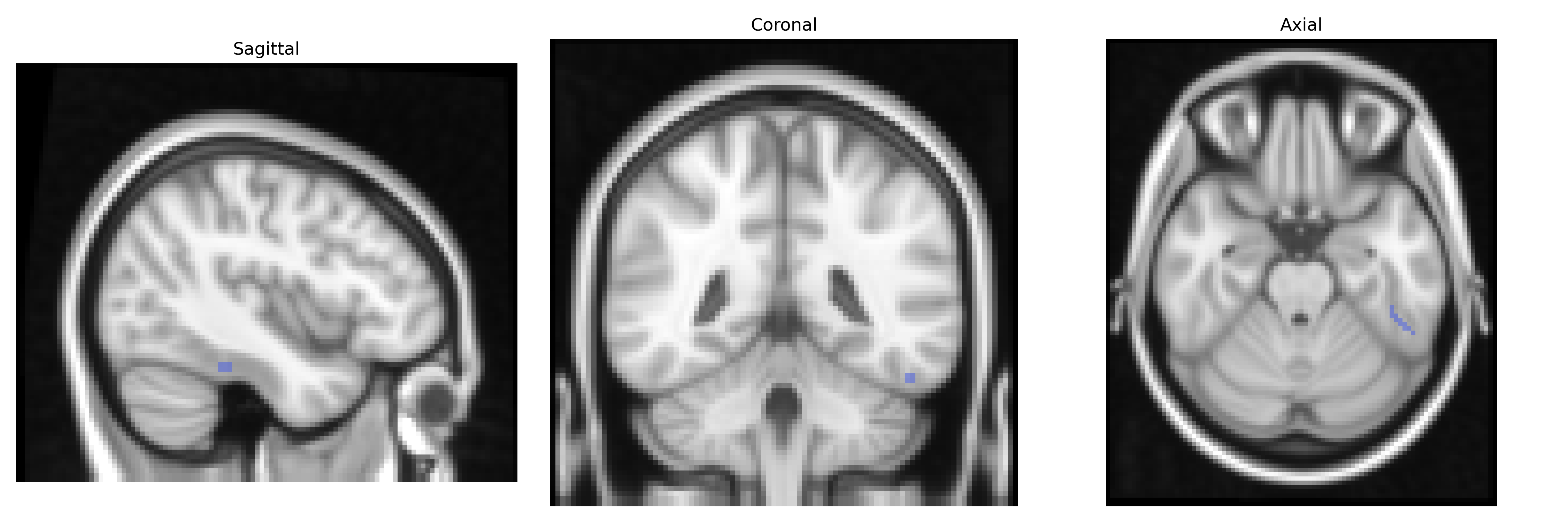
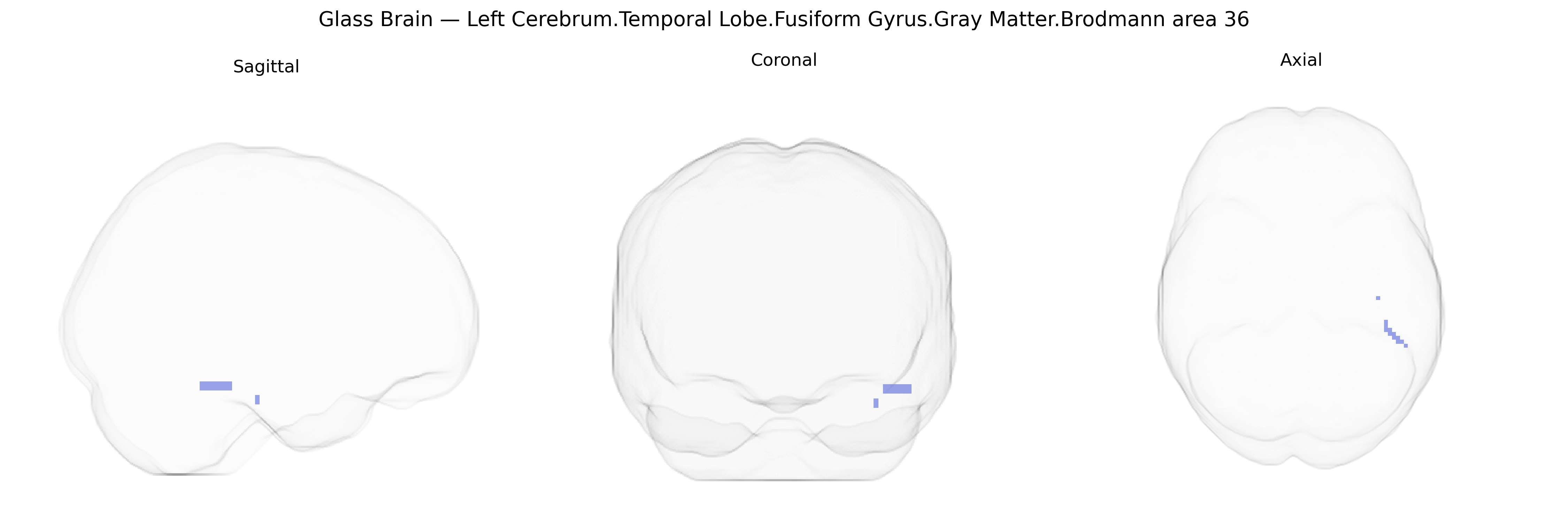
The bilateral Left Cerebrum.Temporal Lobe.Fusiform Gyrus.Gray Matter.Brodmann area 36 corresponds to a ventromedial temporal cortical region within the fusiform gyrus, forming part of the perirhinal/entorhinal cortex complex implicated in higher-order visual processing and declarative memory. Cytoarchitectonically defined as Brodmann area 36, it receives polymodal sensory input, contributes to object recognition and familiarity judgments, and plays a crucial role in associative memory and semantic integration, particularly in linking visual features with stored knowledge. This region is also involved in recognition of complex stimuli such as faces and objects, and lesions or dysfunction in its vicinity have been associated with impairments in visual memory and semantic processing. There is no direct link for Brodmann area 36; a closely related structure is the Perirhinal cortex.
The bilateral left fusiform gyrus gray matter in Brodmann area 36 (BA36), part of the temporal lobe and implicated in visual recognition and higher-order associative processing, has been linked in genetic and imaging-genetic studies to several neurodevelopmental, psychiatric, and cognitive traits. GWAS of cortical structure and surface-based morphometry have identified common variants in genes related to synaptic function, axon guidance, and neurodevelopment (e.g., in or near CADM2, DLG2, and other neuronal signaling loci) that are associated with fusiform/ventral temporal cortex thickness and surface area, suggesting polygenic influences on BA36 morphology. Imaging-genetic analyses in autism spectrum disorder highlight altered fusiform gyrus volume and activation, with risk variants in synaptic and chromatin-remodeling genes (such as those in the 16p11.2 region, SHANK family genes, and other ASD-associated loci) indirectly linked to structural and functional changes in this region. In schizophrenia and bipolar disorder, polygenic risk scores and GWAS-implicated genes involved in glutamatergic signaling and neurodevelopment have been associated with fusiform and adjacent temporal lobe volume reductions, although the specificity to BA36 is limited. Face recognition and social cognition traits show heritable variation with imaging-genetic work pointing to fusiform gyrus involvement, including associations with variants near genes influencing occipitotemporal connectivity, while dyslexia and language-related traits have been linked to left temporal fusiform/BA36 structure in candidate and GWAS-based studies involving neuronal migration and axon guidance genes. Overall, genetic influences on BA36 appear highly polygenic and pleiotropic, contributing to interindividual differences in fusiform gyrus structure and function, and mediating risk for disorders involving social perception, language, and high-level visual processing.
Overview generated by GPT-4o (2026).
Region ID: 136
Hemisphere: bilateral
Atlas: Talairach labels 2mm
Full Quality Version: Download MP4
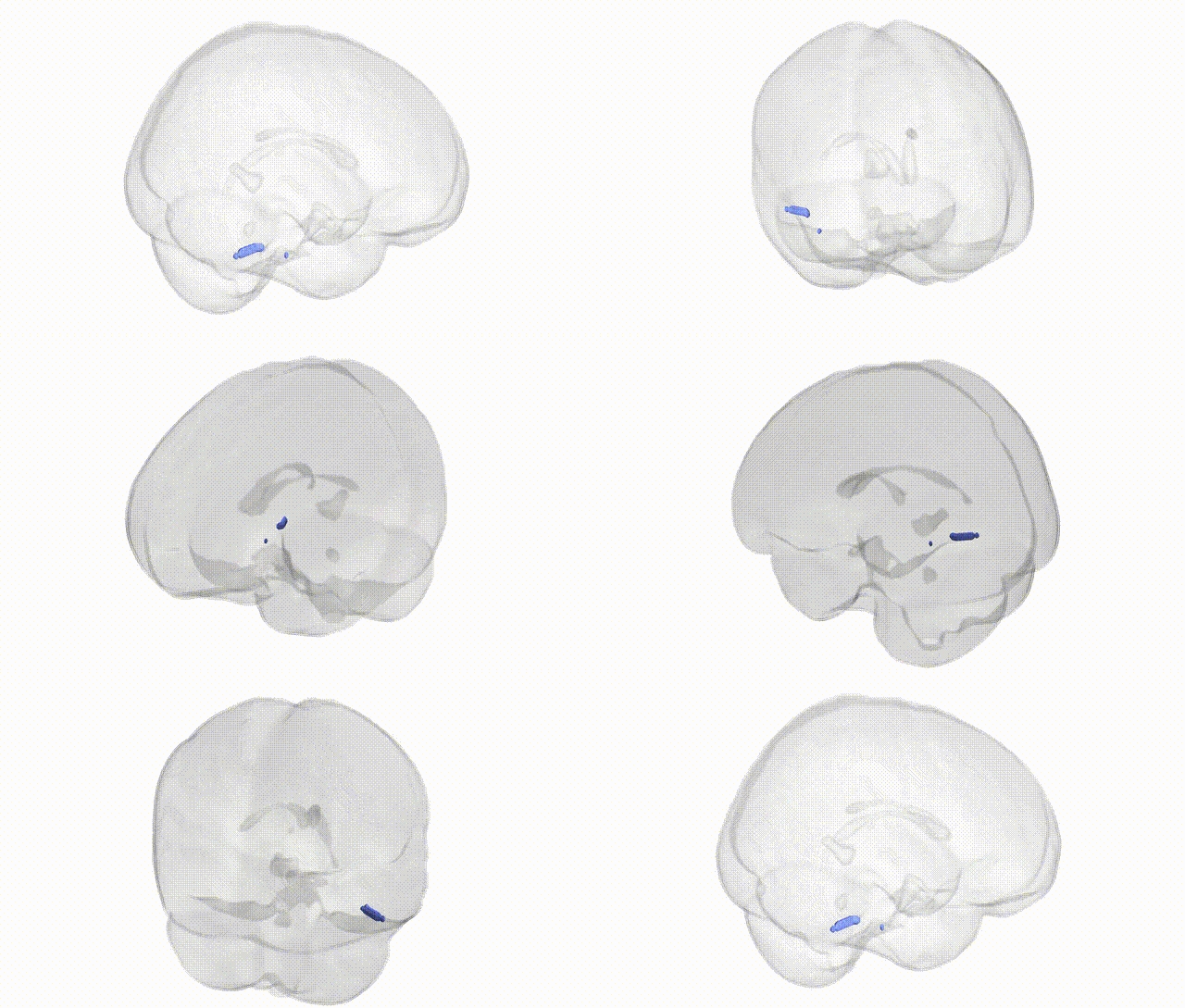
Full Quality Version: Download MP4
Wali Sidiqyar*, Gaurav Rudravaram*, Elyssa M. McMaster, Trent M. Schwartz, Adam M. Saunders, Kurt G. Schilling, Bennett A. Landman "Introducing SPINS: A Shared Public Visualization Library of Neuroanatomical Structures." Medical Imaging with Deep Learning- short paper
This resource is licensed under CC0 1.0 Universal (Public Domain).