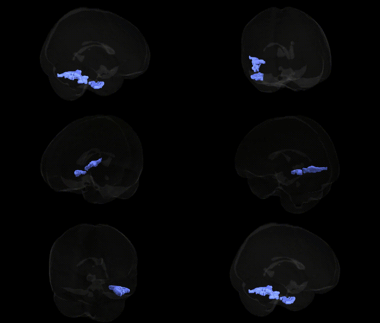
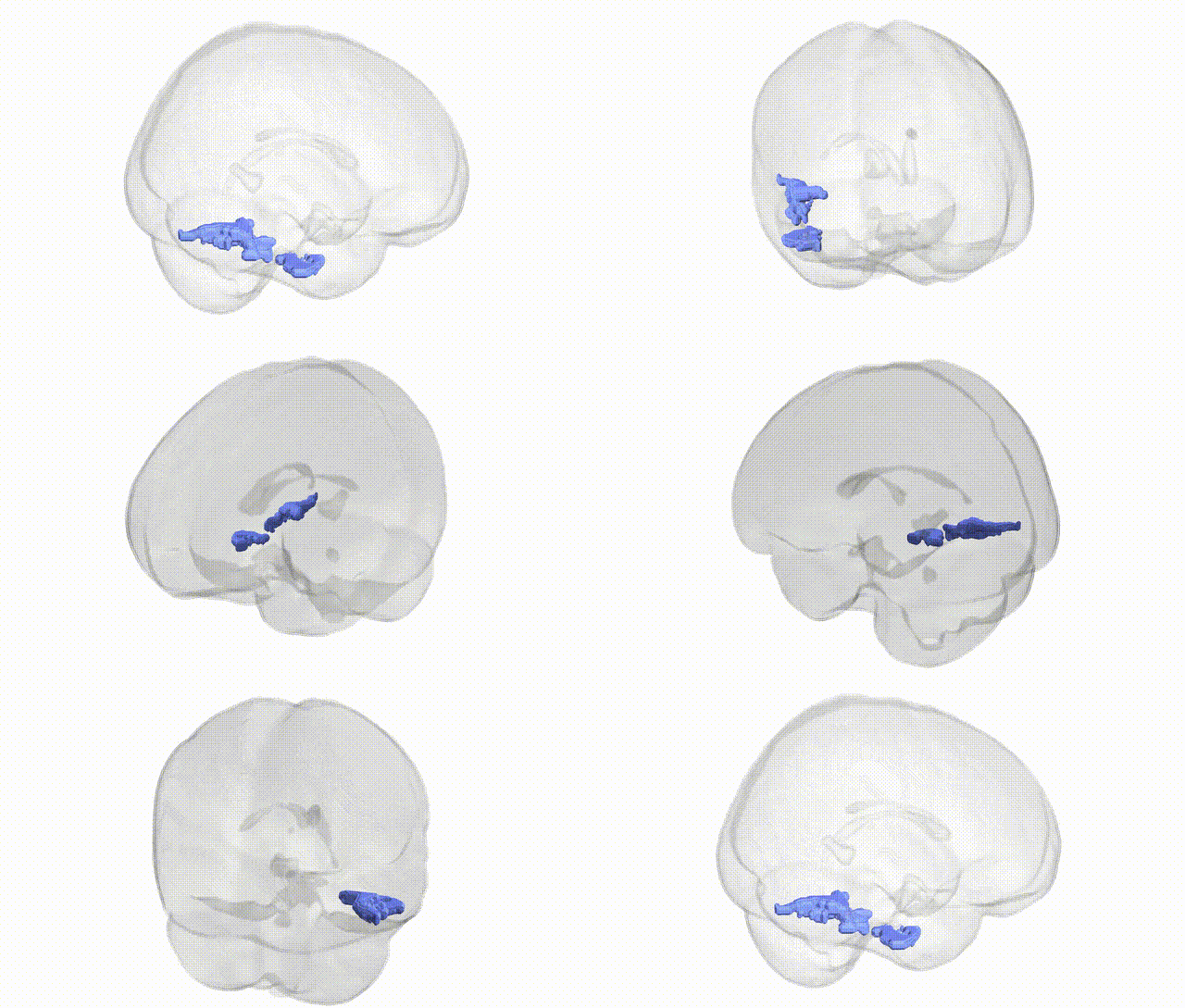
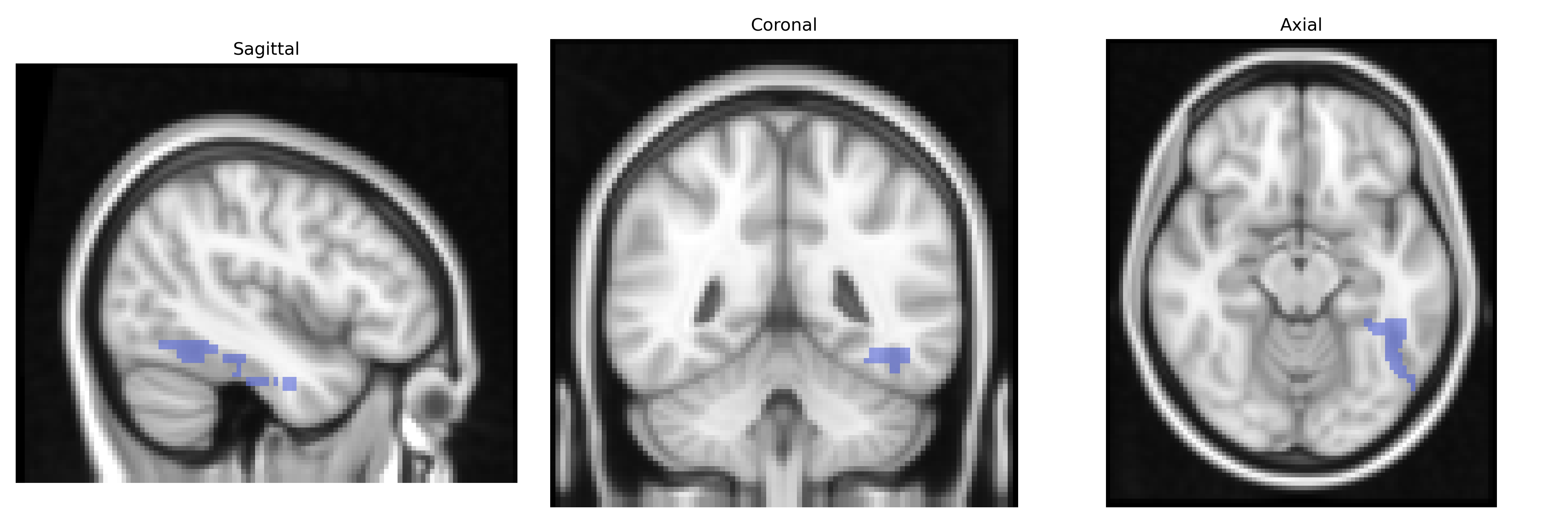
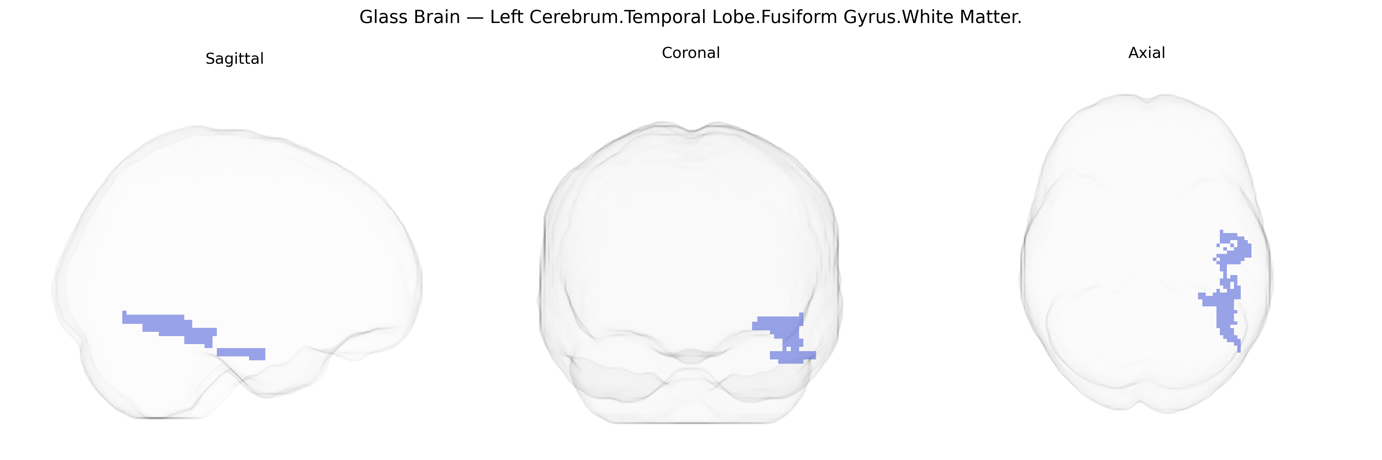
The bilateral Left Cerebrum.Temporal Lobe.Fusiform Gyrus.White Matter corresponds to the subcortical myelinated fiber pathways underlying the fusiform gyrus in the left temporal lobe. This white matter region supports communication between the fusiform cortex—critically involved in high-level visual processing such as face, word, and object recognition—and other cortical and subcortical areas, including occipital visual regions, the inferior temporal cortex, and language-related areas in the dominant hemisphere. Functionally, these fibers form part of long-range association tracts (such as components of the inferior longitudinal fasciculus and possibly the inferior fronto-occipital fasciculus), enabling integration of visual perceptual information with semantic, linguistic, and memory processes. Damage to this white matter can contribute to deficits in visual recognition (e.g., prosopagnosia or alexia) by disrupting connectivity rather than directly injuring fusiform cortical neurons. There is no direct link for the white matter subdivision alone; a related cortical structure is the Fusiform gyrus.
Genetic associations involving the bilateral left cerebrum temporal lobe fusiform gyrus white matter, as defined in the Talairach 2 mm atlas, largely arise from imaging genetics and GWAS of structural and functional MRI phenotypes rather than region-specific single-gene studies. Variants in genes involved in axonal development, synaptic plasticity, and myelination—such as BDNF, NRG1/3, CNTNAP2, and MBP—have been associated with white matter integrity (e.g., fractional anisotropy) in temporal regions that include fusiform gyrus tracts, often via diffusion tensor imaging GWAS meta-analyses. Large consortia (e.g., ENIGMA) have reported heritable variation in temporal lobe and fusiform volumes and white matter microstructure, with polygenic influences overlapping neurodevelopmental and psychiatric risk loci. The fusiform region is repeatedly implicated in disorders affecting face and social perception, language, and visual processing, including autism spectrum disorder, schizophrenia, bipolar disorder, major depression, and developmental dyslexia, where GWAS risk variants and polygenic scores show associations with altered fusiform gray matter and adjacent temporal white matter connectivity. In autism and schizophrenia, risk alleles in synaptic and neurodevelopmental genes (e.g., those near DISC1, DLG2, and SHANK family genes) have been linked to abnormal temporal lobe white matter organization that encompasses fusiform pathways, while dyslexia and language-related GWAS have connected FOXP2-related and other speech–language loci to temporal white matter changes in regions overlapping the fusiform gyrus. Overall, genetic studies support a highly polygenic architecture in which shared neurodevelopmental and myelination-related variants influence fusiform-associated temporal white matter structure and function across multiple cognitive traits and neuropsychiatric disorders.
Overview generated by GPT-4o (2026).
Region ID: 128
Hemisphere: bilateral
Atlas: Talairach labels 2mm
Full Quality Version: Download MP4
Full Quality Version: Download MP4
Wali Sidiqyar*, Gaurav Rudravaram*, Elyssa M. McMaster, Trent M. Schwartz, Adam M. Saunders, Kurt G. Schilling, Bennett A. Landman "Introducing SPINS: A Shared Public Visualization Library of Neuroanatomical Structures." Medical Imaging with Deep Learning- short paper
This resource is licensed under CC0 1.0 Universal (Public Domain).