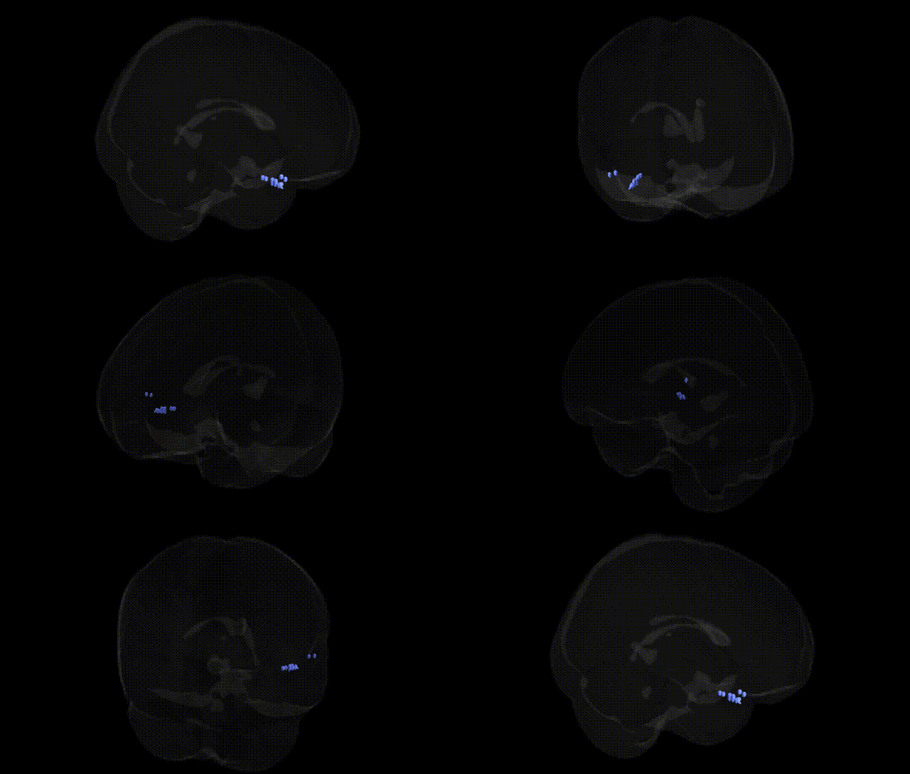
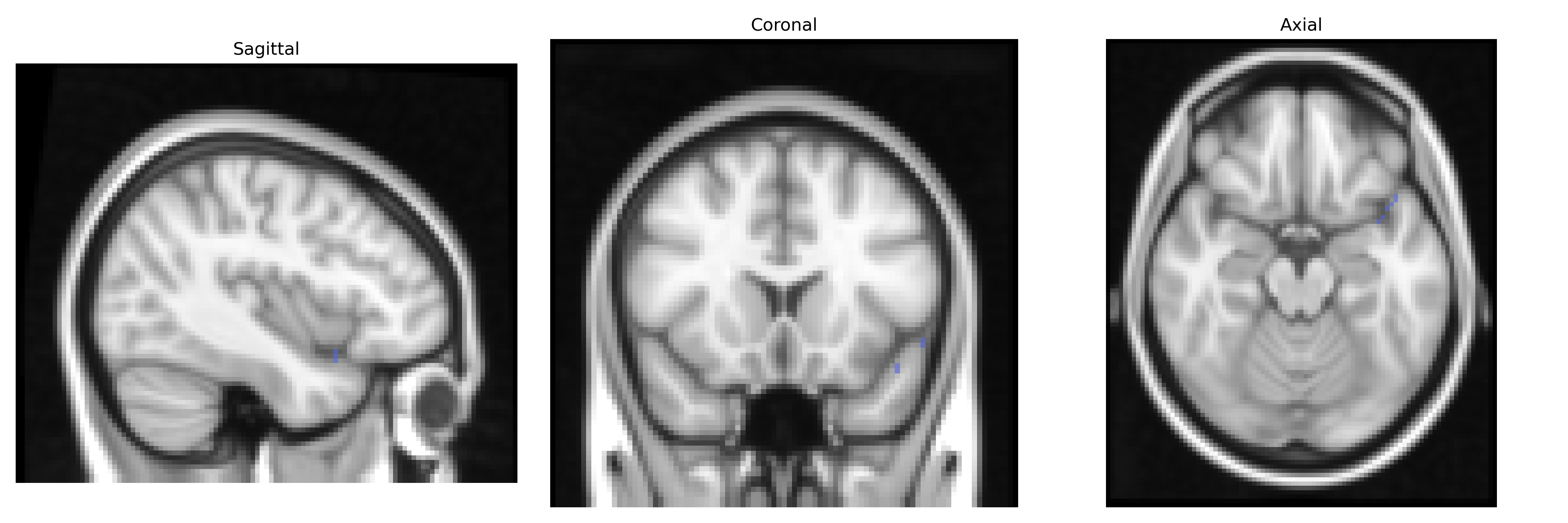
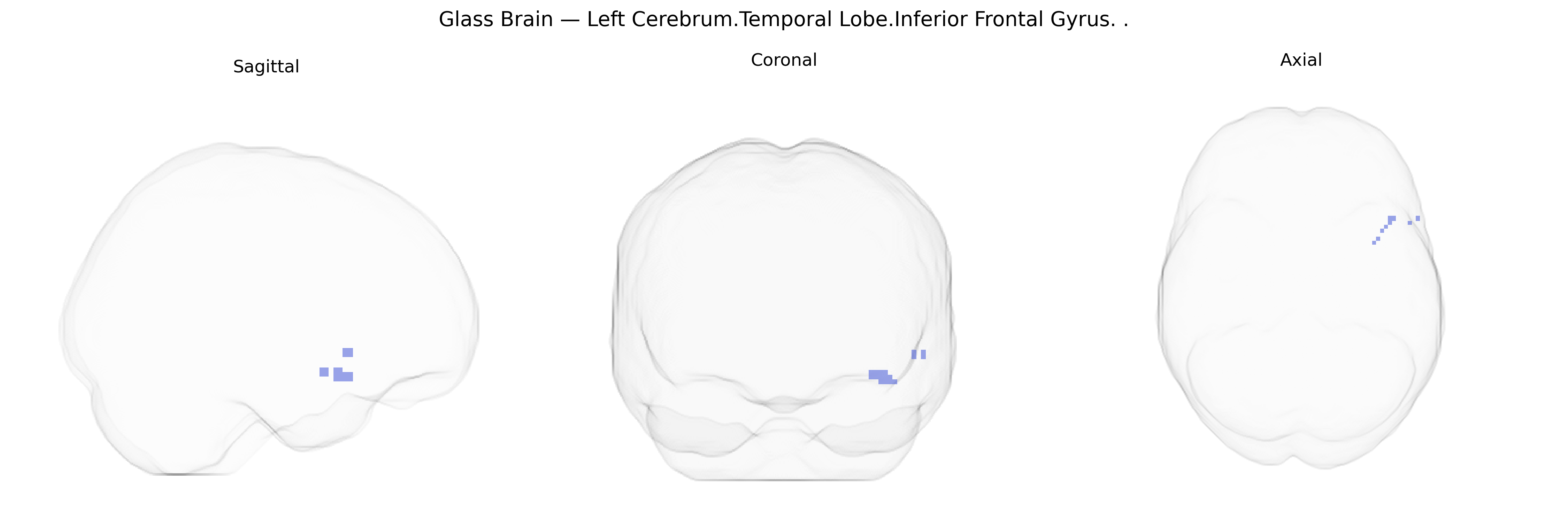
The bilateral Left Cerebrum.Temporal Lobe.Inferior Frontal Gyrus refers to Talairach atlas labeling that spans two distinct cortical regions: the inferior frontal gyrus, located in the frontal lobe along the lateral surface anterior to the precentral gyrus, and adjacent portions of the temporal lobe within the left cerebral hemisphere. The inferior frontal gyrus (particularly its pars opercularis and pars triangularis) forms the core of Broca’s area, critically involved in speech production, syntactic processing, and aspects of language working memory, while neighboring temporal regions contribute to auditory processing, semantic representation, and integration of multimodal language information. Functionally, this combined territory is central to left-lateralized language networks, supporting phonological encoding, lexical access, and higher-order linguistic operations, and is frequently implicated in aphasia, language-related epilepsy, and functional imaging studies of speech and comprehension. There is no single Wikipedia article for the full Talairach label combination; a closely related primary component is Inferior frontal gyrus.
The bilateral Left Cerebrum.Temporal Lobe.Inferior Frontal Gyrus region (Talairach labels 2 mm) falls within the inferior frontal and anterior temporal language network, including territory overlapping Broca’s area, and has been repeatedly implicated in genetic studies of language, cognition, and neuropsychiatric risk. GWAS and imaging–genetics work show that cortical thickness, surface area, and gyrification in inferior frontal and adjacent temporal regions are heritable and associated with common variants, including those near genes involved in neurodevelopment and synaptic function (e.g., FOXP2-related pathways for speech and language, CNTNAP2 for language and autism risk, and multiple loci from ENIGMA consortia affecting frontal and temporal cortical morphology). Variants linked to schizophrenia, bipolar disorder, major depressive disorder, and autism spectrum disorder often show downstream effects on inferior frontal and temporal activation during tasks requiring language, working memory, or social cognition, and structural differences in these regions frequently mediate genetic risk for psychosis and mood disorders in large imaging–genetics studies. Alzheimer’s disease and frontotemporal dementia GWAS (e.g., APOE, MAPT, GRN and others) are associated with atrophy patterns that include the inferior frontal and anterior temporal cortices, and polygenic risk scores for these disorders correlate with reduced volume or thickness in overlapping areas. In addition, GWAS of educational attainment, general cognitive ability, and reading/language skills report associations with genes whose expression and imaging correlates cluster in frontotemporal language circuits, implicating this region as a key anatomical substrate through which polygenic influences on cognition and neurodevelopmental and psychiatric disorders are expressed.
Overview generated by GPT-4o (2026).
Region ID: 232
Hemisphere: bilateral
Atlas: Talairach labels 2mm
Full Quality Version: Download MP4
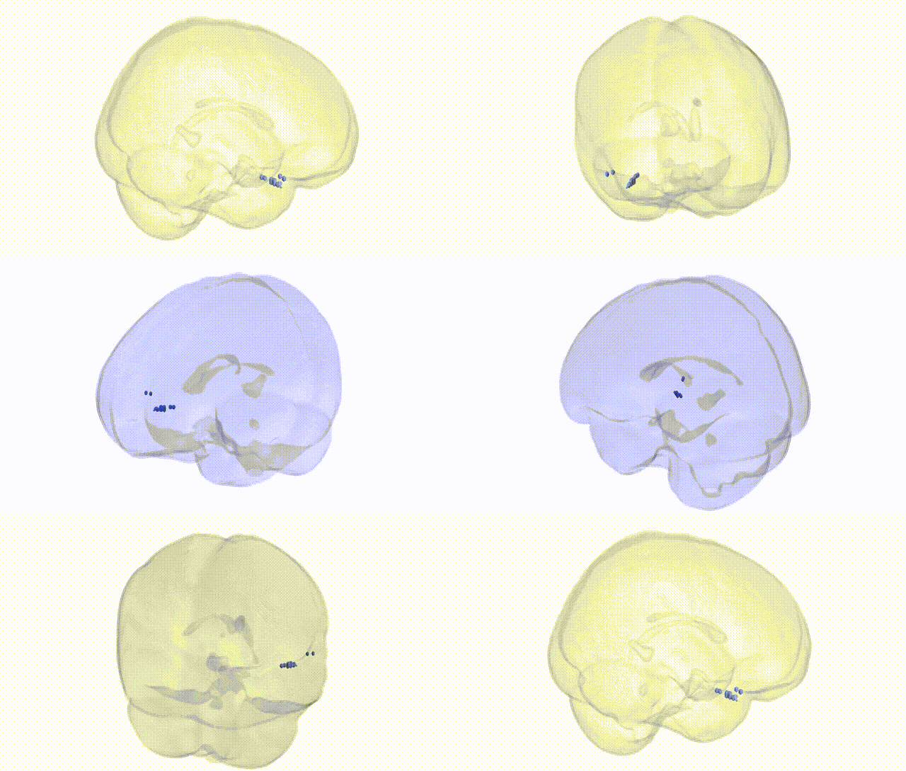
Full Quality Version: Download MP4
Wali Sidiqyar*, Gaurav Rudravaram*, Elyssa M. McMaster, Trent M. Schwartz, Adam M. Saunders, Kurt G. Schilling, Bennett A. Landman "Introducing SPINS: A Shared Public Visualization Library of Neuroanatomical Structures." Medical Imaging with Deep Learning- short paper
This resource is licensed under CC0 1.0 Universal (Public Domain).