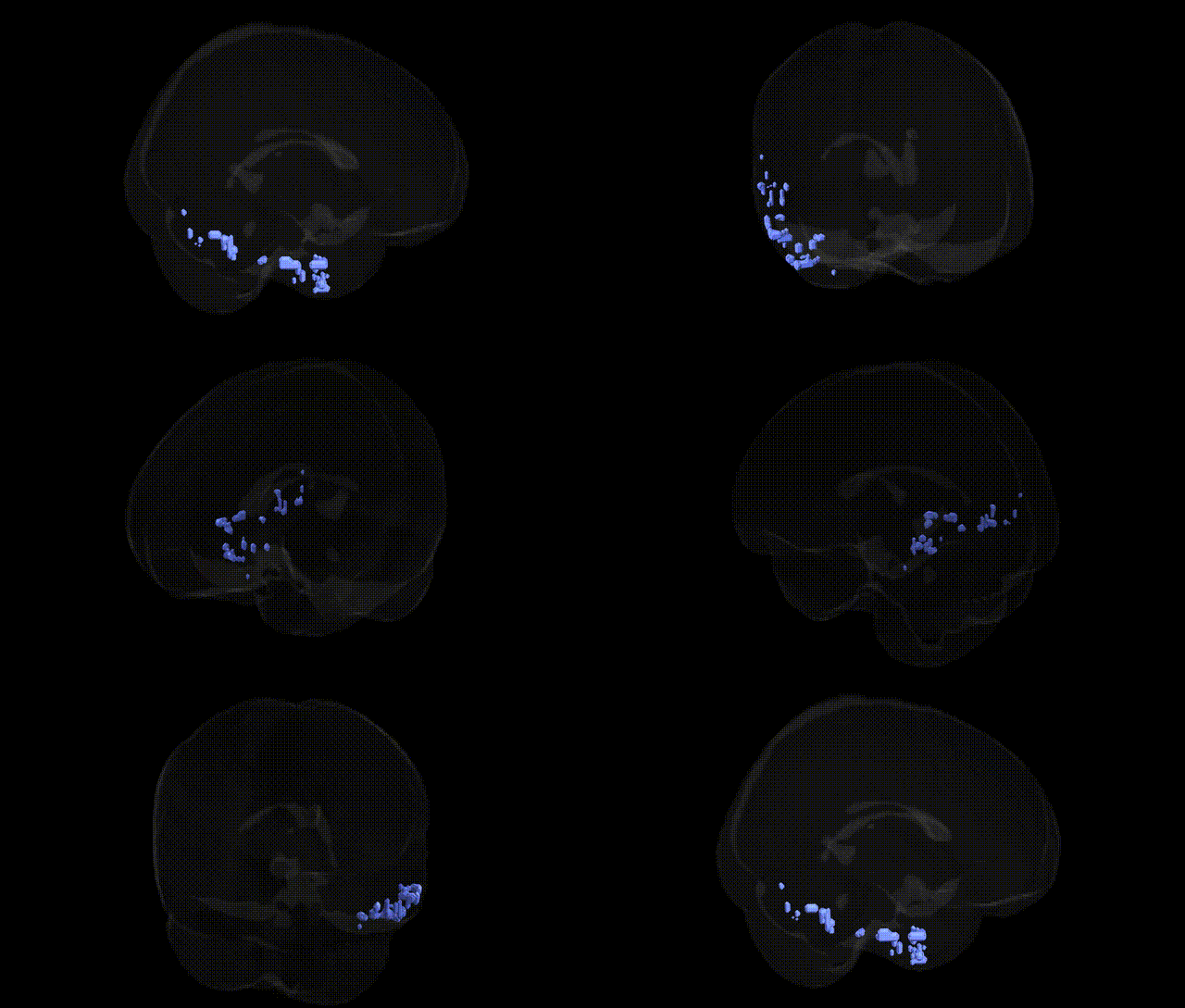
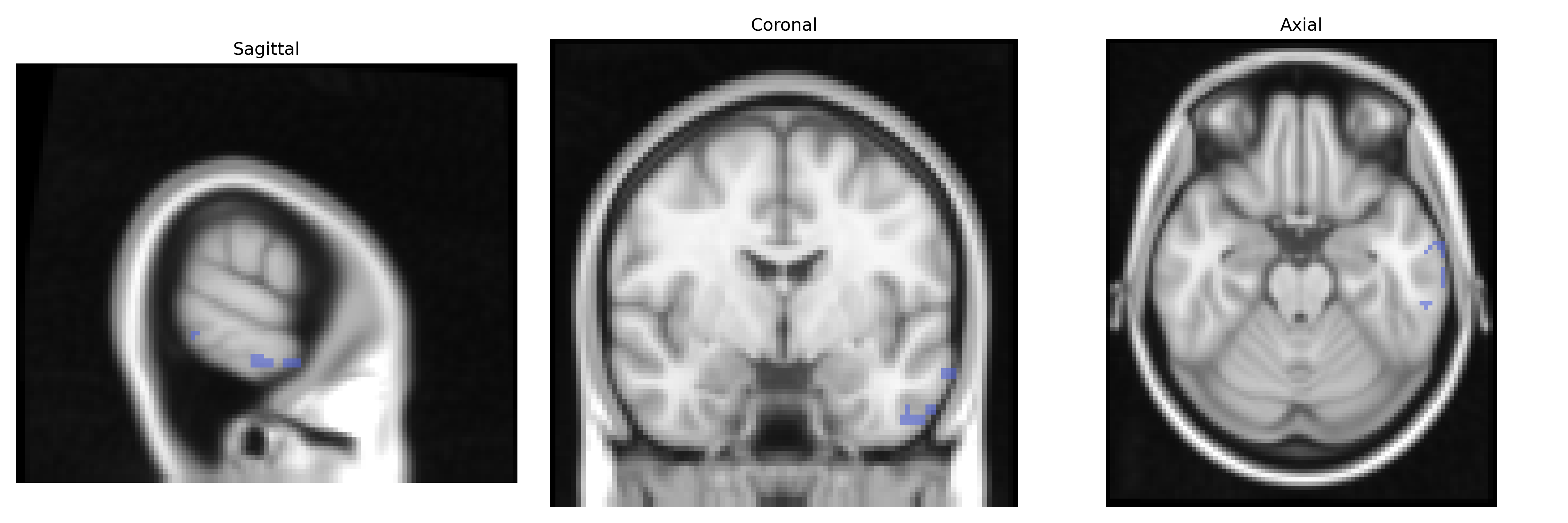
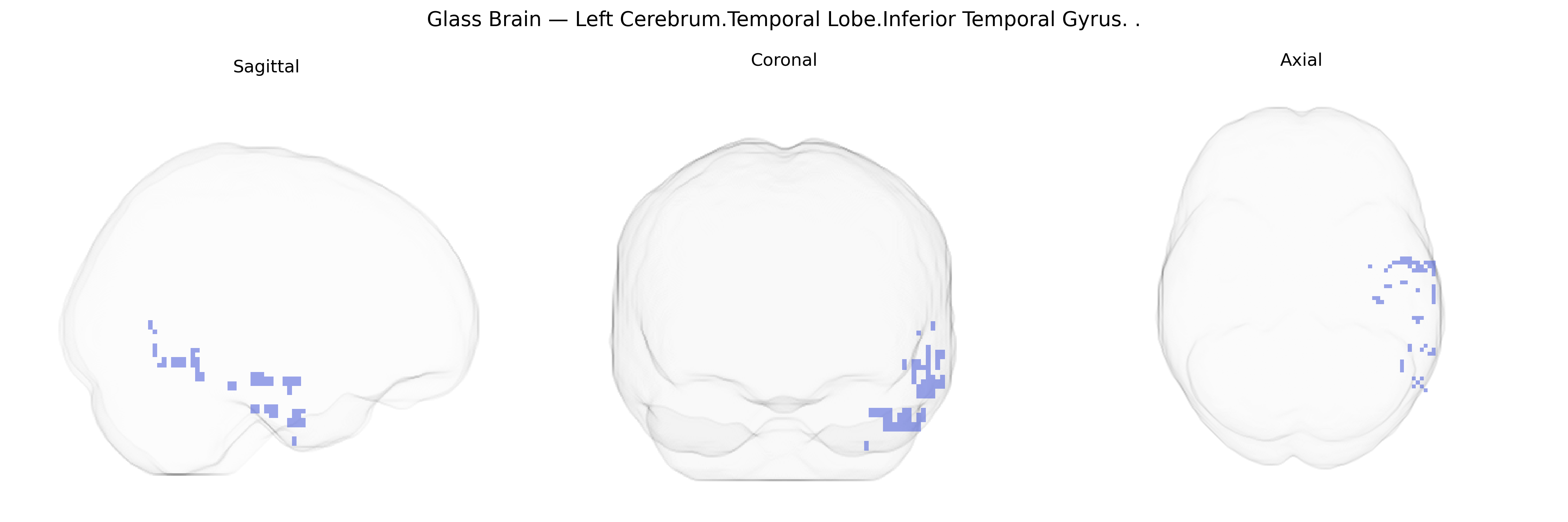
The bilateral Left Cerebrum.Temporal Lobe.Inferior Temporal Gyrus corresponds to cortex located on the ventral aspect of the temporal lobe, lateral to the fusiform gyrus and inferior to the middle temporal gyrus, extending along the rostro-caudal axis of the temporal lobe. This region is primarily associated with high-level visual processing, including object recognition, complex pattern analysis, and aspects of visual memory, and it participates in the ventral “what” visual stream connecting occipital visual areas to anterior temporal and frontal regions. Neuronal populations here are tuned to complex visual features and contribute to the integration of shape, color, and texture information necessary for categorizing and identifying objects, faces, and scenes. There is no direct link for the Inferior Temporal Gyrus; a closely related structure is the Temporal lobe.
The bilateral inferior temporal gyrus (ITG) of the left temporal lobe, as localized in the Talairach 2 mm atlas, has been implicated in genetic studies of brain structure and cognition, particularly through imaging genetics and GWAS of cortical morphology and functional activation. Large-scale GWAS of cortical thickness and surface area (e.g., ENIGMA consortium) have identified associations between ITG morphology and variants near genes involved in neurodevelopment and synaptic function, including loci in or near FOXO3, PAX6, KIAA0586, and other regulators of cortical patterning, though these are generally regionally distributed rather than uniquely specific to the ITG. ITG volume and cortical thickness have shown heritability and genetic correlations with reading ability, face and object recognition performance, and broader measures of visual semantic processing, consistent with the role of this region in higher-order visual cognition. Genetic risk for neurodevelopmental and neuropsychiatric disorders—such as autism spectrum disorder (ASD), schizophrenia, and dyslexia—has been linked to altered structure or activation in inferior temporal regions; for example, ASD- and schizophrenia-associated common variants in genes like CNTNAP2, NRXN1, and DCC have been related to atypical temporal cortical connectivity and morphology in imaging-genetics cohorts. GWAS of Alzheimer’s disease and other dementias have connected risk alleles (e.g., APOE ε4 and variants near BIN1, CLU, and CR1) to accelerated atrophy in temporal association cortices, including the ITG, where early neurodegenerative changes are often observed. Additionally, polygenic scores for educational attainment, general cognitive ability, and language-related traits have shown associations with temporal lobe measures that encompass the inferior temporal gyrus, suggesting shared genetic architecture between this visual association region and higher cognitive functions, although pinpointing effects specifically to the bilateral left ITG remains limited by the spatial resolution and parcellation schemes of current GWAS.
Overview generated by GPT-4o (2026).
Region ID: 9
Hemisphere: bilateral
Atlas: Talairach labels 2mm
Full Quality Version: Download MP4
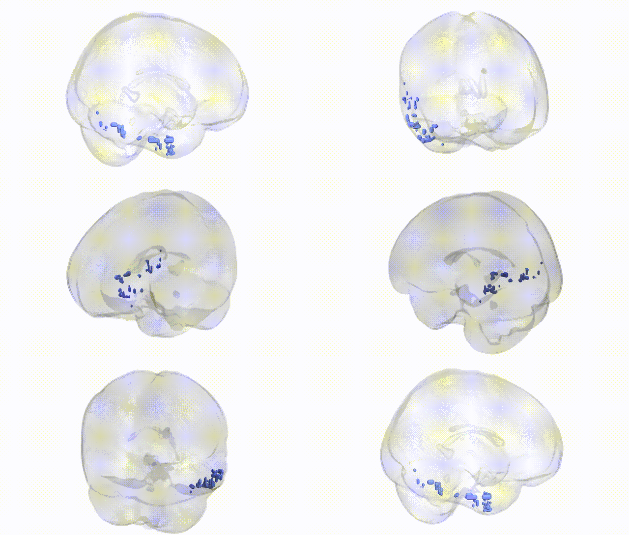
Full Quality Version: Download MP4
Wali Sidiqyar*, Gaurav Rudravaram*, Elyssa M. McMaster, Trent M. Schwartz, Adam M. Saunders, Kurt G. Schilling, Bennett A. Landman "Introducing SPINS: A Shared Public Visualization Library of Neuroanatomical Structures." Medical Imaging with Deep Learning- short paper
This resource is licensed under CC0 1.0 Universal (Public Domain).