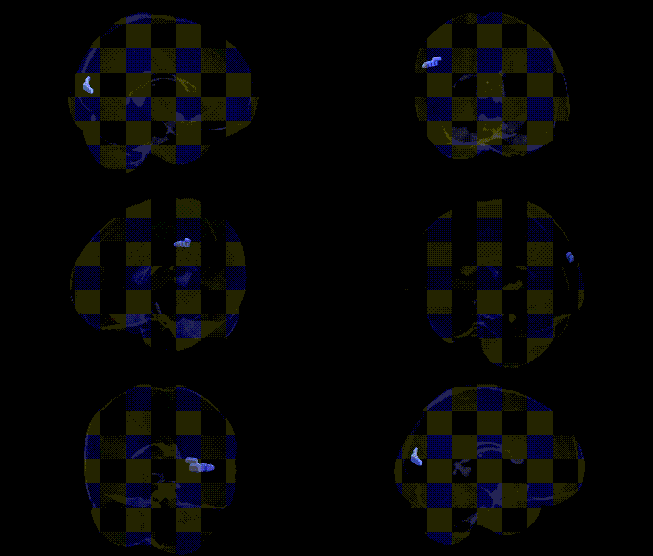
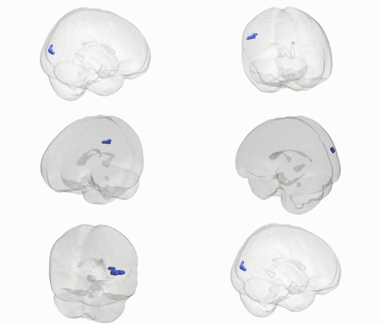
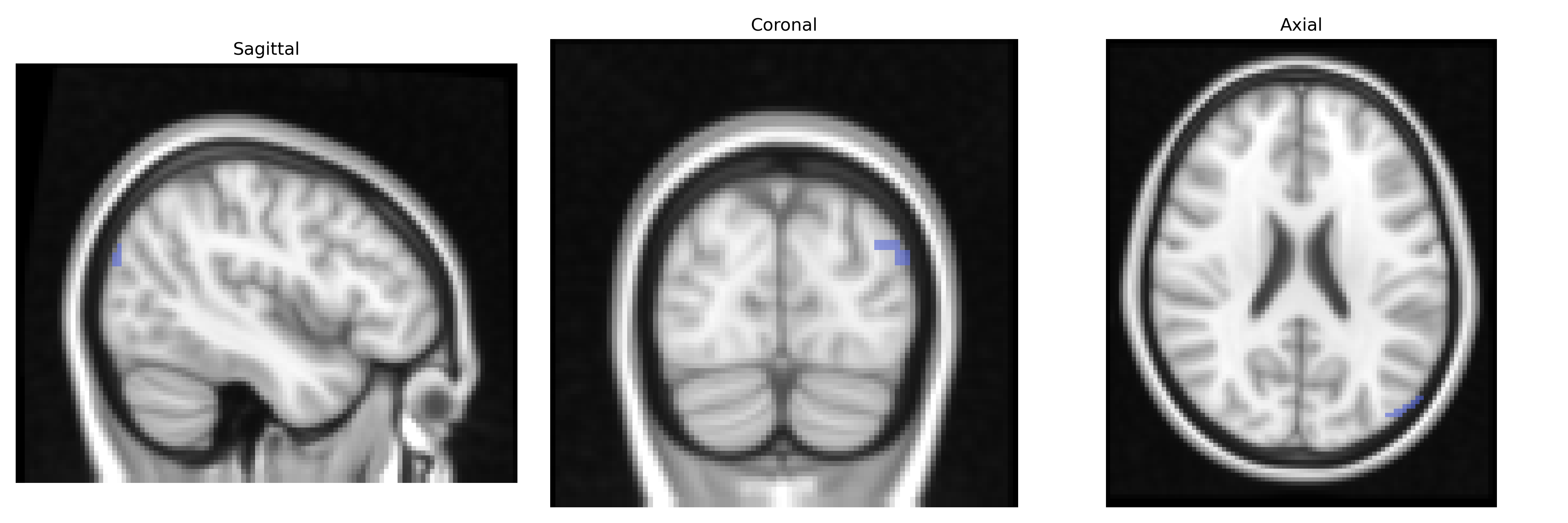
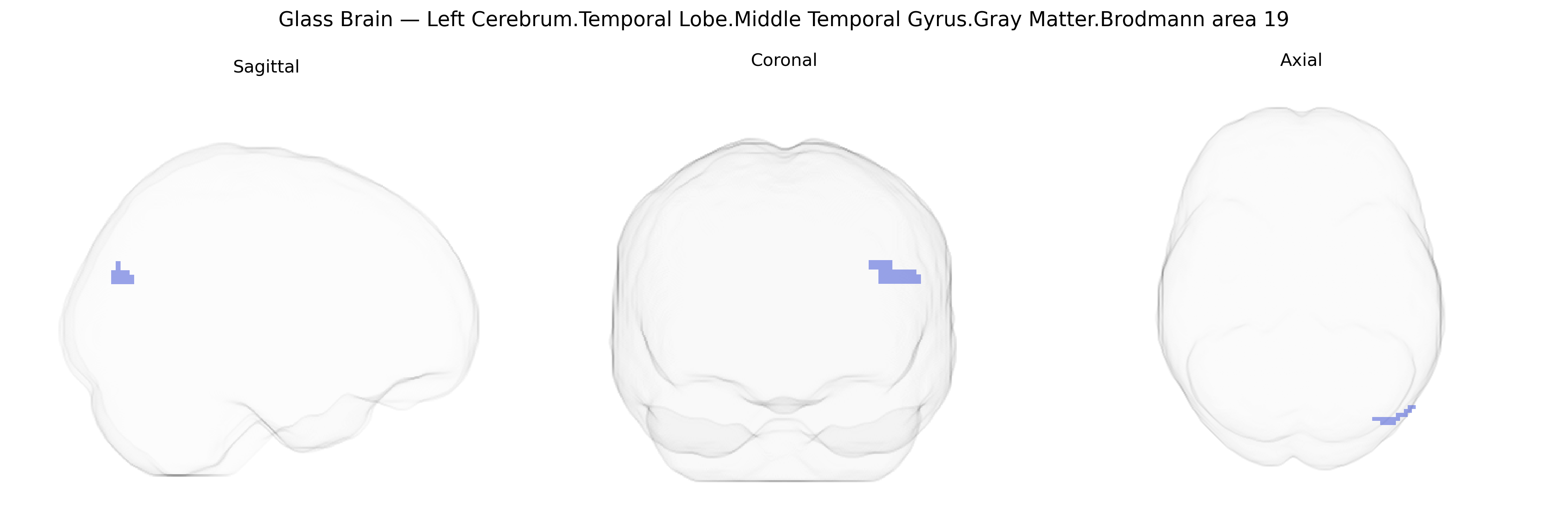
The bilateral Left Cerebrum.Temporal Lobe.Middle Temporal Gyrus.Gray Matter.Brodmann area 19 region, as defined in the Talairach 2 mm Atlas, represents visual association cortex located anatomically within the middle temporal gyrus but functionally assigned to Brodmann area 19, a higher-order visual processing area traditionally centered in the occipital lobe. Neuronal populations in this region are involved in complex visual analysis, including integration of motion, form, and spatial relationships, and in linking visual input to multimodal representations important for perception and recognition. Its gray matter contains pyramidal neurons and interneurons organized into six cortical layers typical of neocortex, mediating cortico-cortical connectivity with primary and secondary visual cortices and temporal association areas. Although the Talairach label specifies a temporal-gyrus localization, the functional designation corresponds to Brodmann area 19.
The bilateral Left Cerebrum.Temporal Lobe.Middle Temporal Gyrus.Gray Matter.Brodmann area 19 (per Talairach labels 2mm Atlas) lies at the intersection of lateral occipitotemporal and posterior temporal cortices and is often functionally treated as part of higher-order visual and language networks rather than as a distinct GWAS target, so genetic associations typically reference overlapping parcels (e.g., middle temporal gyrus, lateral occipital cortex, posterior temporal regions) rather than BA19 specifically. Large neuroimaging GWAS from ENIGMA, UK Biobank, and related consortia have identified multiple common variants associated with cortical thickness and surface area in middle temporal and occipitotemporal regions, frequently implicating genes involved in neurodevelopment and synaptic plasticity (such as CNTNAP2, KIAA0319, DCDC2, and FOXP2 in language/reading-related traits; and general neurodevelopmental loci near MAPT, MEF2C, and MIR137 in cognition and brain morphology). Posterior middle temporal/BA19-related territory is genetically implicated in reading disability/dyslexia and specific language impairment via variants in DYX loci (e.g., DYX1C1, ROBO1), with imaging-genetics studies linking these alleles to altered gray matter and functional activation in temporo-occipital language/visual word form areas. GWAS of schizophrenia, bipolar disorder, and major depression have repeatedly found polygenic risk to correlate with structural and functional alterations in bilateral middle temporal and occipitotemporal cortices, suggesting that many pleiotropic psychiatric risk loci (CACNA1C, ZNF804A, GRM3, among others) may have downstream effects on BA19-adjacent networks. Additionally, polygenic scores for general cognitive ability, educational attainment, and reading performance are associated with variability in cortical metrics and activation in this region during language and visual processing tasks, reinforcing the view that BA19 middle temporal territory is a convergence zone where widespread neurodevelopmental and cognitive risk variants exert measurable structural and functional effects.
Overview generated by GPT-4o (2026).
Region ID: 818
Hemisphere: bilateral
Atlas: Talairach labels 2mm
Full Quality Version: Download MP4
Full Quality Version: Download MP4
Wali Sidiqyar*, Gaurav Rudravaram*, Elyssa M. McMaster, Trent M. Schwartz, Adam M. Saunders, Kurt G. Schilling, Bennett A. Landman "Introducing SPINS: A Shared Public Visualization Library of Neuroanatomical Structures." Medical Imaging with Deep Learning- short paper
This resource is licensed under CC0 1.0 Universal (Public Domain).