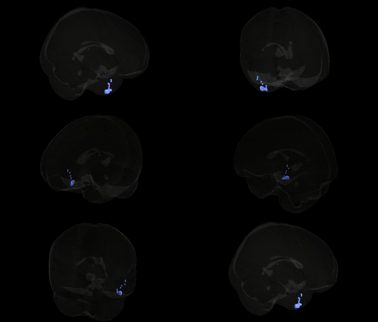
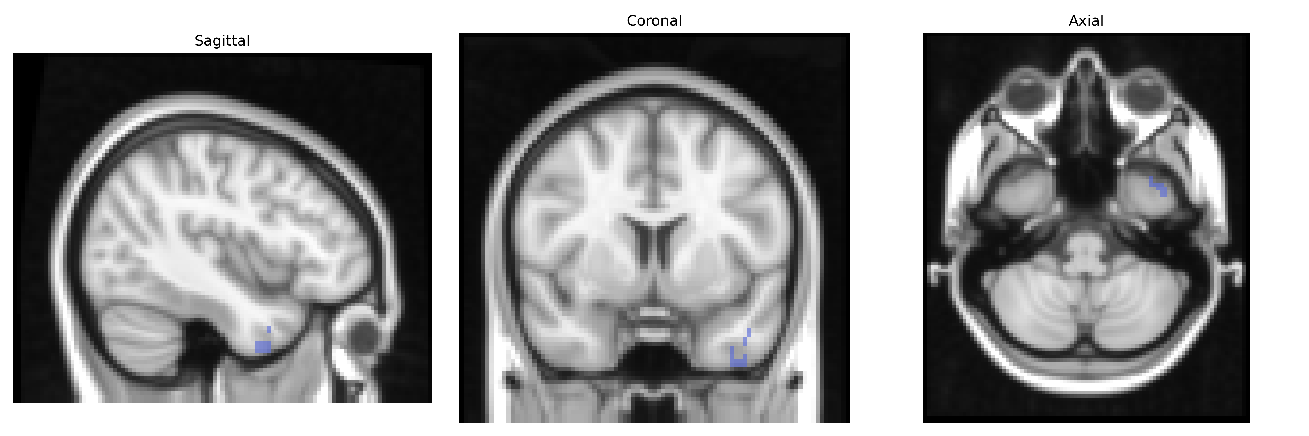
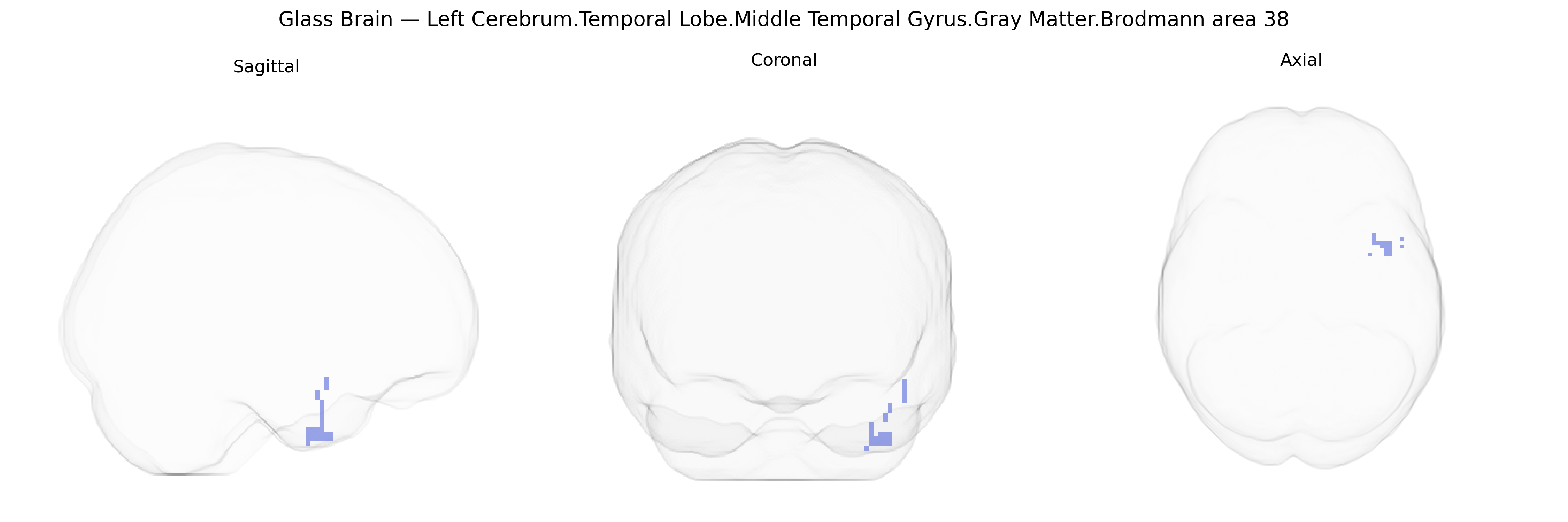
Bilateral Left Cerebrum.Temporal Lobe.Middle Temporal Gyrus.Gray Matter.Brodmann area 38 corresponds to gray matter in the temporal pole region of the middle temporal gyrus within the left cerebral hemisphere, encompassing the most anterior part of the temporal lobe. Brodmann area 38 is implicated in high-level multimodal integration, including processing of complex social and emotional stimuli, semantic memory, and aspects of language and autobiographical memory, due to its extensive connectivity with limbic, frontal, and other temporal regions. Cytoarchitectonically, it is characterized by a granular cortical structure consistent with association cortex, and functionally it contributes to integrating auditory, visual, and affective information within the anterior temporal network. There is no direct Wikipedia article for “Brodmann area 38”; a closely related structure is the Temporal pole.
The bilateral left temporal pole (Brodmann area 38) of the middle temporal gyrus, corresponding to the Talairach “Left Cerebrum.Temporal Lobe.Middle Temporal Gyrus.Gray Matter.Brodmann area 38” label, has been implicated in genetic studies of social cognition, language, and neuropsychiatric disorders, though most findings are indirect via imaging genetics and GWAS of cortical structure or related phenotypes rather than region-specific association tests. Variants in genes such as CNTNAP2, FOXP2, and DCDC2 have been linked to language and reading abilities and to structural or functional differences in anterior temporal and middle temporal regions that encompass BA38. Large-scale GWAS of cortical thickness and surface area (e.g., ENIGMA consortium studies) have identified common variants influencing temporal lobe morphometry, including loci near genes involved in neurodevelopment, synaptic signaling, and axon guidance, with temporal pole thickness or volume showing heritability and shared genetic influences with cognitive performance and educational attainment. Imaging genetics work in autism spectrum disorder, schizophrenia, and frontotemporal dementia has associated risk alleles in genes such as GRIN2A, C4A, and MAPT with altered temporal pole and adjacent anterior temporal cortex structure or connectivity, consistent with the region’s role in semantic memory and social-emotional processing. Although GWAS typically do not isolate BA38 specifically, convergent evidence from structural and functional MRI studies combined with genetic data suggests that common and rare variants affecting language networks, semantic processing, and social behavior exert measurable influences on the anatomy and function of this temporal pole/middle temporal gyrus region.
Overview generated by GPT-4o (2026).
Region ID: 33
Hemisphere: bilateral
Atlas: Talairach labels 2mm
Full Quality Version: Download MP4
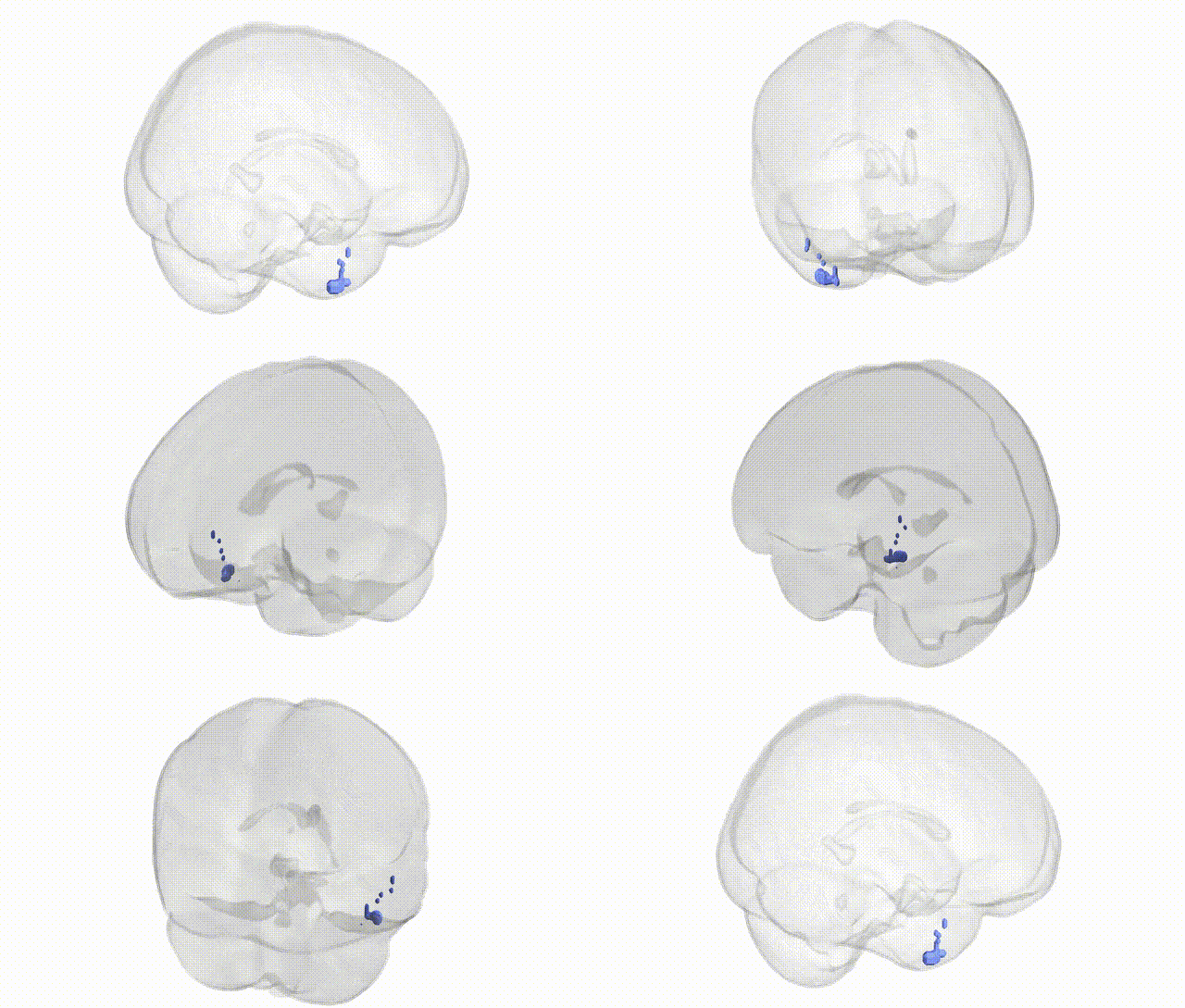
Full Quality Version: Download MP4
Wali Sidiqyar*, Gaurav Rudravaram*, Elyssa M. McMaster, Trent M. Schwartz, Adam M. Saunders, Kurt G. Schilling, Bennett A. Landman "Introducing SPINS: A Shared Public Visualization Library of Neuroanatomical Structures." Medical Imaging with Deep Learning- short paper
This resource is licensed under CC0 1.0 Universal (Public Domain).