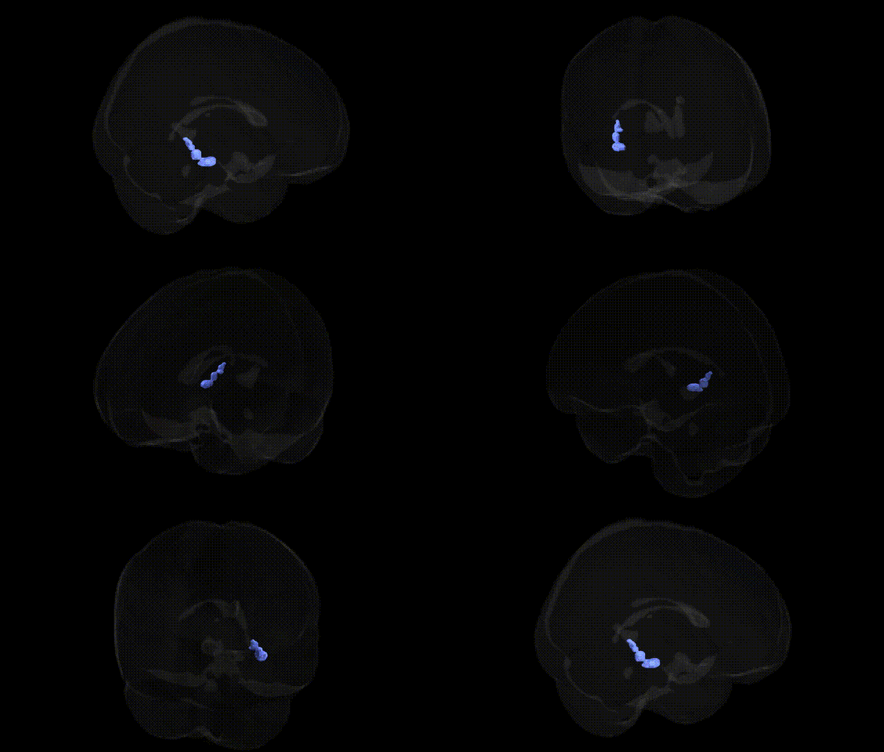
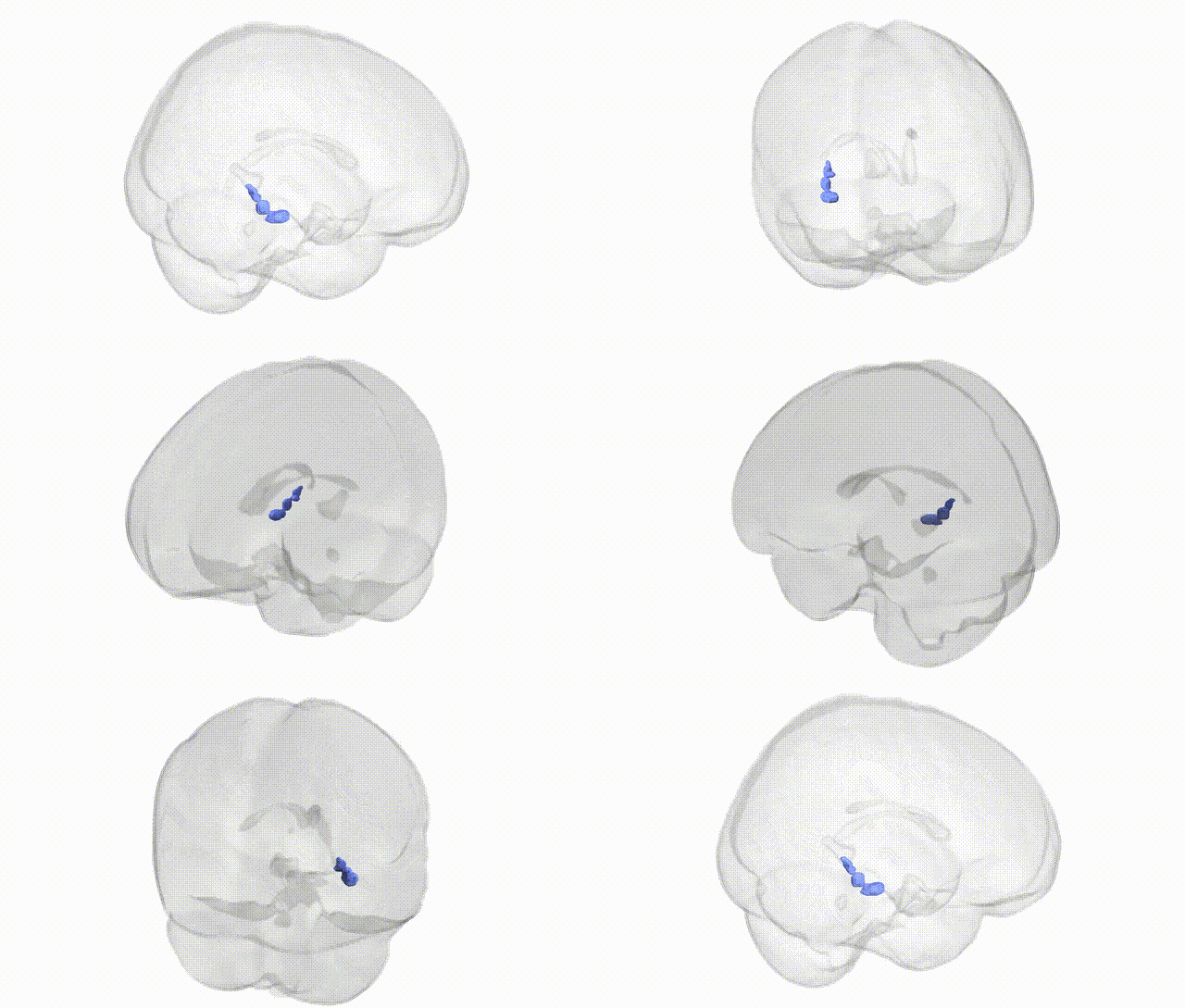
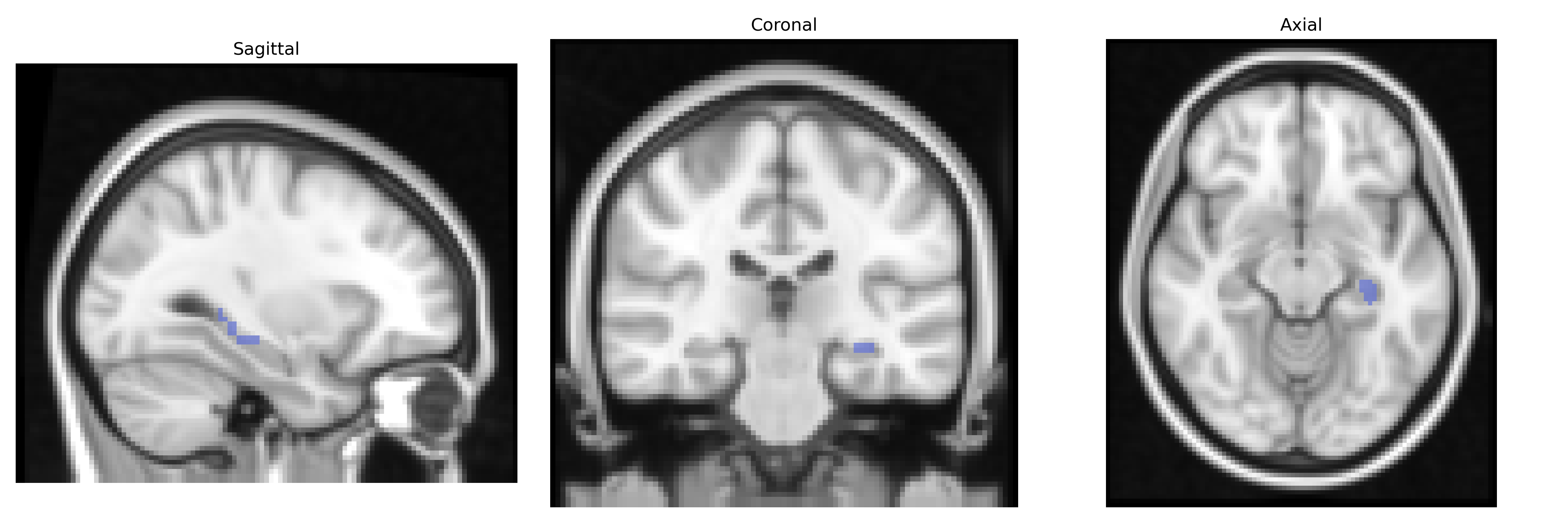
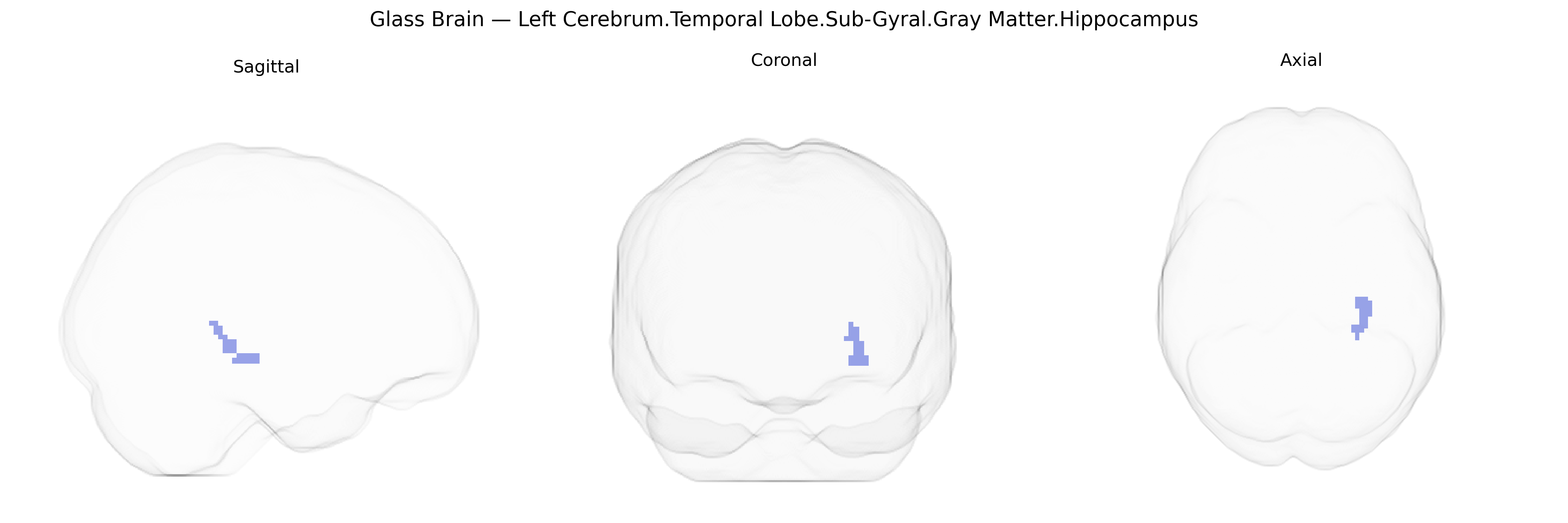
The bilateral Left Cerebrum.Temporal Lobe.Sub-Gyral.Gray Matter.Hippocampus corresponds to the paired hippocampal formations located deep within the medial temporal lobes, beneath the cortical gyri. This archicortical structure is composed primarily of gray matter and includes subfields such as CA1–CA4 and the dentate gyrus, interconnected with the entorhinal cortex and other limbic regions. The hippocampus is crucial for the consolidation of episodic and declarative memories, spatial navigation, and contextual processing, and it plays a key role in neuroplasticity through mechanisms like long-term potentiation. Pathology in this region is strongly associated with temporal lobe epilepsy, Alzheimer’s disease–related memory impairment, and stress-related alterations in emotional and cognitive function. Hippocampus
The bilateral left temporal lobe sub-gyral gray matter encompassing the hippocampus, as defined in the Talairach 2 mm atlas, corresponds largely to hippocampal and adjacent medial temporal structures that have been extensively implicated in genetic studies of cognition, neuropsychiatric disease, and neurodegeneration. GWAS of hippocampal volume and medial temporal gray matter have repeatedly identified variants in genes related to neurodevelopment, synaptic function, and neurodegenerative pathways, including strong associations with APOE (particularly ε4) and loci in/near BIN1, CLU, PICALM, and CR1 in the context of Alzheimer’s disease risk and hippocampal atrophy. Common variants in BDNF, notably Val66Met, have been linked to hippocampal structure and memory performance, while genes such as KIBRA (WWC1), COMT, and GRIN2B have shown associations with episodic memory and temporal lobe activation in imaging-genetics studies. Large-scale GWAS and imaging-genetics consortia (e.g., ENIGMA) have also identified polygenic influences on hippocampal volume involving multiple loci across the genome, some overlapping with schizophrenia and major depressive disorder risk loci, consistent with reduced hippocampal and temporal lobe gray matter volumes observed in these disorders. Additional associations involve stress- and HPA-axis–related genes (e.g., FKBP5, NR3C1) in relation to hippocampal structure and PTSD, as well as genes impacting neurogenesis and plasticity that modulate vulnerability to cognitive decline, dementia, and mood disorders, indicating that this hippocampal-temporal sub-gyral region is a key anatomical substrate through which diverse genetic variants exert effects on memory, emotion regulation, and disease susceptibility.
Overview generated by GPT-4o (2026).
Region ID: 337
Hemisphere: bilateral
Atlas: Talairach labels 2mm
Full Quality Version: Download MP4
Full Quality Version: Download MP4
Wali Sidiqyar*, Gaurav Rudravaram*, Elyssa M. McMaster, Trent M. Schwartz, Adam M. Saunders, Kurt G. Schilling, Bennett A. Landman "Introducing SPINS: A Shared Public Visualization Library of Neuroanatomical Structures." Medical Imaging with Deep Learning- short paper
This resource is licensed under CC0 1.0 Universal (Public Domain).