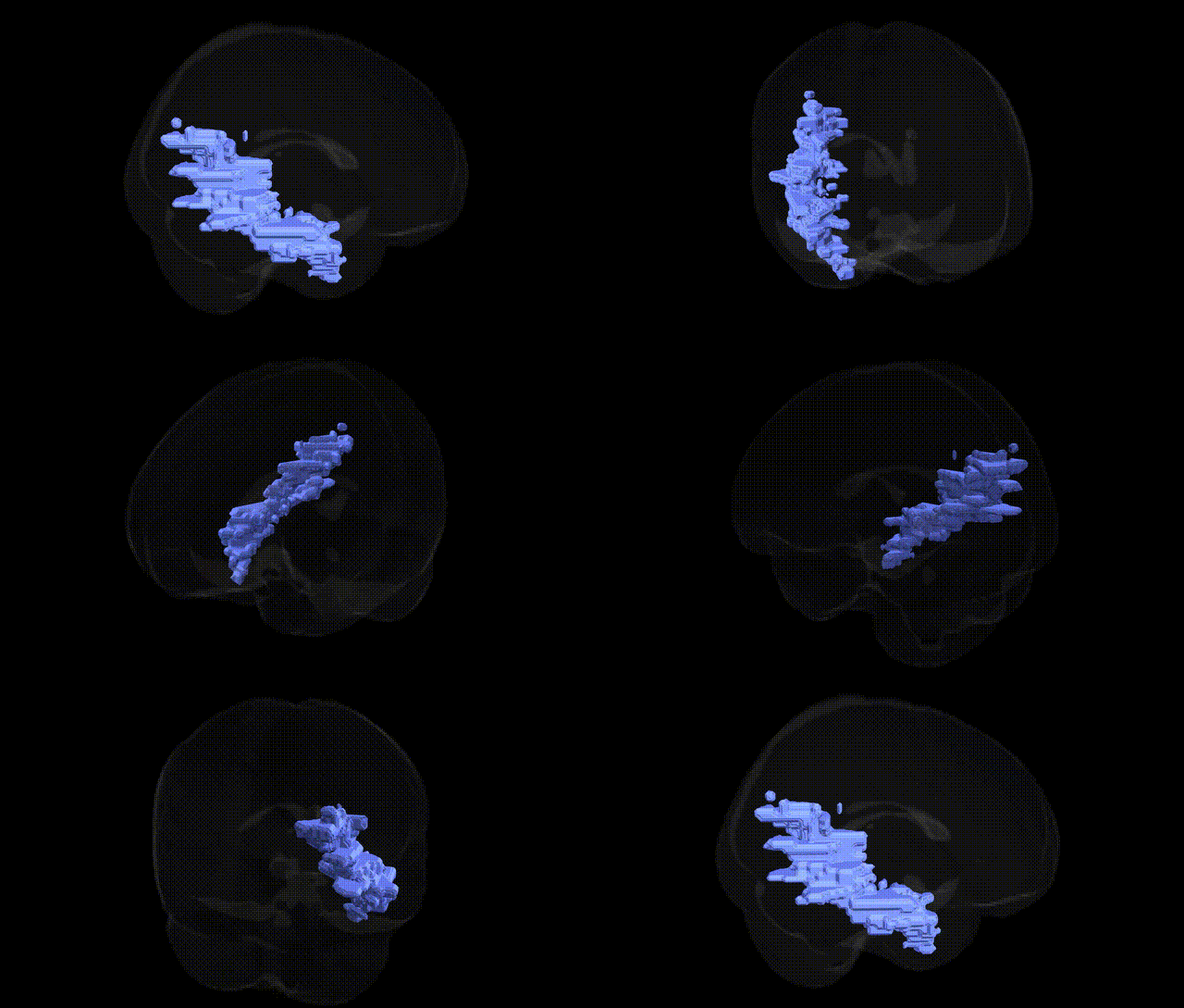
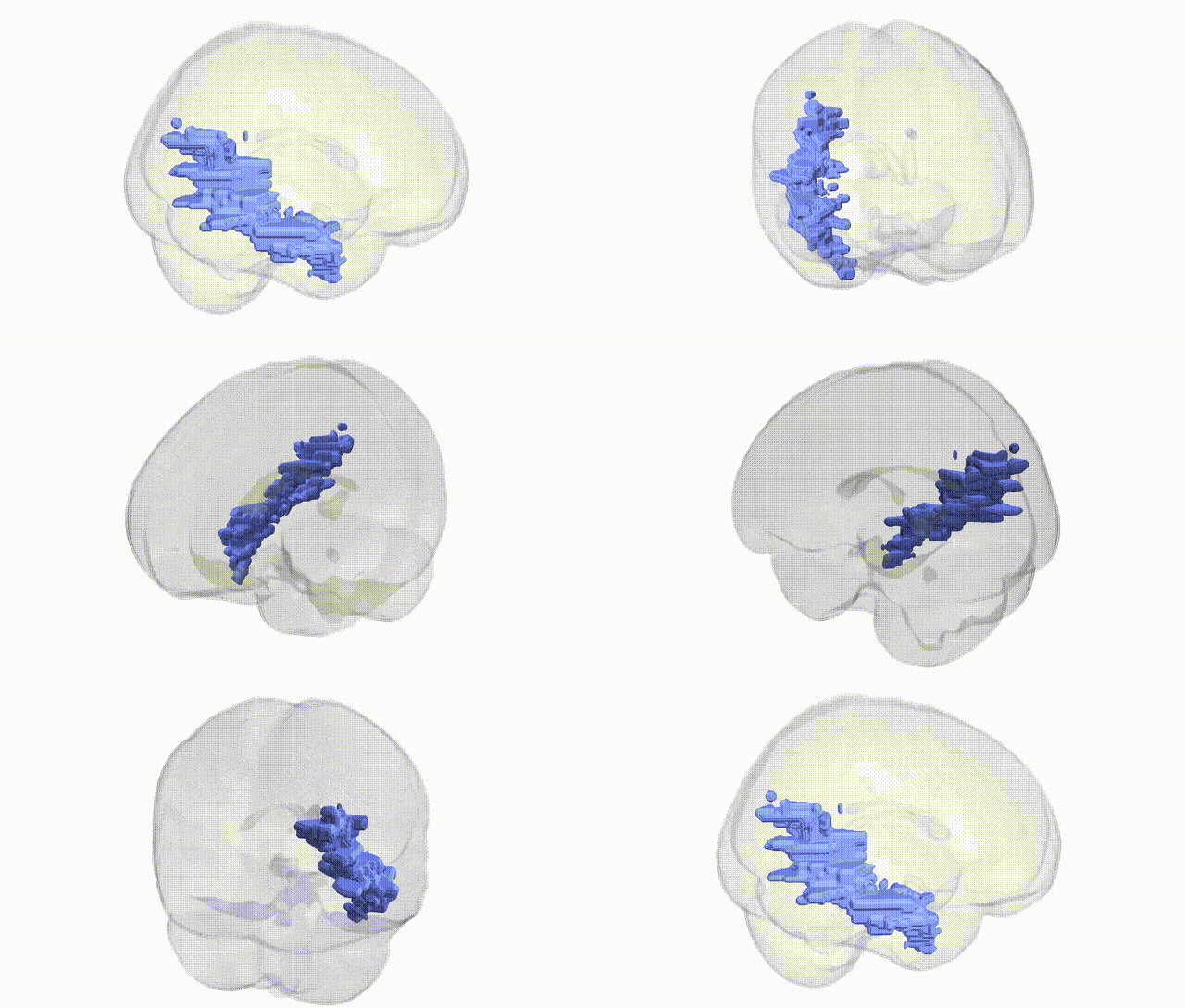
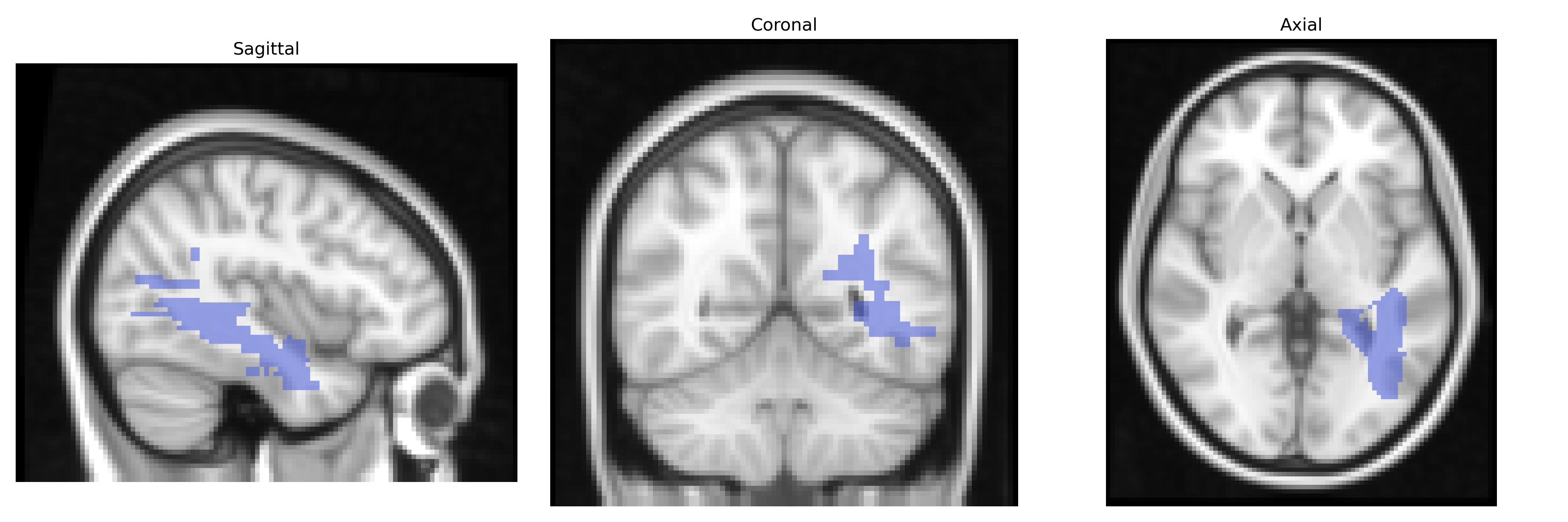
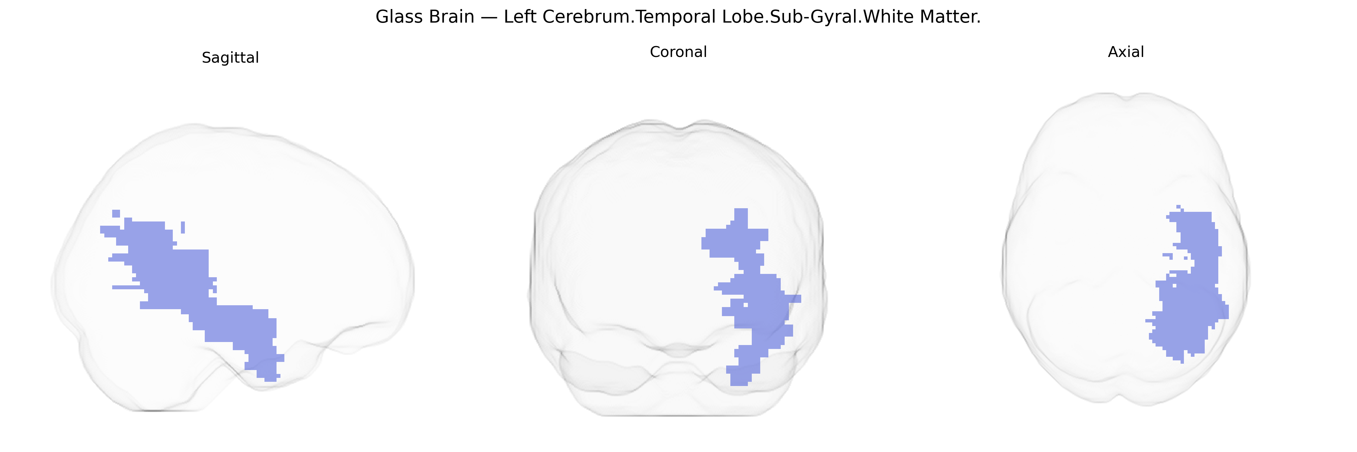
The bilateral Left Cerebrum.Temporal Lobe.Sub-Gyral.White Matter refers to the deep white matter tracts lying beneath the cortical gyri of the left temporal lobe. These sub-gyral fibers consist primarily of myelinated axons that interconnect temporal cortical regions with other parts of the cerebrum, including frontal, parietal, and limbic areas, and contribute to major pathways involved in language processing, auditory perception, semantic memory, and aspects of emotion. Functionally, this white matter supports efficient communication between temporal cortical areas such as the superior, middle, and inferior temporal gyri and subcortical structures, enabling integration of auditory, visual, and mnemonic information. There is no direct Wikipedia article for this specific Talairach label; a closely related structure is the Temporal lobe.
Genetic associations with the bilateral Left Cerebrum.Temporal Lobe.Sub-Gyral.White Matter region, as defined in the Talairach 2 mm atlas, are largely inferred from studies of temporal lobe white matter more generally, rather than this specific micro-anatomical label. Genome-wide association studies (GWAS) of diffusion MRI measures, such as fractional anisotropy and mean diffusivity in temporal lobe white matter tracts (including inferior longitudinal fasciculus and uncinate fasciculus), have implicated variants near genes involved in axonal guidance, myelination, and synaptic function, including loci near CSMD1, CNTNAP2, NRG1, and genes in the ERBB and neurodevelopmental pathways. Temporal lobe white matter integrity has been genetically linked to risk for schizophrenia, bipolar disorder, major depressive disorder, autism spectrum disorder, and epilepsy, with several psychiatric GWAS indicating that polygenic risk scores for these disorders correlate with reduced temporal lobe white matter integrity. Alzheimer’s disease and other dementias show genetic associations (e.g., APOE, CLU, PICALM, BIN1) with temporal lobe white matter changes, reflecting vulnerability of medial and lateral temporal networks. Additionally, GWAS of cognitive traits (general intelligence, memory performance, reading ability, language-related skills) and brain structural measures have identified loci that influence temporal lobe volume and connectivity, suggesting that genetic variation in neurodevelopmental and myelination pathways contributes to individual differences in the microstructure of temporal sub-gyral white matter.
Overview generated by GPT-4o (2026).
Region ID: 51
Hemisphere: bilateral
Atlas: Talairach labels 2mm
Full Quality Version: Download MP4
Full Quality Version: Download MP4
Wali Sidiqyar*, Gaurav Rudravaram*, Elyssa M. McMaster, Trent M. Schwartz, Adam M. Saunders, Kurt G. Schilling, Bennett A. Landman "Introducing SPINS: A Shared Public Visualization Library of Neuroanatomical Structures." Medical Imaging with Deep Learning- short paper
This resource is licensed under CC0 1.0 Universal (Public Domain).