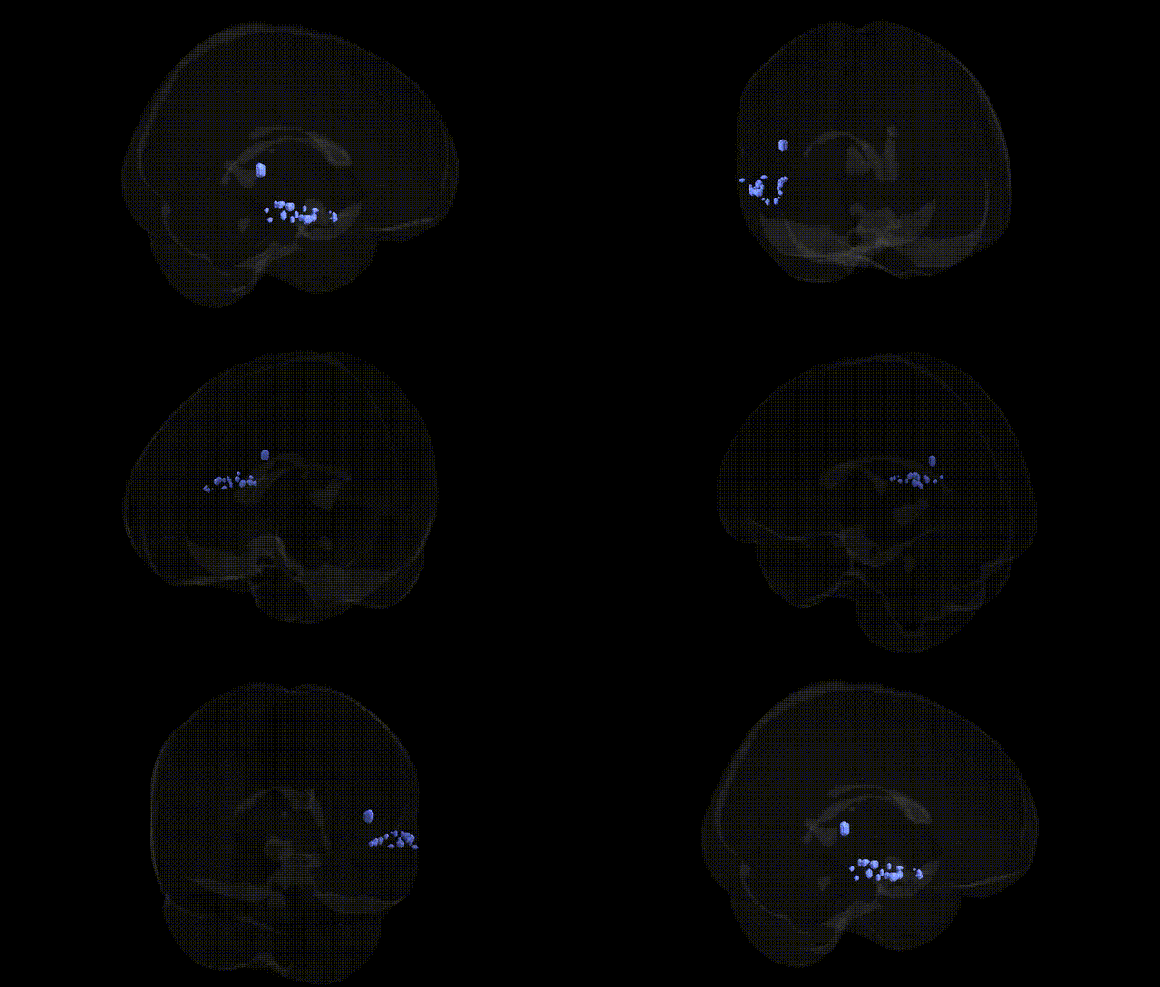
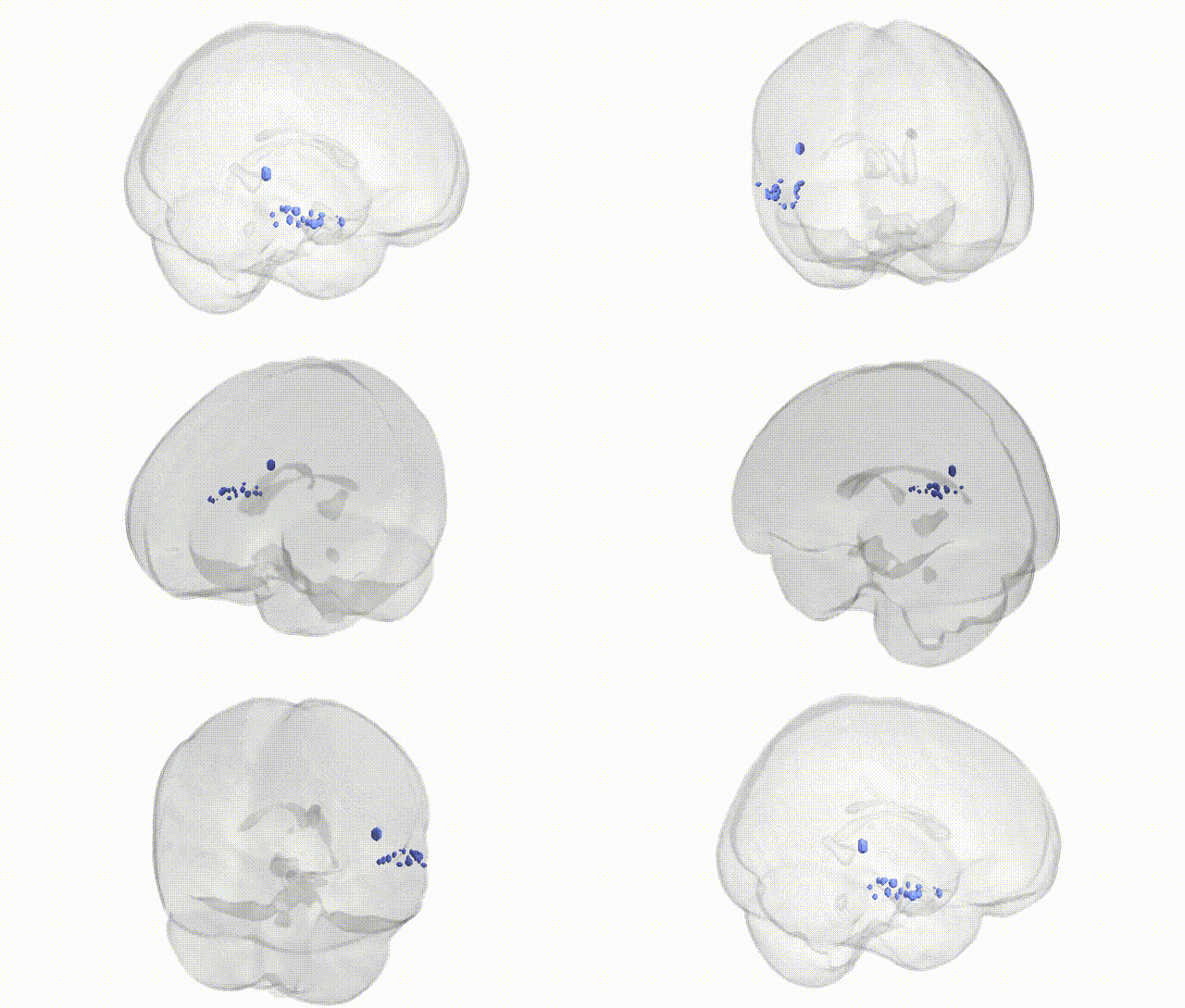
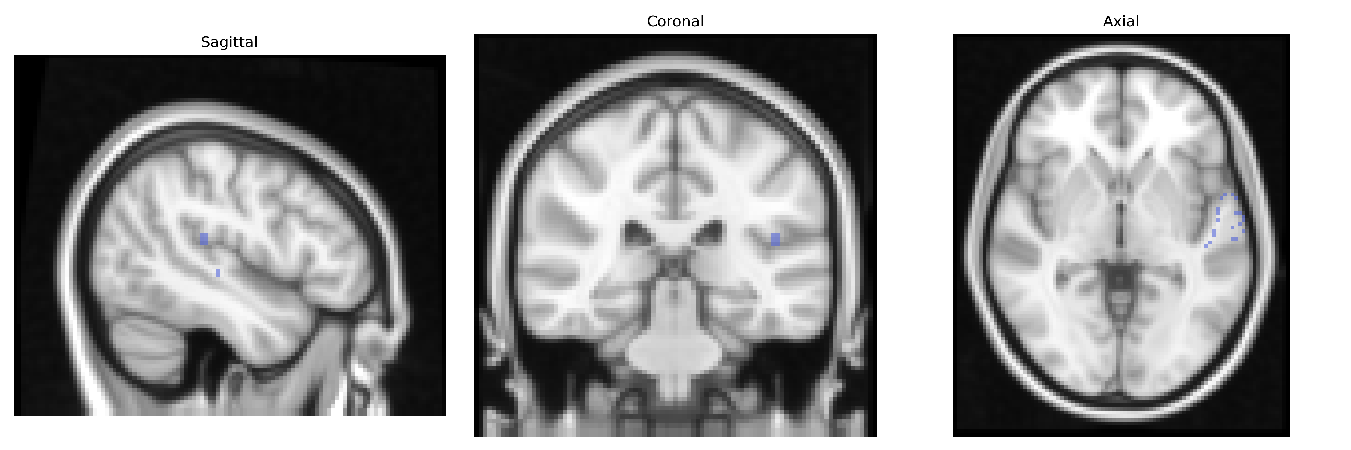
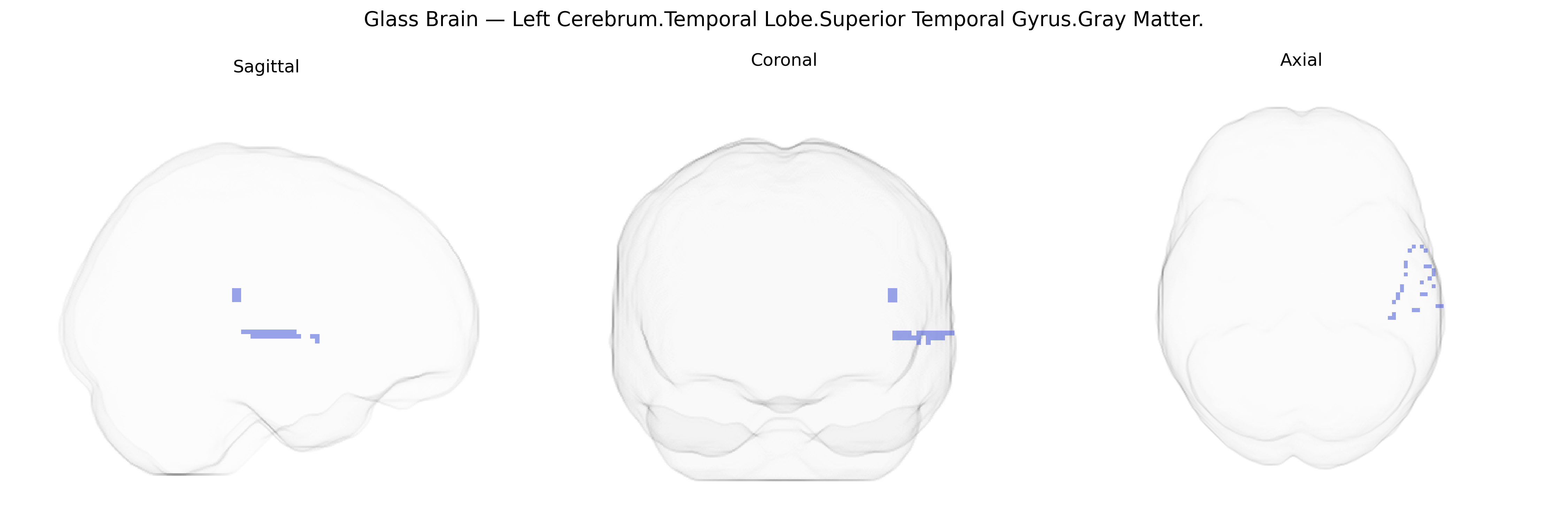
The bilateral Left Cerebrum.Temporal Lobe.Superior Temporal Gyrus.Gray Matter corresponds to cortical tissue in the superior temporal gyrus of the left temporal lobe, a key region for auditory processing and language-related functions. This gray matter zone encompasses primary and secondary auditory areas that decode sound features such as pitch and intensity, and contributes critically to speech perception and phonological analysis, often overlapping with or bordering Wernicke’s area in the posterior portion. It participates in integration of auditory input with semantic and syntactic processing, supports aspects of social cognition through analysis of vocal prosody and communicative signals, and interfaces with adjacent temporal and parietal regions for multimodal sensory integration. There is no direct Wikipedia article for this exact Talairach label; see the related structure Superior temporal gyrus.
Genetic associations involving the bilateral Left Cerebrum.Temporal Lobe.Superior Temporal Gyrus.Gray Matter (as defined in Talairach 2 mm atlases) primarily emerge from imaging genetics and GWAS of cortical thickness, surface area, and functional activation in language and auditory regions. Large-scale neuroimaging GWAS (e.g., ENIGMA, UK Biobank) have identified variants near genes such as DCDC2, KIAA0319, CNTNAP2, FOXP2, and ROBO1 that influence superior temporal gyrus morphology or activation and are implicated in language processing and reading abilities, including dyslexia and specific language impairment. FOXP2 and CNTNAP2 variants have been linked to structural and functional alterations in left superior temporal areas underlying speech perception and production. GWAS of schizophrenia, bipolar disorder, and autism spectrum disorder have associated polygenic risk scores and specific loci (e.g., in CACNA1C, GRIN2A, complement component genes such as C4) with gray matter reductions and altered connectivity in superior temporal regions, often in relation to auditory hallucinations and social communication deficits. Imaging genetics work on Alzheimer’s disease risk genes (notably APOE ε4) shows associations with atrophy and functional changes in temporal lobe structures including superior temporal gyrus, reflecting vulnerability of language and semantic networks. Additional studies link common variation in genes affecting synaptic plasticity and glutamatergic transmission (e.g., BDNF Val66Met, GRM3, NRGN) to superior temporal gyrus measures and to traits such as verbal memory, musical aptitude, and susceptibility to psychosis-related auditory perceptual anomalies, highlighting this region as a convergence point for genetic influences on language, auditory processing, and higher-order social cognition.
Overview generated by GPT-4o (2026).
Region ID: 532
Hemisphere: bilateral
Atlas: Talairach labels 2mm
Full Quality Version: Download MP4
Full Quality Version: Download MP4
Wali Sidiqyar*, Gaurav Rudravaram*, Elyssa M. McMaster, Trent M. Schwartz, Adam M. Saunders, Kurt G. Schilling, Bennett A. Landman "Introducing SPINS: A Shared Public Visualization Library of Neuroanatomical Structures." Medical Imaging with Deep Learning- short paper
This resource is licensed under CC0 1.0 Universal (Public Domain).