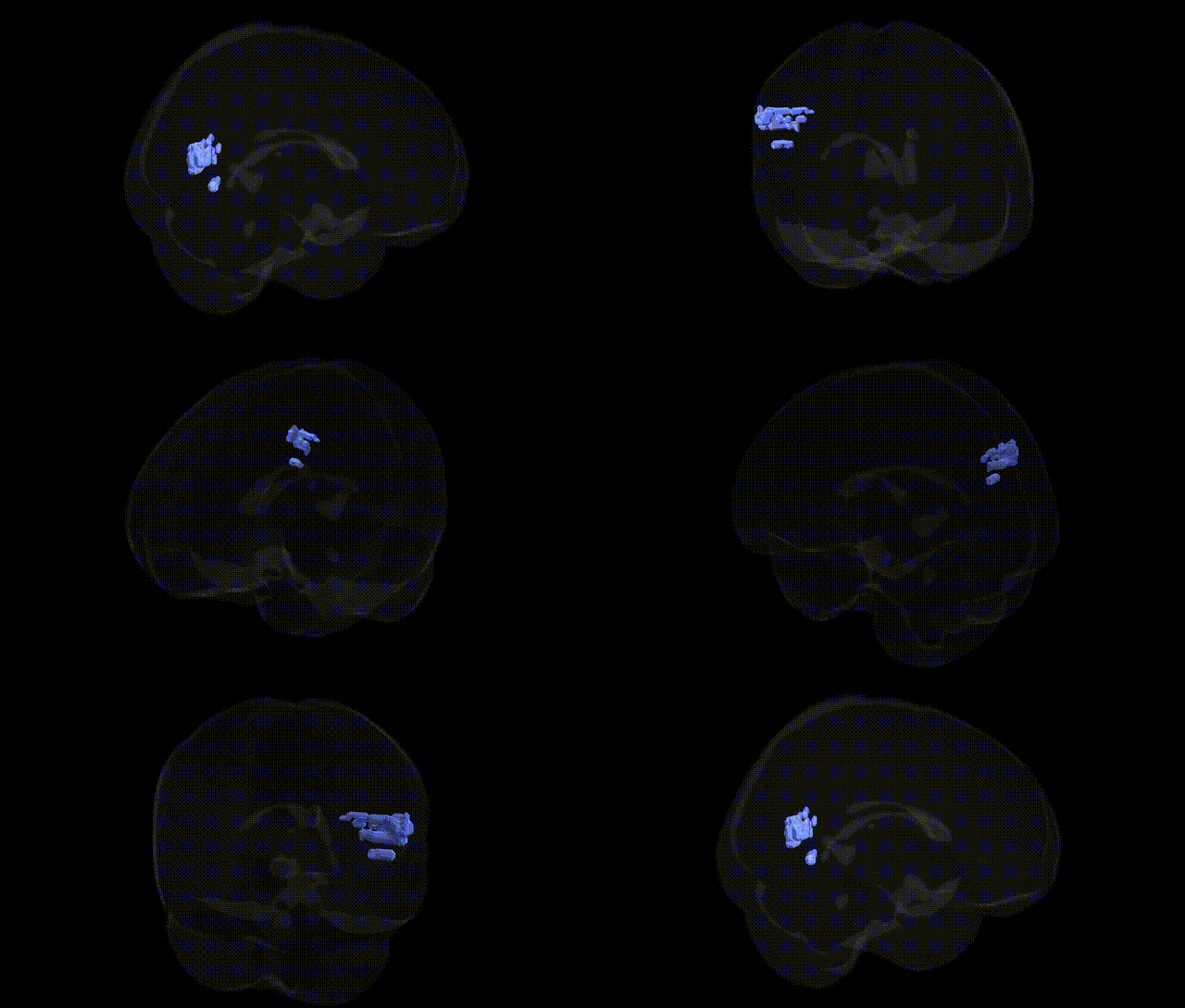
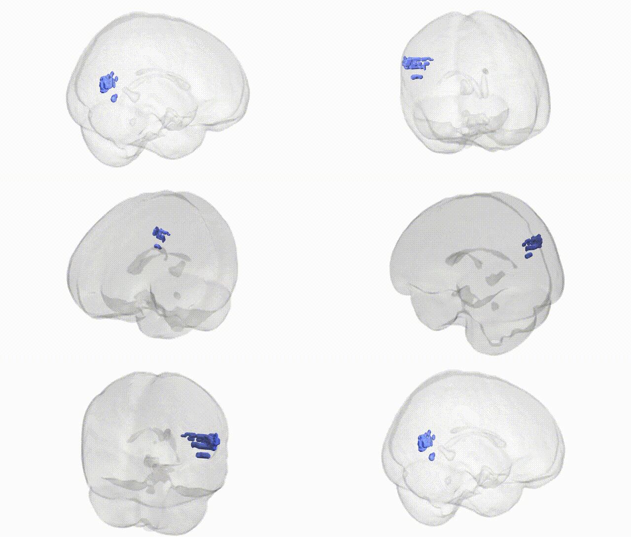
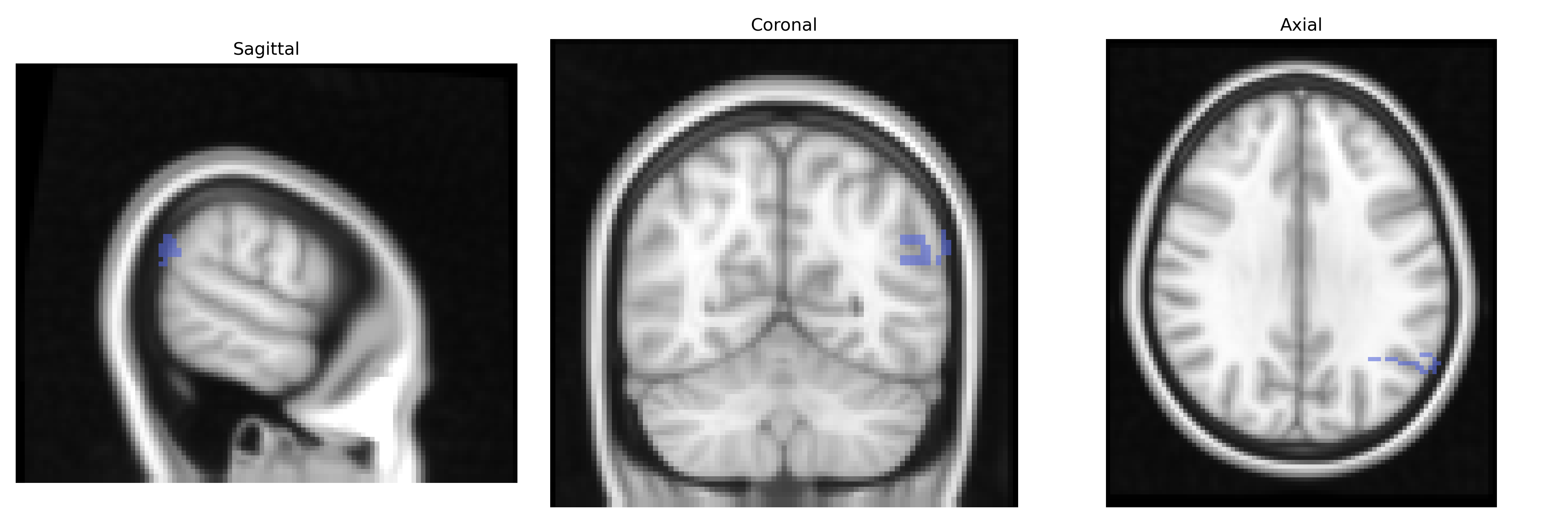
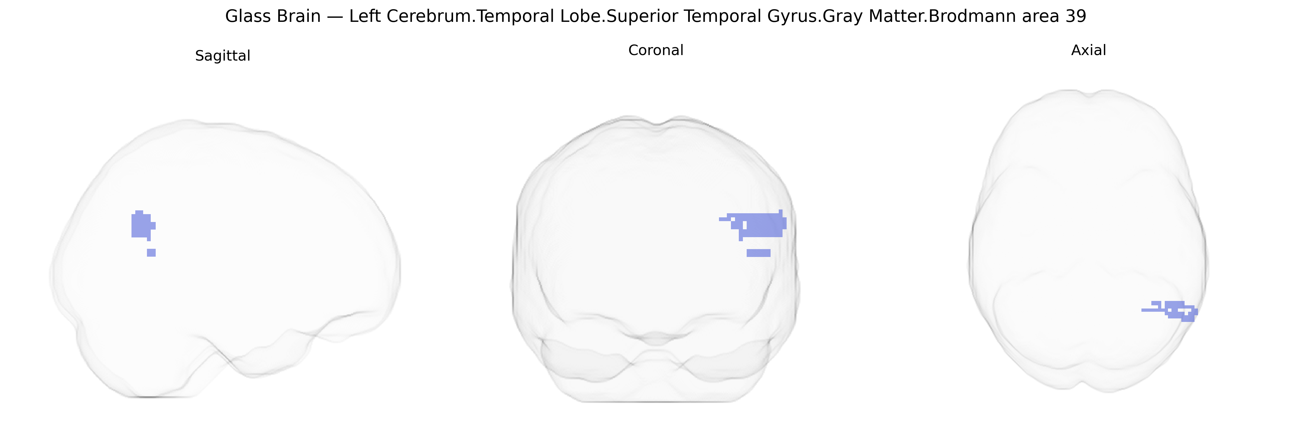
The bilateral Left Cerebrum.Temporal Lobe.Superior Temporal Gyrus.Gray Matter.Brodmann area 39 corresponds primarily to the left angular gyrus region, situated at the junction of the temporal, parietal, and occipital lobes, though the Talairach labeling here emphasizes its extension into the superior temporal gyrus. This heteromodal association cortex integrates auditory, visual, and somatosensory information and is critically involved in language-related processes such as reading, semantic processing, and aspects of comprehension, as well as number processing, spatial cognition, and theory of mind. Functionally, left BA39 is a key component of the dominant-hemisphere language network, contributes to the default mode and semantic networks, and participates in complex conceptual and symbolic operations; damage in this area can be associated with alexia, agraphia, acalculia, and certain aphasic syndromes.
Angular gyrus (Brodmann area 39)
The bilateral left superior temporal gyrus (STG) gray matter in Brodmann area 39, as defined in the Talairach labels 2 mm Atlas, overlaps substantially with the angular gyrus and temporo‑parietal association cortex and has been repeatedly implicated in genetic studies of language, reading, social cognition, and psychiatric risk. GWAS and imaging‑genetics work show that polygenic scores for schizophrenia, bipolar disorder, and major depression are associated with altered cortical thickness and surface area in the STG/BA39 region, with risk variants in genes such as CACNA1C, ZNF804A, GRIN2A, and common complement component 4 (C4) haplotypes contributing to volume and connectivity differences in temporal association cortex. Multiple dyslexia and reading-disorder loci (e.g., KIAA0319, DCDC2, ROBO1) have been linked to structural and functional variation in posterior temporal and temporo‑parietal regions encompassing BA39, consistent with the region’s role in phonological and orthographic processing. FOXP2 and CNTNAP2 variants, associated with speech and language impairment, have also been tied to altered activation and morphology in left temporal language circuits including the STG. In autism spectrum conditions, GWAS-based polygenic risk and variants in synaptic and neuronal adhesion genes (e.g., NRXN1, SHANK3, CNTNAP2) relate to atypical development and connectivity of the superior temporal and temporo‑parietal junction, supporting its role in social perception and theory of mind. Large-scale GWAS of cortical morphology (e.g., ENIGMA, UK Biobank) indicate that BA39/STG cortical thickness and surface area are highly polygenic, with numerous common variants of small effect distributed across the genome, and are genetically correlated with cognitive ability, educational attainment, and neuropsychiatric liability, underscoring this region as a key genetically modulated hub in language and higher-order association networks.
Overview generated by GPT-4o (2026).
Region ID: 708
Hemisphere: bilateral
Atlas: Talairach labels 2mm
Full Quality Version: Download MP4
Full Quality Version: Download MP4
Wali Sidiqyar*, Gaurav Rudravaram*, Elyssa M. McMaster, Trent M. Schwartz, Adam M. Saunders, Kurt G. Schilling, Bennett A. Landman "Introducing SPINS: A Shared Public Visualization Library of Neuroanatomical Structures." Medical Imaging with Deep Learning- short paper
This resource is licensed under CC0 1.0 Universal (Public Domain).