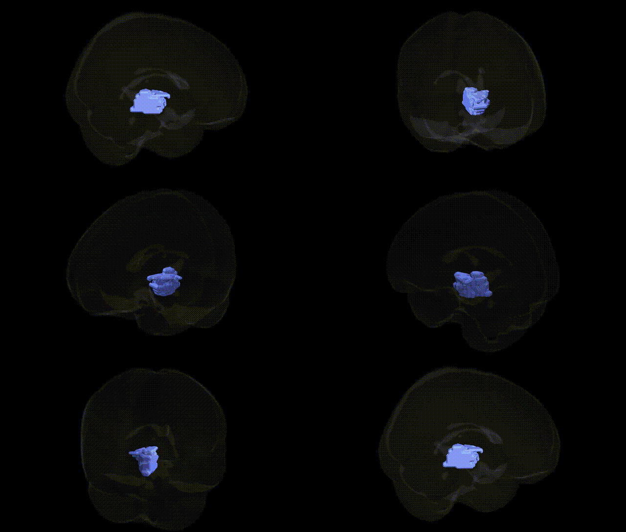
The bilateral right brainstem midbrain region, as identified in the Talairach 2 mm Atlas, corresponds to the dorsal portion of the upper brainstem located rostral to the pons and caudal to the diencephalon, comprising structures such as the tectum (including the superior and inferior colliculi), tegmentum, and parts of the cerebral peduncles. Functionally, the midbrain plays key roles in visual and auditory reflex pathways, motor control, modulation of pain, arousal, and autonomic regulation, integrating ascending sensory and descending motor signals between the spinal cord, brainstem, and forebrain. Although there is no direct Wikipedia article specifically titled “Right Brainstem Midbrain,” this region is encompassed within the broader midbrain structure: Midbrain.
The bilateral right brainstem midbrain region, as defined in the Talairach 2 mm Atlas, has been implicated indirectly in genetic studies through its roles in dopaminergic, serotonergic, and motor pathways rather than via region-specific GWAS hits. Genetic variants in dopaminergic system genes (e.g., DRD2, DRD3, COMT) and serotonergic genes (e.g., SLC6A4, HTR1A, HTR2A) influence midbrain circuits involving the substantia nigra and ventral tegmental area, contributing to risk for Parkinson’s disease, schizophrenia, substance use disorders, and mood disorders. GWAS of Parkinson’s disease highlight loci such as SNCA, LRRK2, GBA, and MAPT, which affect survival and function of midbrain dopaminergic neurons, while imaging genetics studies link COMT and DRD2 polymorphisms to variation in midbrain activation during reward and cognitive tasks. Structural and functional MRI GWAS have associated polygenic risk for schizophrenia, ADHD, and major depression with alterations in brainstem and midbrain volumes or connectivity, although these findings are typically reported at broader brainstem or basal ganglia levels rather than as precise Talairach midbrain coordinates. Overall, genetic associations involving this region are mediated through neurotransmitter-related genes and neurodegeneration or neuropsychiatric risk loci that influence midbrain circuitry, rather than through GWAS findings uniquely localized to the bilateral right midbrain in the Talairach atlas.
Overview generated by GPT-4o (2026).
Region ID: 216
Hemisphere: bilateral
Atlas: Talairach labels 2mm
Full Quality Version: Download MP4
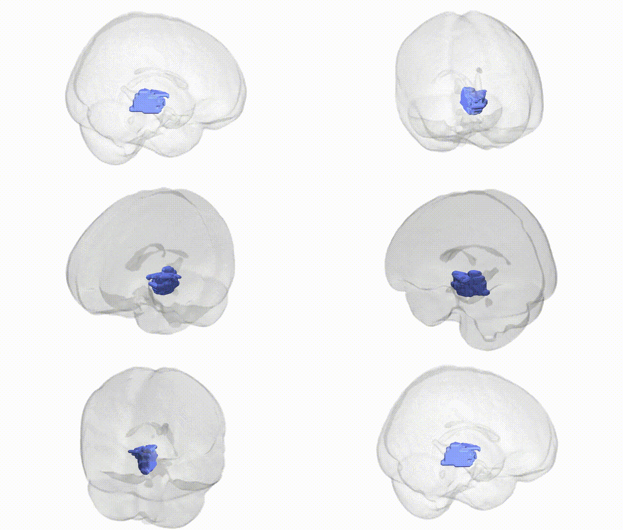
Full Quality Version: Download MP4
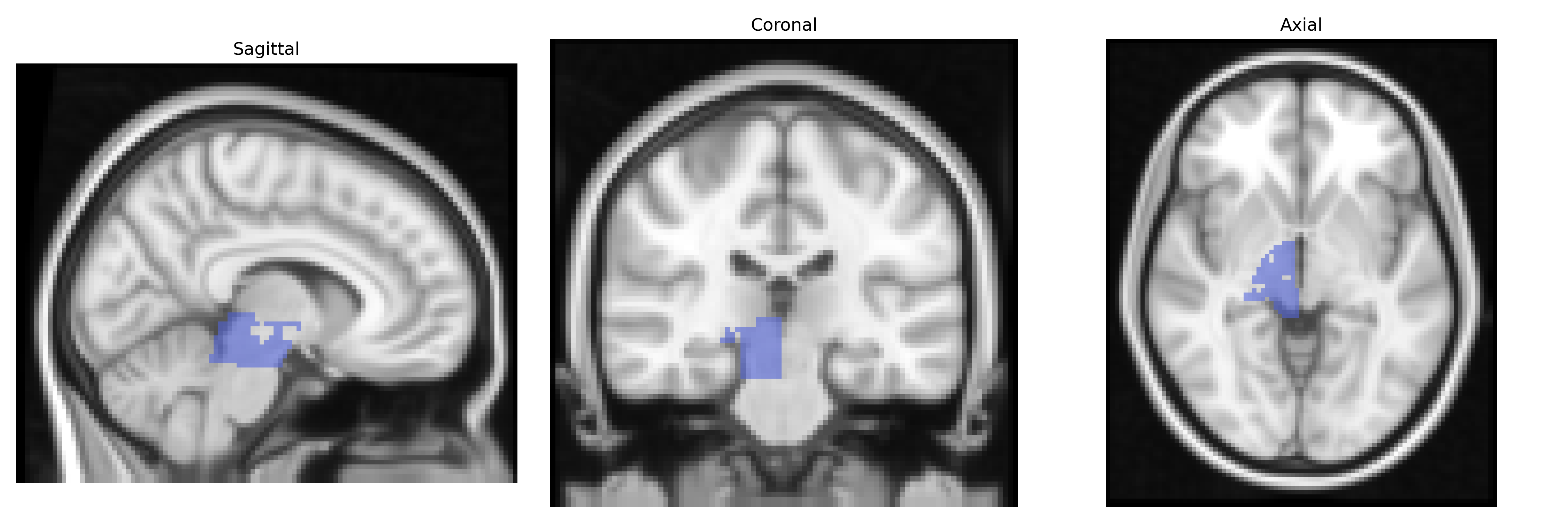
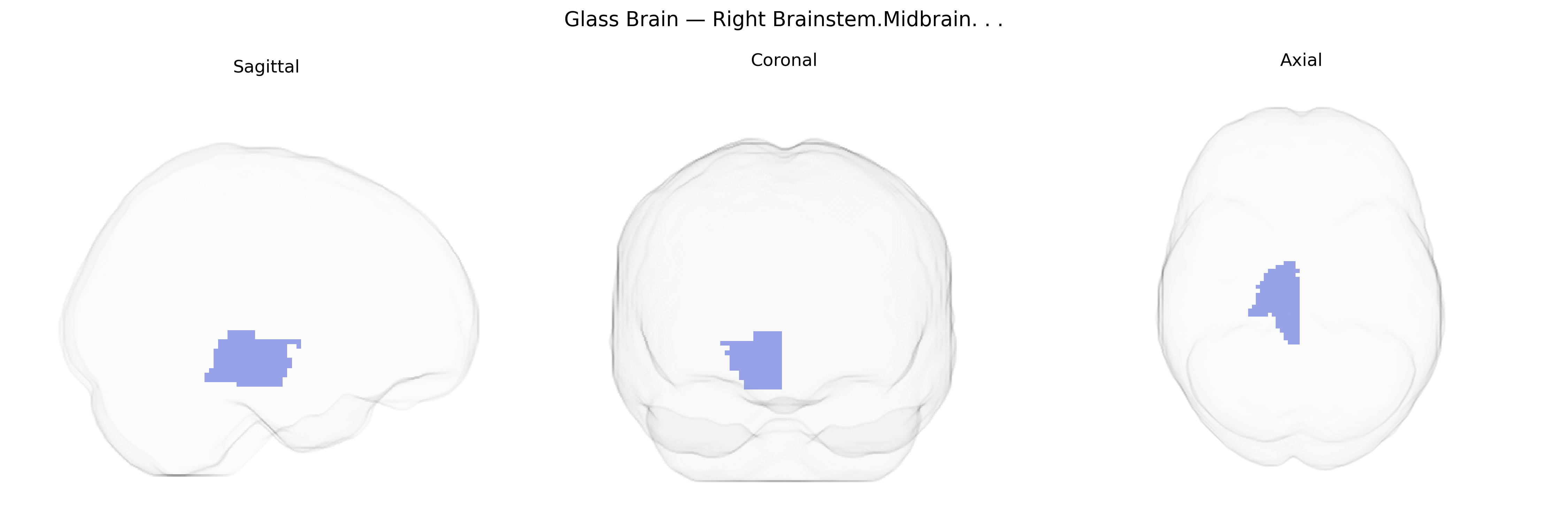
Wali Sidiqyar*, Gaurav Rudravaram*, Elyssa M. McMaster, Trent M. Schwartz, Adam M. Saunders, Kurt G. Schilling, Bennett A. Landman "Introducing SPINS: A Shared Public Visualization Library of Neuroanatomical Structures." Medical Imaging with Deep Learning- short paper
This resource is licensed under CC0 1.0 Universal (Public Domain).