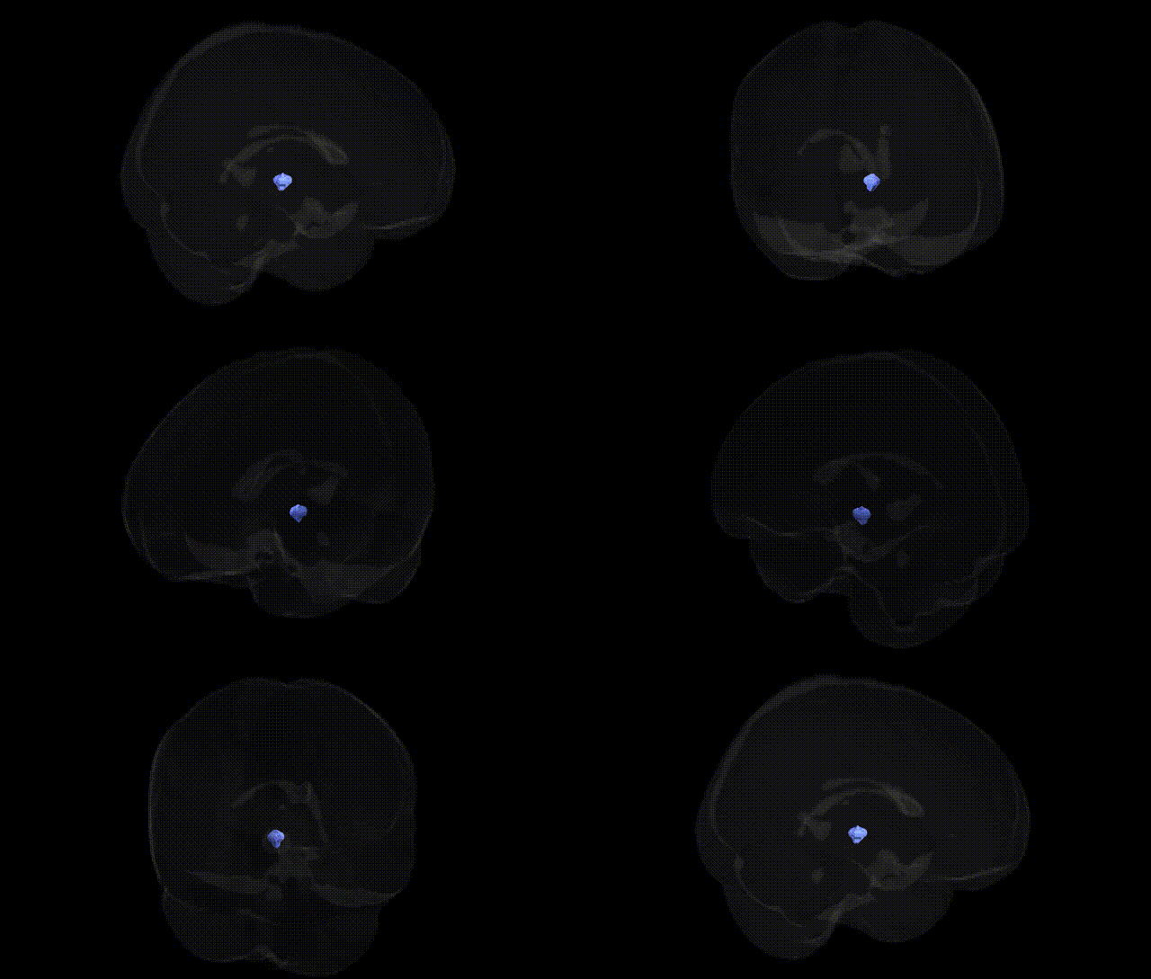
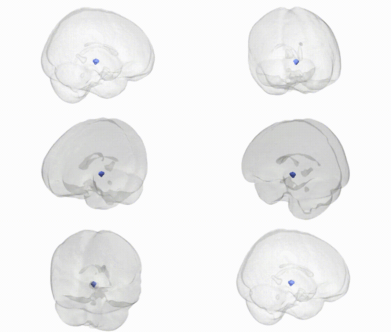
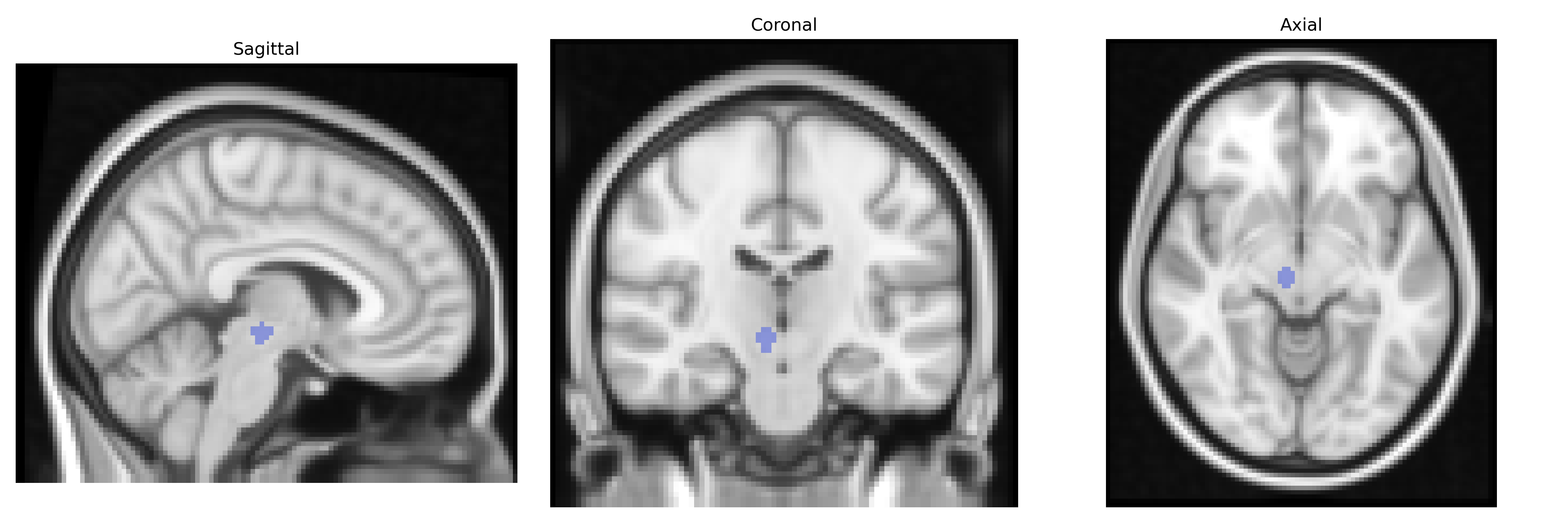
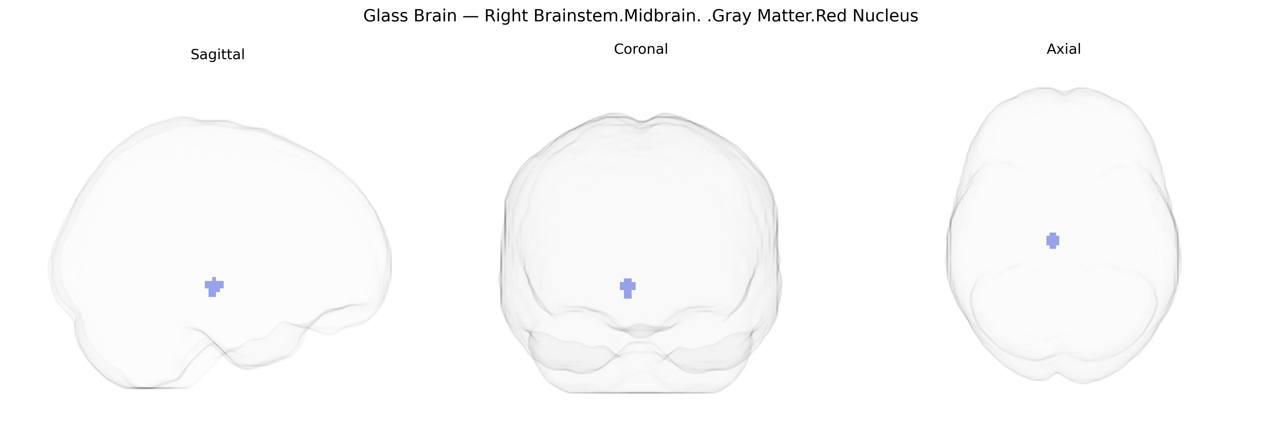
The bilateral Right Brainstem.Midbrain.Gray Matter.Red Nucleus corresponds to the paired red nuclei located in the rostral midbrain tegmentum, forming a key relay in motor coordination pathways. Composed of magnocellular and parvocellular neuronal populations, this gray matter structure receives major afferents from the contralateral cerebellar interposed nuclei and the ipsilateral motor cortex, and sends efferent projections via the rubrospinal tract to the spinal cord and via rubro-olivary and rubro-thalamic pathways to the inferior olive and motor-related thalamic nuclei. Functionally, the red nucleus contributes to limb movement control, motor learning, and coordination, particularly influencing flexor musculature and fine-tuning cerebellar output, with additional roles in sensorimotor integration and some aspects of locomotor control. In humans, its descending motor role is reduced compared with other mammals, but it remains an important component of the extrapyramidal motor system and a landmark in midbrain imaging and stereotactic atlases. Red nucleus
The red nucleus, a midbrain gray matter structure involved in motor coordination, has been implicated in several genetically influenced disorders and traits, although direct GWAS hits specifically localized to the bilateral right red nucleus in the Talairach 2 mm atlas are rare. Neuroimaging genetics studies using voxel-based morphometry and diffusion measures have shown that common variants affecting motor and cerebellar pathways—for example in genes such as CACNA1A, ATXN1/2/3, and other spinocerebellar ataxia loci—can alter midbrain and brainstem structures including the red nucleus in ataxias and tremor disorders. In Parkinson’s disease and essential tremor, loci such as LRRK2, SNCA, GBA, and ET-associated variants (e.g., in LINGO1 and STK32B) are linked to structural and functional changes in the cerebello–thalamo–red nucleus–cortical motor network, though most imaging focuses on substantia nigra and cerebellum rather than the red nucleus specifically. GWAS of motor performance, gait, and coordination, as well as deep brain stimulation outcome studies, indirectly implicate the red nucleus as part of genetically modulated motor circuits, with polygenic risk scores for movement disorders sometimes correlating with midbrain volume or signal changes. In multiple sclerosis and other neuroinflammatory conditions, risk variants in HLA-DRB1 and other immune-related genes have been associated with lesion burden and atrophy in brainstem regions that encompass the red nucleus. Overall, genetic associations typically involve broad motor and brainstem networks rather than isolated red nucleus loci, and evidence for trait- or disorder-linked variation maps onto this region via network-level imaging-genetic analyses rather than region-specific GWAS signals.
Overview generated by GPT-4o (2026).
Region ID: 344
Hemisphere: bilateral
Atlas: Talairach labels 2mm
Full Quality Version: Download MP4
Full Quality Version: Download MP4
Wali Sidiqyar*, Gaurav Rudravaram*, Elyssa M. McMaster, Trent M. Schwartz, Adam M. Saunders, Kurt G. Schilling, Bennett A. Landman "Introducing SPINS: A Shared Public Visualization Library of Neuroanatomical Structures." Medical Imaging with Deep Learning- short paper
This resource is licensed under CC0 1.0 Universal (Public Domain).