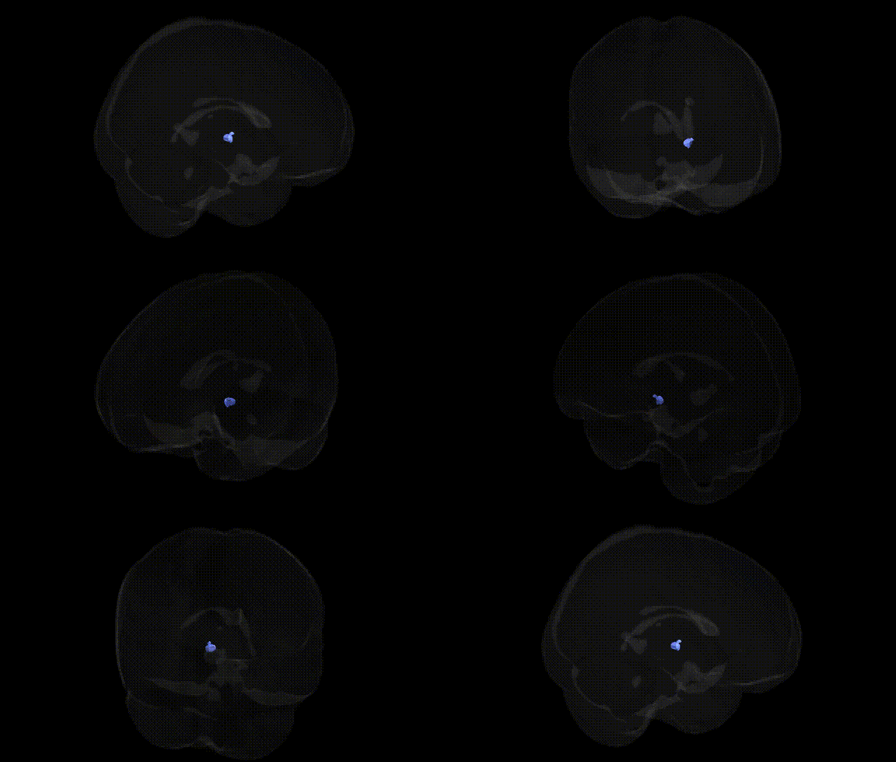
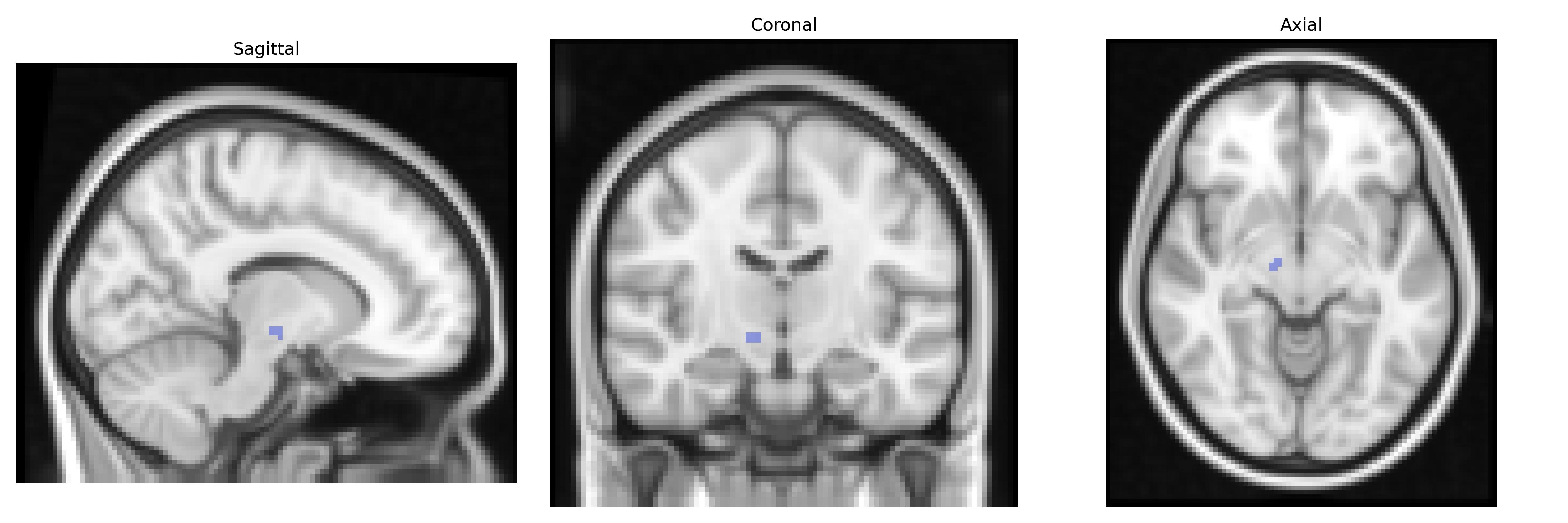
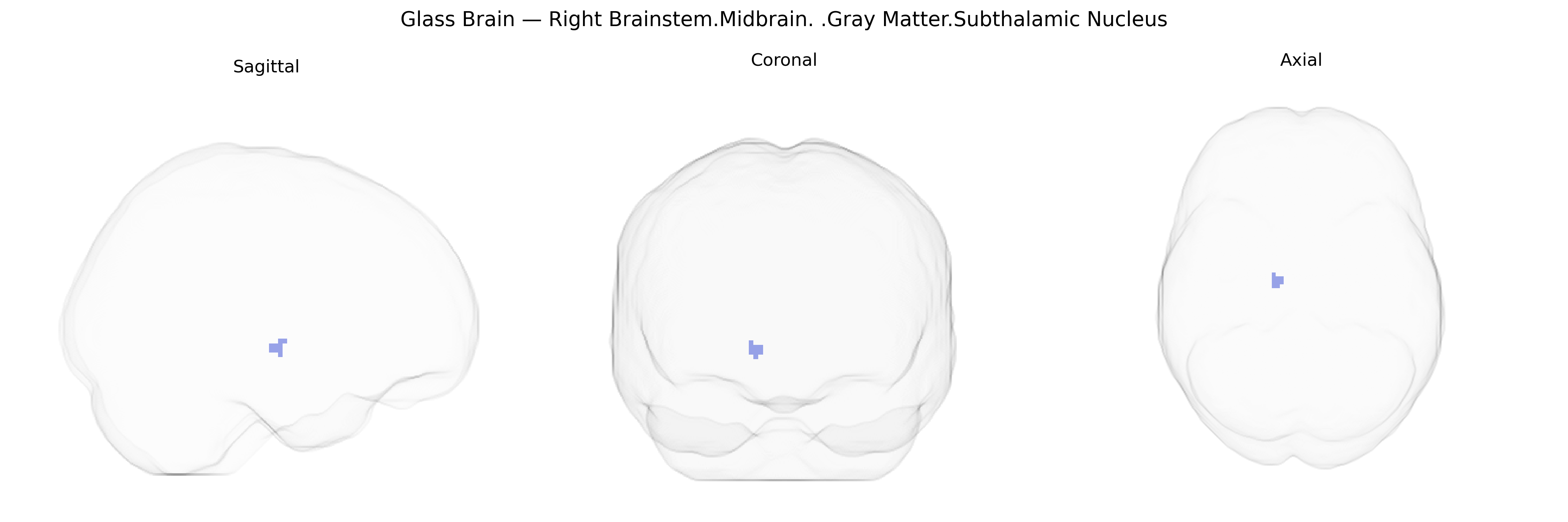
The bilateral Right Brainstem.Midbrain.Gray Matter.Subthalamic Nucleus, as referenced in the Talairach 2 mm Atlas, corresponds to the subthalamic nucleus located within the subthalamus of the ventral diencephalon, adjacent to the midbrain. This small, lens-shaped gray matter structure lies inferior to the thalamus and medial to the internal capsule, and is a key component of the basal ganglia circuitry. It receives glutamatergic input from the cerebral cortex (via the hyperdirect pathway) and the external segment of the globus pallidus, and sends excitatory projections to the internal segment of the globus pallidus and the substantia nigra pars reticulata, thereby modulating motor output. Functionally, it is critically involved in the regulation of movement, motor inhibition, and aspects of cognitive and limbic processing; pathological changes or lesions in this nucleus are associated with movement disorders such as hemiballismus and Parkinson’s disease, and it is a standard target for deep brain stimulation in advanced Parkinsonian therapy.
Subthalamic nucleus
The subthalamic nucleus, a key basal ganglia structure within the midbrain region, is strongly implicated in genetically influenced motor and neuropsychiatric disorders rather than in large-scale GWAS of common traits, which rarely resolve specifically to this nucleus due to imaging resolution limits. Monogenic forms of Parkinson’s disease (e.g., mutations in SNCA, LRRK2, PARK2, PINK1, DJ-1, GBA) and risk variants identified by PD GWAS (such as MAPT and multiple loci near immune and lysosomal genes) converge on basal ganglia circuitry for which the subthalamic nucleus is a critical relay, and deep brain stimulation of this nucleus is guided in part by these genetically defined PD subtypes. Variants in genes affecting glutamatergic and GABAergic transmission (e.g., GRIN2A, GAD1) and dopaminergic pathways (e.g., DRD2, COMT) have been linked via neuroimaging genetics and circuit models to altered activity in cortico–subthalamic–pallidal loops relevant to impulse control, obsessive-compulsive disorder, and addiction. Neurodevelopmental and neuropsychiatric GWAS (schizophrenia, ADHD, OCD) often implicate genes regulating synaptic plasticity and basal ganglia development (e.g., CACNA1C, DCC, NRXN1), which influence functional connectivity and morphology of the broader subthalamic circuitry, though specific gray matter volume or functional activation associations localized precisely to “bilateral Right Brainstem.Midbrain.Gray Matter.Subthalamic Nucleus” in the Talairach 2 mm atlas are not commonly reported. Overall, genetic findings point to the subthalamic nucleus as a crucial node through which risk variants for movement disorders and compulsive/impulsive traits exert circuit-level effects, even when the nucleus is not the primary locus of structural GWAS hits.
Overview generated by GPT-4o (2026).
Region ID: 454
Hemisphere: bilateral
Atlas: Talairach labels 2mm
Full Quality Version: Download MP4
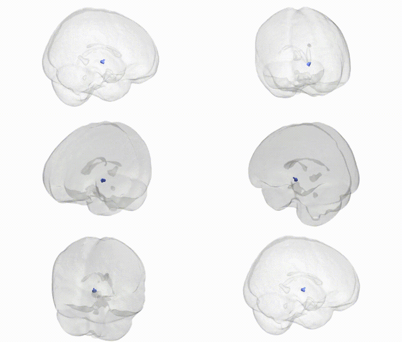
Full Quality Version: Download MP4
Wali Sidiqyar*, Gaurav Rudravaram*, Elyssa M. McMaster, Trent M. Schwartz, Adam M. Saunders, Kurt G. Schilling, Bennett A. Landman "Introducing SPINS: A Shared Public Visualization Library of Neuroanatomical Structures." Medical Imaging with Deep Learning- short paper
This resource is licensed under CC0 1.0 Universal (Public Domain).