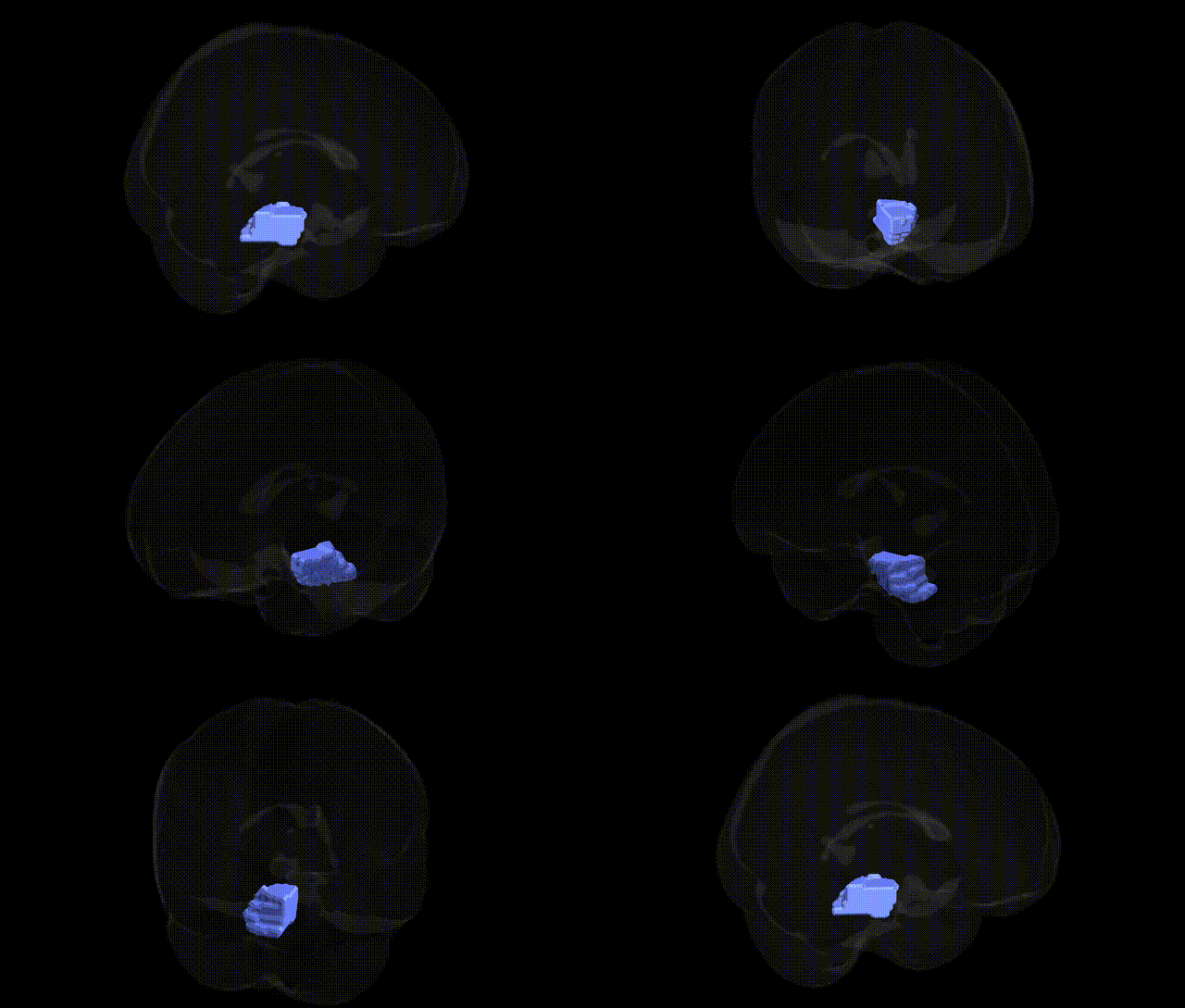
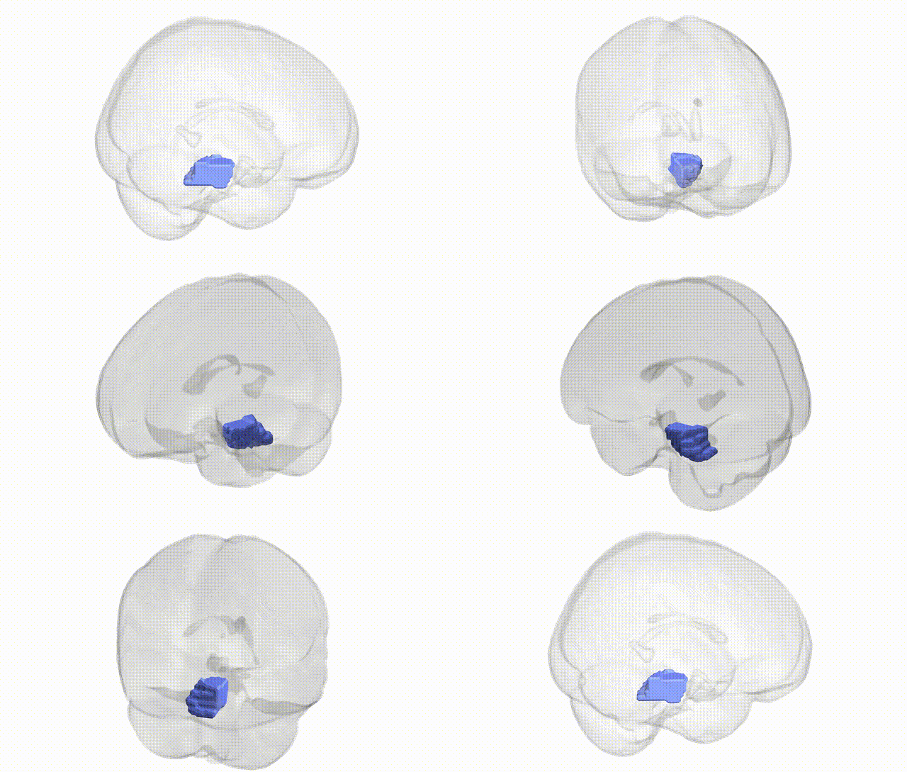
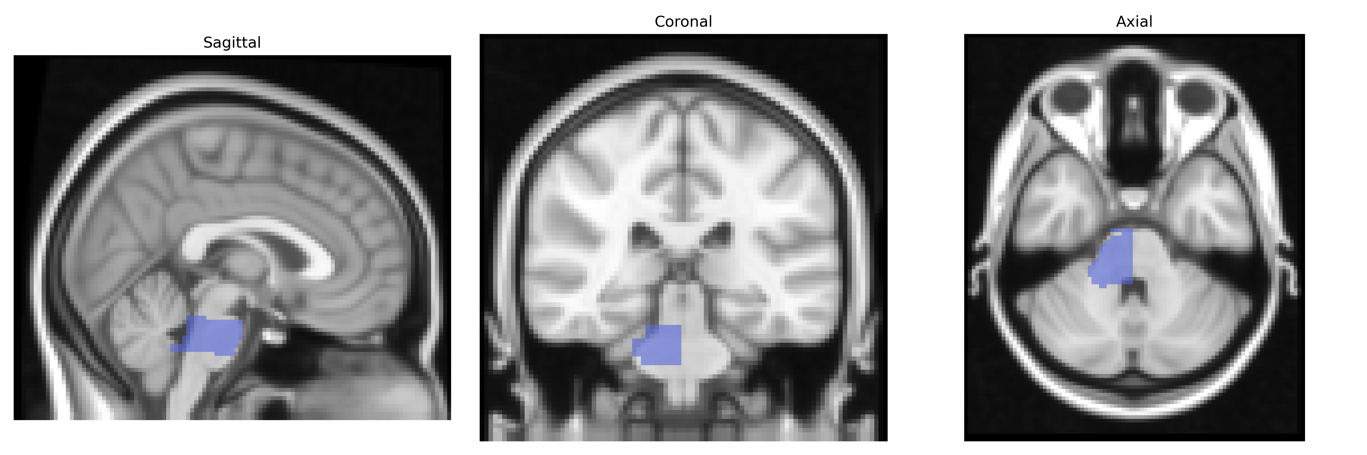
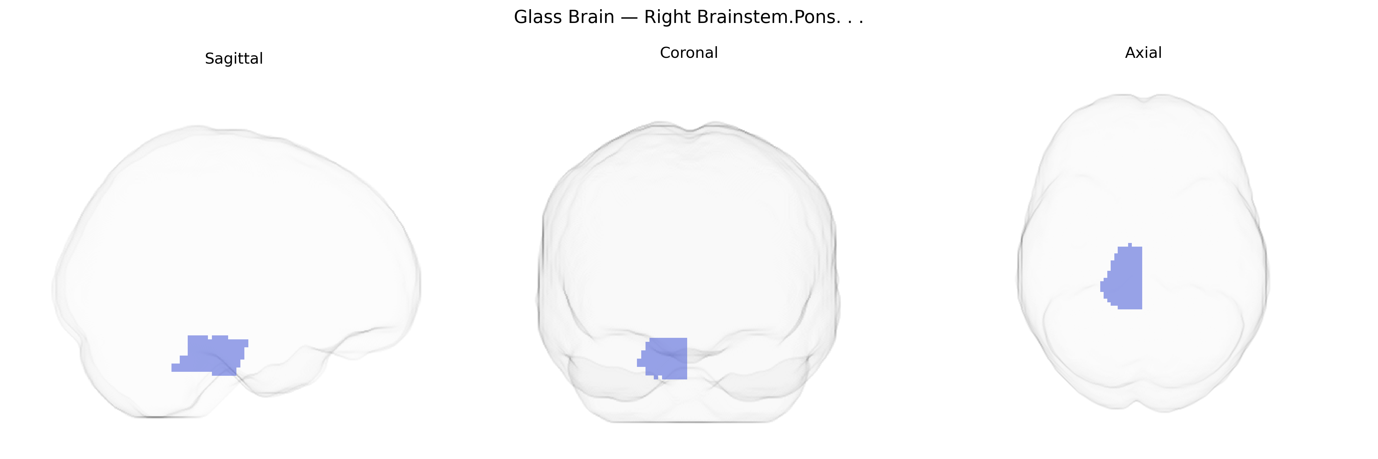
The bilateral right brainstem pons is a segment of the brainstem located rostral to the medulla and caudal to the midbrain, characterized by prominent transverse pontine fibers and the pontine nuclei that relay information from the cerebral cortex to the cerebellum. It contains ascending and descending white matter tracts, including portions of the corticospinal, corticobulbar, and somatosensory pathways, as well as key cranial nerve nuclei (notably V, VI, VII, and part of VIII), which mediate facial sensation and motor control, lateral eye movements, facial expression, lacrimation, salivation, and aspects of hearing and balance. The pons plays a central role in regulating sleep and arousal through interactions with the reticular formation, contributes to autonomic functions such as respiration through pontine respiratory centers, and participates in motor coordination via its dense connections with the cerebellum.
The bilateral right brainstem pons, as defined in the Talairach 2 mm atlas, is not typically used as a primary locus in genetic association studies, but several GWAS and neuroimaging–genetics analyses implicate pontine structure and function in relation to specific traits and disorders. Variants in genes involved in myelination and axonal integrity (e.g., CNTNAP2, NRG1, and oligodendrocyte-related loci) have been associated with overall brainstem and white matter microstructure, sometimes including pontine regions on diffusion MRI. Large-scale imaging GWAS (such as UK Biobank-based studies) have reported associations between common variants near genes related to neurodevelopment and vascular regulation and volumetric measures or microstructural properties of the brainstem, including the pons, although these findings are usually reported for “brainstem” rather than specifically lateralized pontine subregions. Clinically, the pons is centrally involved in disorders such as multiple sclerosis, neuromyelitis optica, hereditary ataxias (e.g., spinocerebellar ataxias with pontine involvement), and certain monogenic leukodystrophies, where causal variants affect widespread CNS structures but often produce characteristic pontine pathology. GWAS of stroke and small-vessel disease have identified loci (e.g., near HDAC9, PITX2) that increase risk of brainstem infarcts, including pontine strokes, though these are vascular rather than region-specific neurodevelopmental associations. Overall, genetic links to the right pontine region are indirect, reflecting genes that influence global brainstem development, white matter integrity, or cerebrovascular risk, rather than genes uniquely tied to this specific Talairach-defined area.
Overview generated by GPT-4o (2026).
Region ID: 72
Hemisphere: bilateral
Atlas: Talairach labels 2mm
Full Quality Version: Download MP4
Full Quality Version: Download MP4
Wali Sidiqyar*, Gaurav Rudravaram*, Elyssa M. McMaster, Trent M. Schwartz, Adam M. Saunders, Kurt G. Schilling, Bennett A. Landman "Introducing SPINS: A Shared Public Visualization Library of Neuroanatomical Structures." Medical Imaging with Deep Learning- short paper
This resource is licensed under CC0 1.0 Universal (Public Domain).