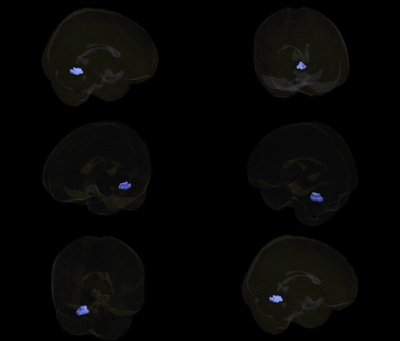
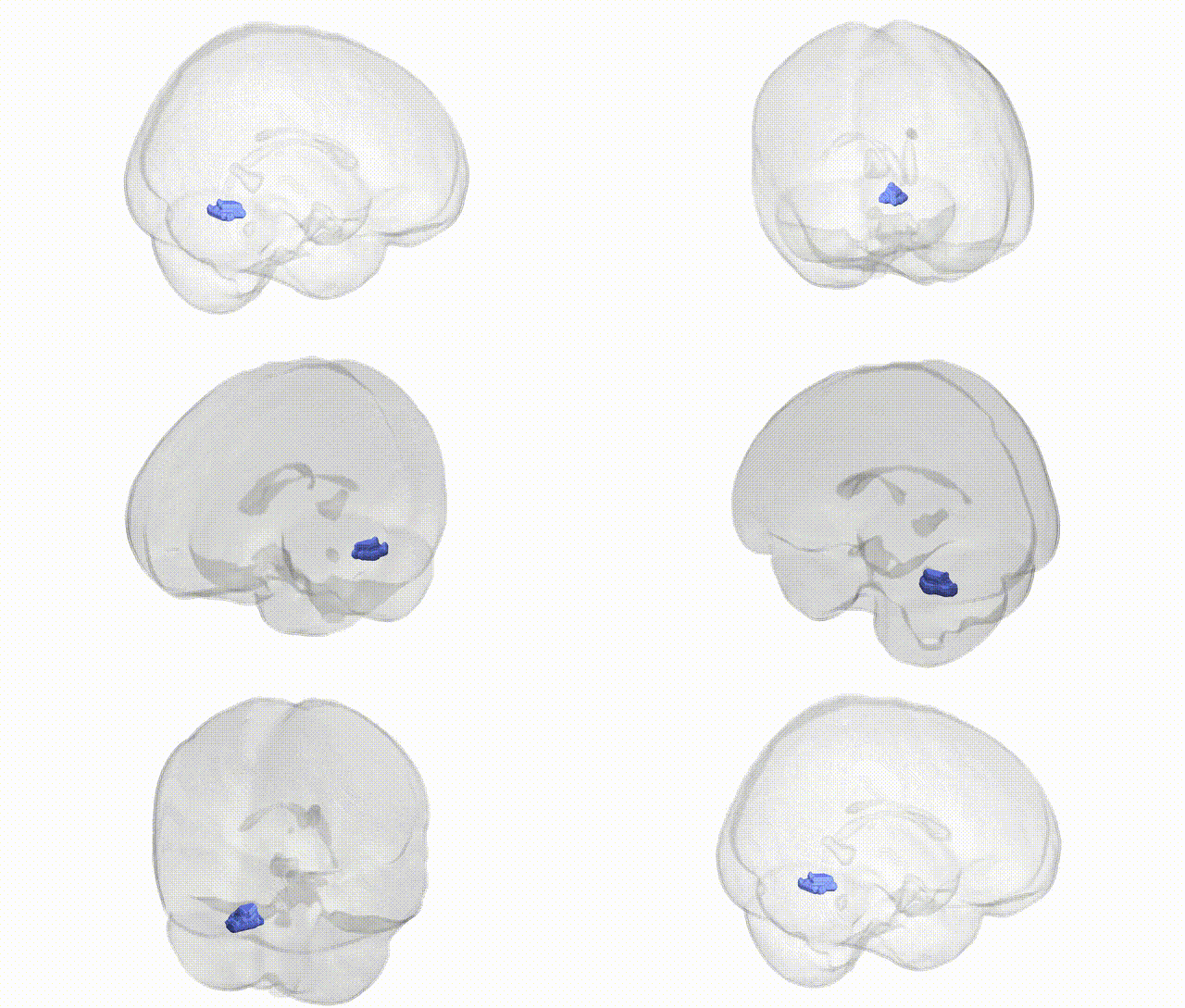
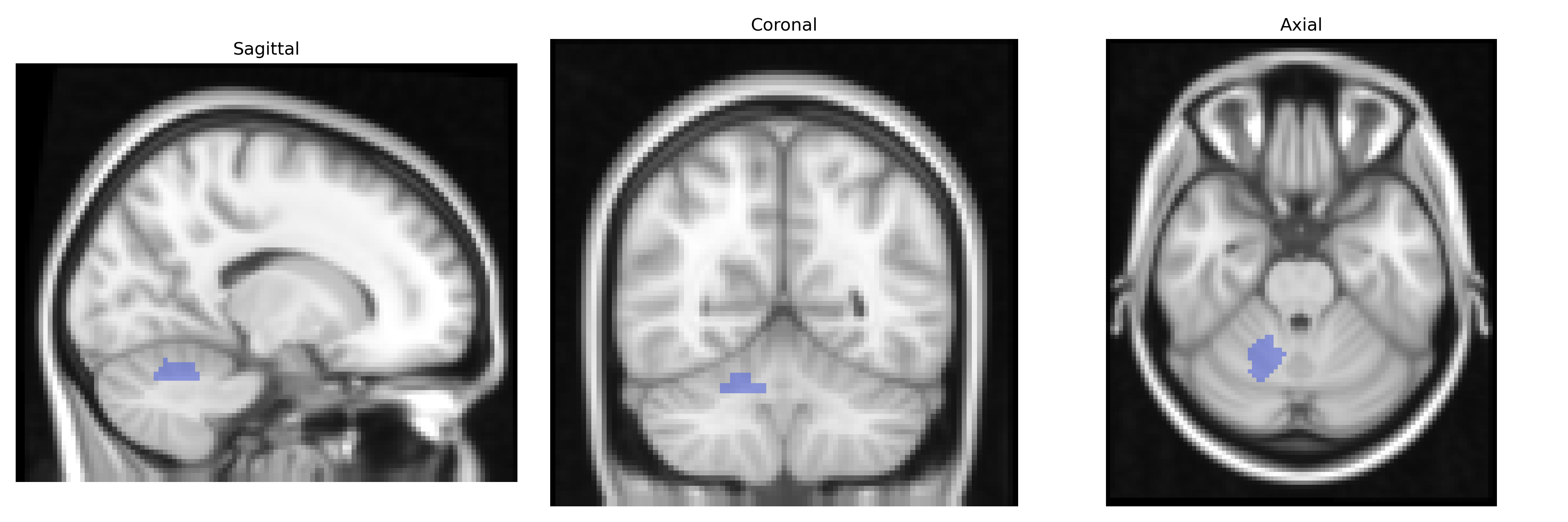
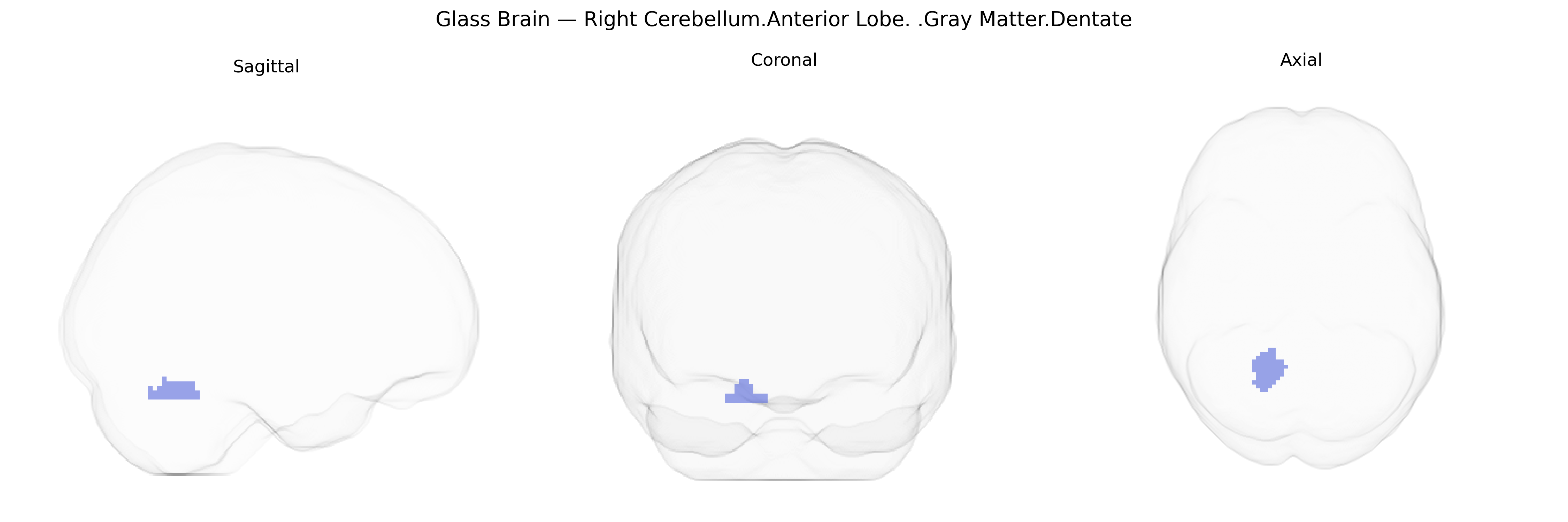
The bilateral Right Cerebellum, Anterior Lobe, Gray Matter, Dentate region corresponds to gray matter within the dentate nucleus of the cerebellum, localized to the anterior lobe on the right side as defined in the Talairach 2 mm Atlas. The dentate nucleus is the largest and most lateral of the deep cerebellar nuclei and serves as a major output hub of the cerebellar cortex, integrating proprioceptive and motor information and relaying it primarily via the superior cerebellar peduncle to the thalamus and motor-related cortical areas. Functionally, this region is implicated in fine motor coordination, timing and precision of movements, motor learning, and, through its extensive cerebello-thalamo-cortical connections, aspects of cognitive processing such as planning and executive control. There is no direct link for this composite atlas label; a related structure is the Dentate nucleus.
The bilateral right cerebellar anterior lobe gray matter (including the dentate nucleus) has been implicated in genetic studies largely through cerebellar volume and structure GWAS and through imaging genetics of neuropsychiatric and motor-related traits, rather than region-specific studies using Talairach labels. Large-scale GWAS of cerebellar volume and cortical morphology (e.g., ENIGMA, UK Biobank) have identified multiple loci—commonly involving genes such as KIAA0586, PAX3, NID2, and loci near FOXC1, SH2B1, and other neurodevelopmental and synaptic genes—associated with global and regional cerebellar volumes that encompass the anterior lobe and dentate. Variants influencing cerebellar structure have been linked to neurodevelopmental and psychiatric conditions including autism spectrum disorder, schizophrenia, bipolar disorder, and major depressive disorder, as well as cognitive traits like general intelligence and motor coordination, with polygenic scores for these traits often correlating with gray matter density or volume in anterior cerebellar regions. Mendelian and rare variant conditions affecting the dentate and anterior lobe—such as spinocerebellar ataxias (e.g., mutations in ATXN1, ATXN2, ATXN3, CACNA1A), Joubert and related cerebellar malformation syndromes, and certain dystonia and tremor syndromes (e.g., variants in TOR1A, HTRA2, and CACNA1B)—also show structural changes involving this territory. Although GWAS rarely isolate the “right cerebellum, anterior lobe, gray matter, dentate” as a discrete outcome, convergent evidence from whole-brain morphometric GWAS, disease-associated imaging genetics, and monogenic cerebellar disorders supports a genetic architecture in which neurodevelopmental, synaptic, ion channel, and axon guidance genes modulate the structure and function of this region, contributing to risk across movement, cognitive, and affective phenotypes.
Overview generated by GPT-4o (2026).
Region ID: 118
Hemisphere: bilateral
Atlas: Talairach labels 2mm
Full Quality Version: Download MP4
Full Quality Version: Download MP4
Wali Sidiqyar*, Gaurav Rudravaram*, Elyssa M. McMaster, Trent M. Schwartz, Adam M. Saunders, Kurt G. Schilling, Bennett A. Landman "Introducing SPINS: A Shared Public Visualization Library of Neuroanatomical Structures." Medical Imaging with Deep Learning- short paper
This resource is licensed under CC0 1.0 Universal (Public Domain).