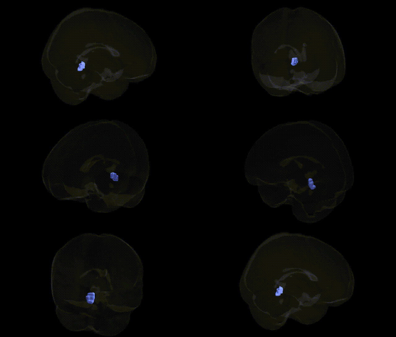
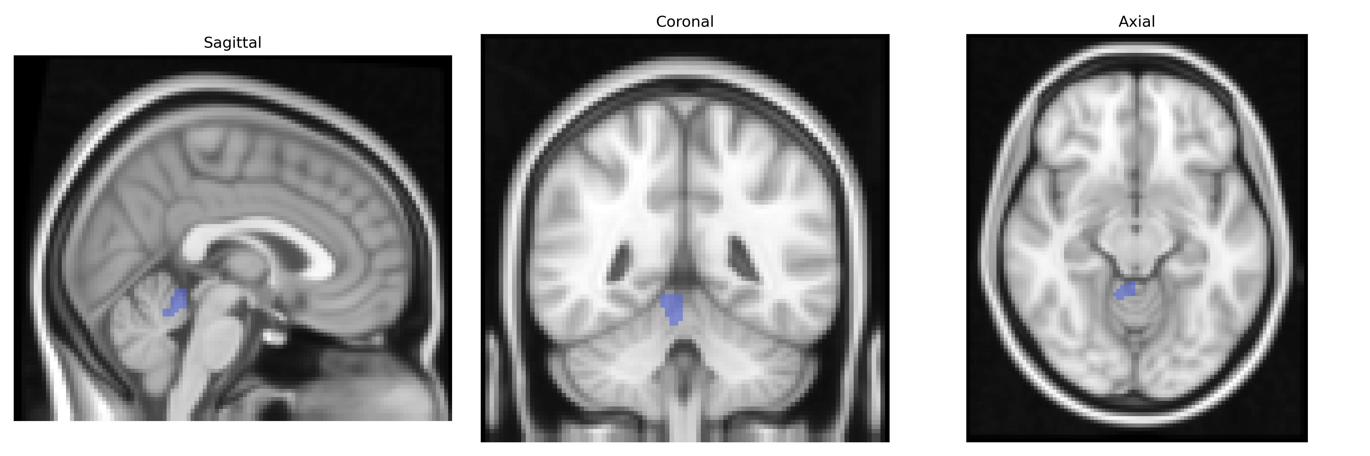
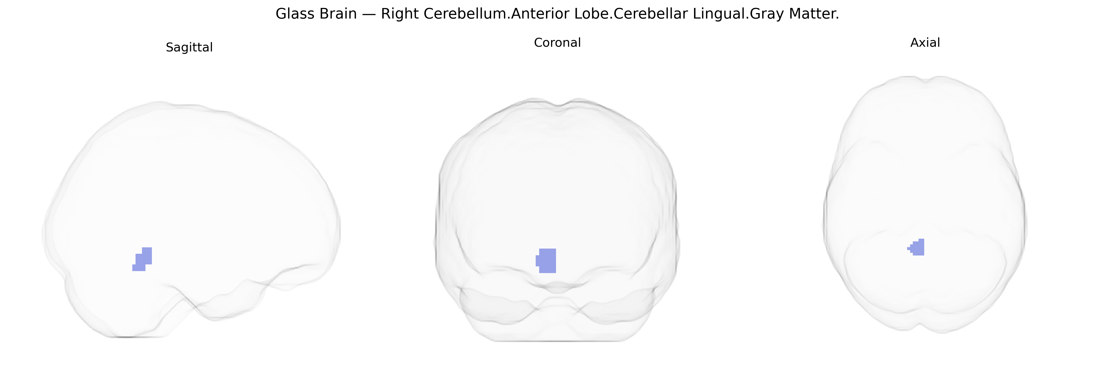
The bilateral Right Cerebellum.Anterior Lobe.Cerebellar Lingual.Gray Matter corresponds to the lingula of the cerebellum, a small tongue-shaped folium located on the superior surface of the anterior lobe, lying close to the midline and bordering the primary fissure. As part of the cerebellar cortex gray matter, the lingula participates in the fine-tuning of motor execution, posture, and coordination by integrating proprioceptive input and modulating descending motor signals. It is structurally continuous with adjacent anterior lobe regions and functionally linked to sensorimotor cerebellar circuits that contribute to precise limb and trunk movements. There is no direct link for the cerebellar lingula; a closely related structure is the Cerebellum.
The bilateral Right Cerebellum.Anterior Lobe.Cerebellar Lingual.Gray Matter, as defined in the Talairach 2 mm atlas, falls within cerebellar regions that have been repeatedly implicated in imaging genetics and GWAS of brain structure, although few studies target this label specifically. Large-scale neuroimaging GWAS (e.g., ENIGMA, UK Biobank) have identified multiple loci associated with cerebellar gray matter volume and cortical thickness—commonly including variants near KIAA0586, PAX6, RELN, DCC, and genes involved in neurodevelopment and synaptic organization—which show effects across anterior cerebellar lobules that likely encompass lingual-related portions. Cerebellar gray matter in this territory has been indirectly linked, via genetic correlations and polygenic scores, to cognitive performance, general intelligence, educational attainment, motor coordination, and emotion regulation, and to risk for neurodevelopmental and psychiatric conditions such as autism spectrum disorder, schizophrenia, ADHD, and mood disorders, where risk variants (e.g., in CACNA1C, GRM3, FOXP1, and glutamatergic or calcium-channel genes) show associations with cerebellar morphometry or activation. Additionally, rare variant and Mendelian ataxia genes (e.g., ATM, CACNA1A, ITPR1, and various spinocerebellar ataxia loci) contribute to structural alterations in anterior cerebellar gray matter, including the lingual-related region, through cerebellar degeneration and hypoplasia. However, current evidence maps primarily to broader cerebellar lobules rather than this precise Talairach-defined subregion, so genetic associations are best viewed as reflecting general cerebellar anatomy and function rather than region-specific GWAS signals.
Overview generated by GPT-4o (2026).
Region ID: 212
Hemisphere: bilateral
Atlas: Talairach labels 2mm
Full Quality Version: Download MP4
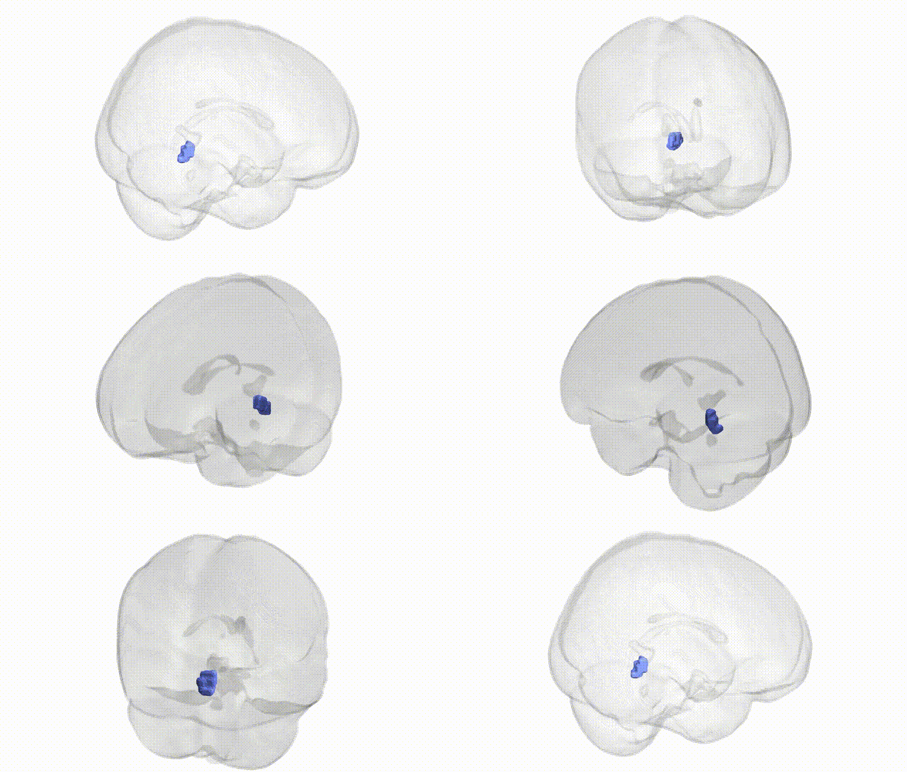
Full Quality Version: Download MP4
Wali Sidiqyar*, Gaurav Rudravaram*, Elyssa M. McMaster, Trent M. Schwartz, Adam M. Saunders, Kurt G. Schilling, Bennett A. Landman "Introducing SPINS: A Shared Public Visualization Library of Neuroanatomical Structures." Medical Imaging with Deep Learning- short paper
This resource is licensed under CC0 1.0 Universal (Public Domain).