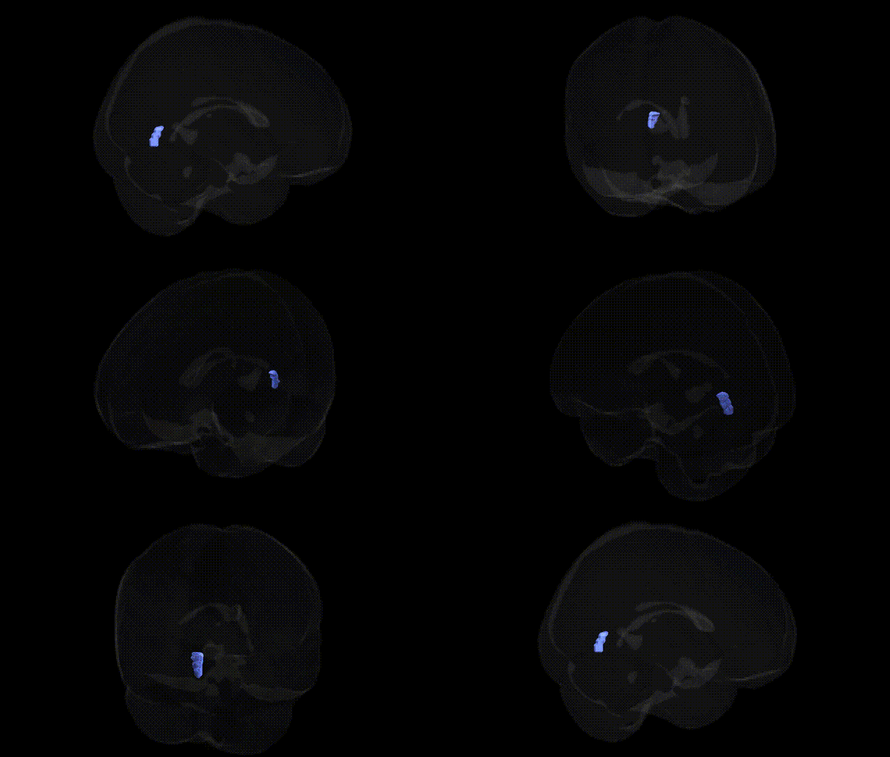
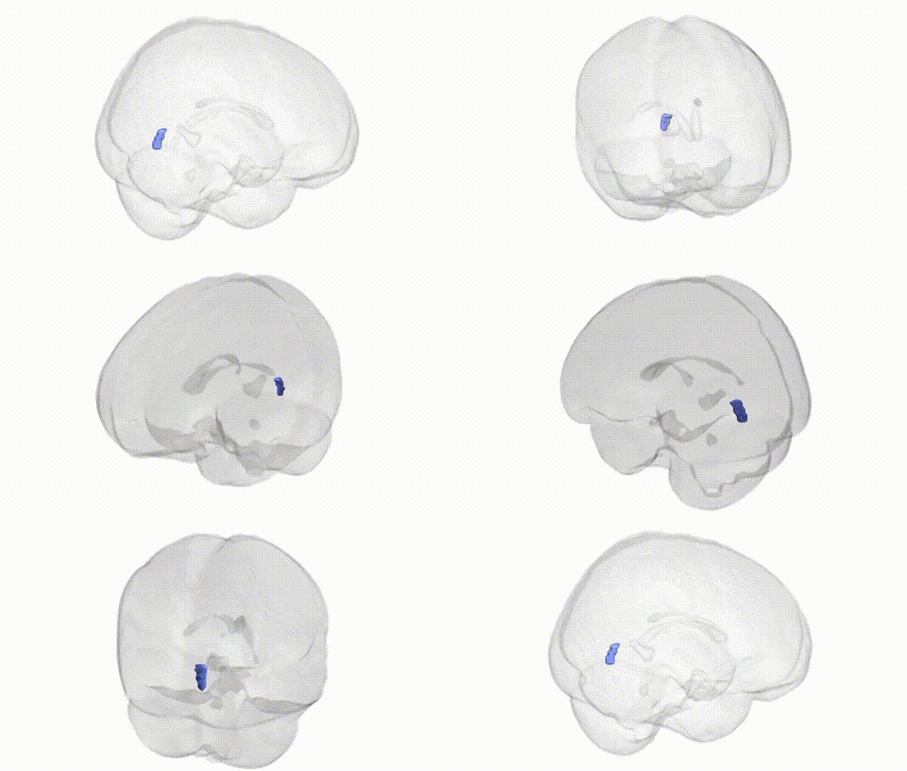
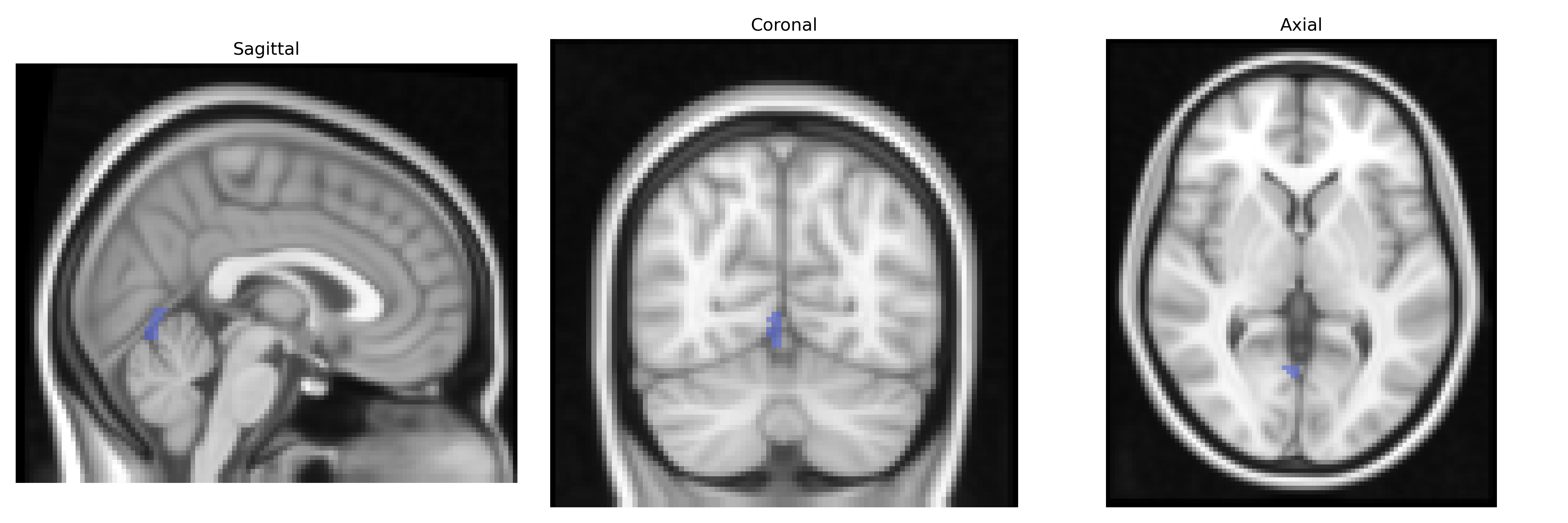
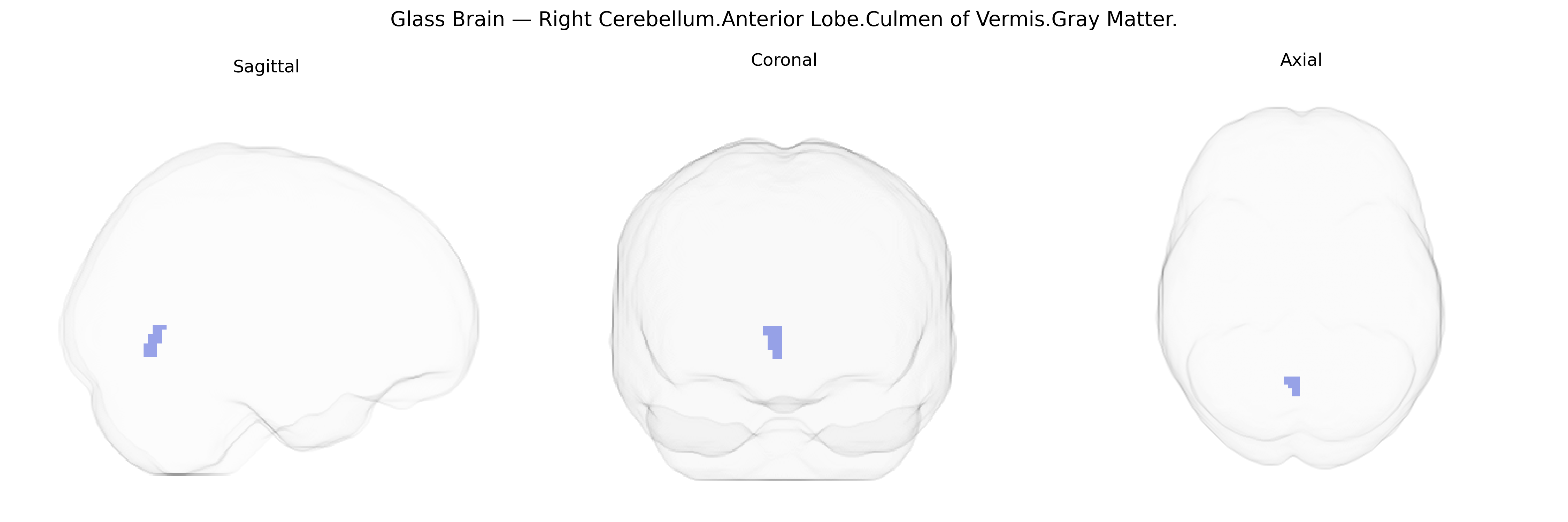
The bilateral Right Cerebellum.Anterior Lobe.Culmen of Vermis.Gray Matter refers to gray matter within the culmen, a lobule located in the anterior lobe of the cerebellar vermis, with Talairach coordinates extending bilaterally but labeled on the right in the atlas. The culmen is part of the spinocerebellum and plays a key role in the regulation and coordination of posture, gait, and limb movements by integrating proprioceptive input and modulating descending motor pathways. Functionally, this region contributes to fine-tuning ongoing motor activity, maintaining balance, and ensuring smooth execution of voluntary movements. Microcircuitry in this gray matter includes Purkinje cells, granule cells, and interneurons organized into folia that project output via the deep cerebellar nuclei, especially the fastigial and interposed nuclei. There is no direct Wikipedia article for this specific subregion; see the related structure Cerebellar vermis.
Genetic associations involving the cerebellar anterior lobe and vermis culmen gray matter—though rarely targeted at the fine-grained Talairach 2 mm parcel level—emerge primarily from imaging-genetics and GWAS of cerebellar volume, cortical thickness, and functional connectivity, as well as clinical studies of cerebellar-related disorders. Large neuroimaging GWAS (e.g., ENIGMA, UK Biobank–based studies) have shown that cerebellar volumes are substantially heritable and linked to multiple loci across the genome, with replicated signals in genes involved in neurodevelopment, synaptic function, and cell adhesion (such as CADM2, KIAA0586, and variants near FOXP genes), though these findings are typically reported at lobular or whole-cerebellum resolution rather than specifically the culmen of vermis. Cerebellar vermis abnormalities and gray matter variation have been associated with genetic risk for autism spectrum disorder, schizophrenia, bipolar disorder, and ADHD, including carriers of rare copy number variants (e.g., 22q11.2 deletions) and pathogenic variants in genes such as RELN, SHANK3, and TSC2 that broadly affect cerebellar development and connectivity. GWAS of psychiatric and cognitive traits (intelligence, educational attainment, neuroticism, depression, and anxiety) consistently show polygenic scores that correlate with structural and functional metrics in midline cerebellar regions, including the vermis, suggesting that thousands of common variants with small effect sizes jointly influence gray matter in this area. In motor and coordination disorders, genes implicated in spinocerebellar ataxias (e.g., ATXN1, ATXN2, ATXN3, CACNA1A) and cerebellar hypoplasia syndromes contribute to structural changes encompassing the anterior lobe and vermis, although not restricted to this specific culmen subregion. Overall, current genetic evidence supports a highly polygenic architecture for cerebellar anterior vermis gray matter, with overlapping risk variants for neurodevelopmental, psychiatric, and motor phenotypes influencing this region, but no single gene or locus uniquely or definitively tied to the bilateral Right Cerebellum.Anterior Lobe.Culmen of Vermis.Gray Matter parcel as defined in the Talairach 2 mm atlas.
Overview generated by GPT-4o (2026).
Region ID: 322
Hemisphere: bilateral
Atlas: Talairach labels 2mm
Full Quality Version: Download MP4
Full Quality Version: Download MP4
Wali Sidiqyar*, Gaurav Rudravaram*, Elyssa M. McMaster, Trent M. Schwartz, Adam M. Saunders, Kurt G. Schilling, Bennett A. Landman "Introducing SPINS: A Shared Public Visualization Library of Neuroanatomical Structures." Medical Imaging with Deep Learning- short paper
This resource is licensed under CC0 1.0 Universal (Public Domain).