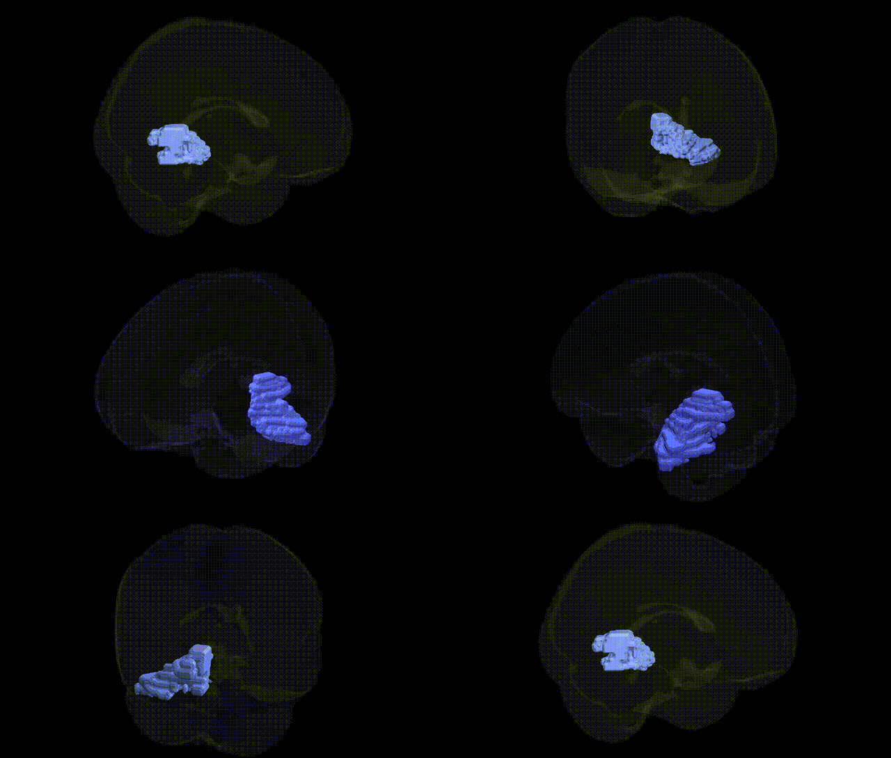
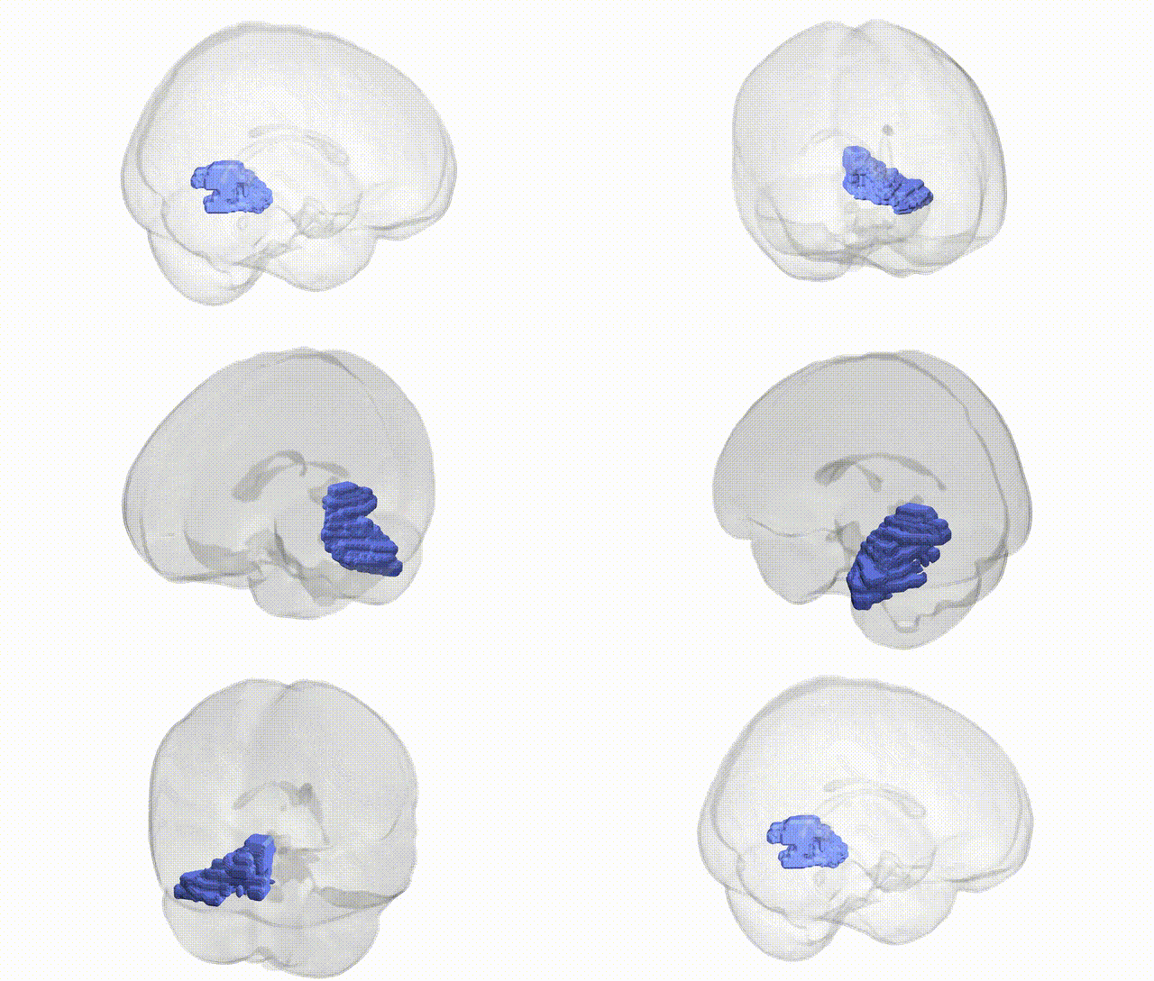
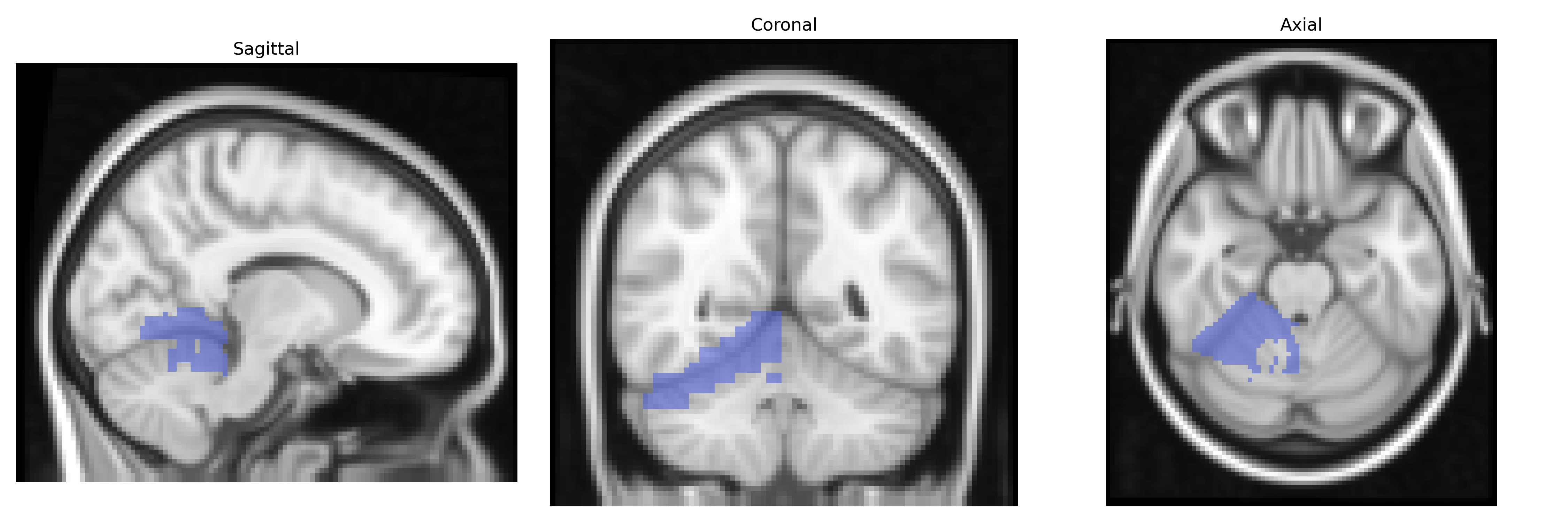
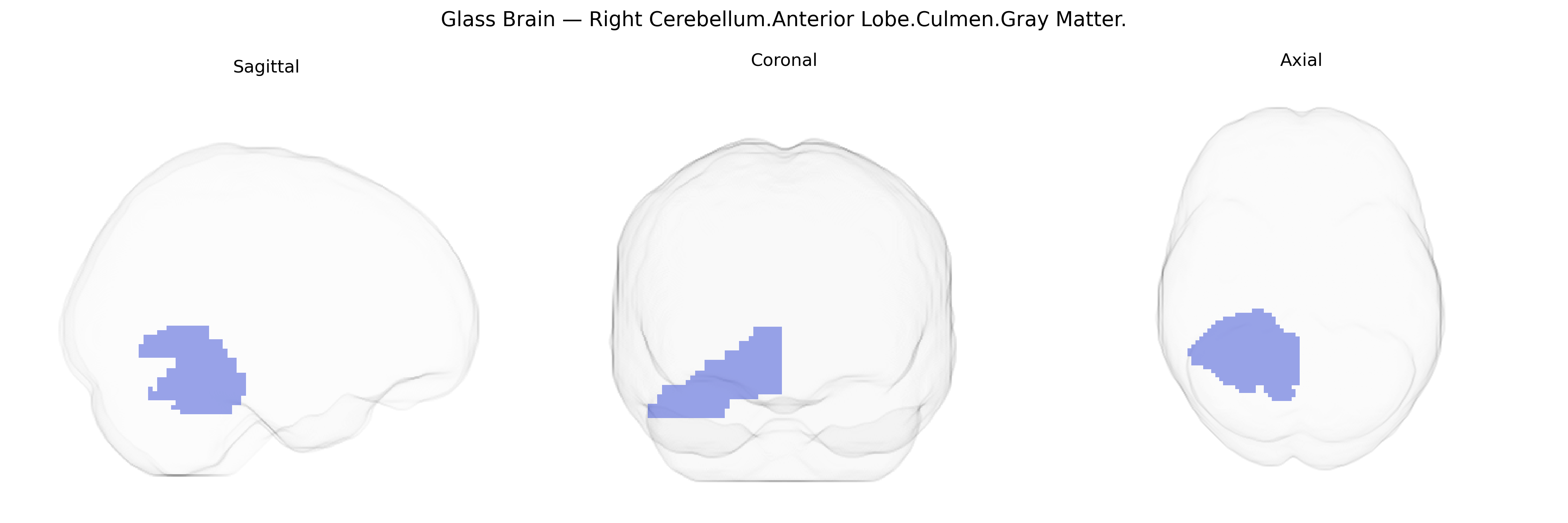
The bilateral Right Cerebellum.Anterior Lobe.Culmen.Gray Matter corresponds to gray matter within the culmen, a lobule of the anterior lobe of the cerebellum located on the superior surface of the cerebellar vermis and extending into the adjacent right hemisphere. This region participates in cerebellar circuitry involved in the coordination and timing of voluntary movements, postural control, and aspects of sensorimotor integration. Functionally, it receives input from spinal, vestibular, and cortical pathways and contributes to fine-tuning motor output via projections through the deep cerebellar nuclei to thalamic and cortical motor areas. The culmen is structurally part of the anterior lobe, which is generally associated with regulation of muscle tone and limb movement, and damage to this region can lead to gait and stance ataxia and impaired coordination. There is no direct link for the culmen itself; a closely related structure is the cerebellum: Cerebellum.
The bilateral right cerebellar anterior lobe (culmen, gray matter) has been implicated in several genetic and GWAS-based findings, primarily through large-scale imaging genetics studies that link common variants to regional cerebellar volume, cortical thickness, and functional connectivity. Genome-wide analyses of brain structure have repeatedly identified loci in and around KIAA0586 (also known as TALPID3), RELN, DLG4, and FOXP2 as associated with cerebellar development and morphology, with some effects mapped to anterior cerebellar regions including the culmen. Variants in genes involved in synaptic scaffolding (e.g., SHANK family genes), glutamatergic signaling (GRM and GRID genes), and neurodevelopmental pathways (e.g., AUTS2, CNTNAP2) have been associated with cerebellar structural or functional differences that often include the anterior lobe and are observed in disorders such as autism spectrum disorder, schizophrenia, and developmental coordination disorder. Cerebellar gray matter in anterior lobules has been linked in GWAS to general cognitive ability, educational attainment, and motor function, suggesting polygenic effects on cerebellar contributions to cognition and movement. In addition, risk loci for spinocerebellar ataxias and other hereditary ataxias, although often identified through rare-variant or Mendelian approaches rather than GWAS, involve genes (e.g., ATXN1–3, CACNA1A) whose pathogenic variants lead to degenerative changes prominently affecting anterior cerebellar regions including the culmen, reinforcing a genetic basis for vulnerability and variation in this specific cerebellar territory.
Overview generated by GPT-4o (2026).
Region ID: 89
Hemisphere: bilateral
Atlas: Talairach labels 2mm
Full Quality Version: Download MP4
Full Quality Version: Download MP4
Wali Sidiqyar*, Gaurav Rudravaram*, Elyssa M. McMaster, Trent M. Schwartz, Adam M. Saunders, Kurt G. Schilling, Bennett A. Landman "Introducing SPINS: A Shared Public Visualization Library of Neuroanatomical Structures." Medical Imaging with Deep Learning- short paper
This resource is licensed under CC0 1.0 Universal (Public Domain).