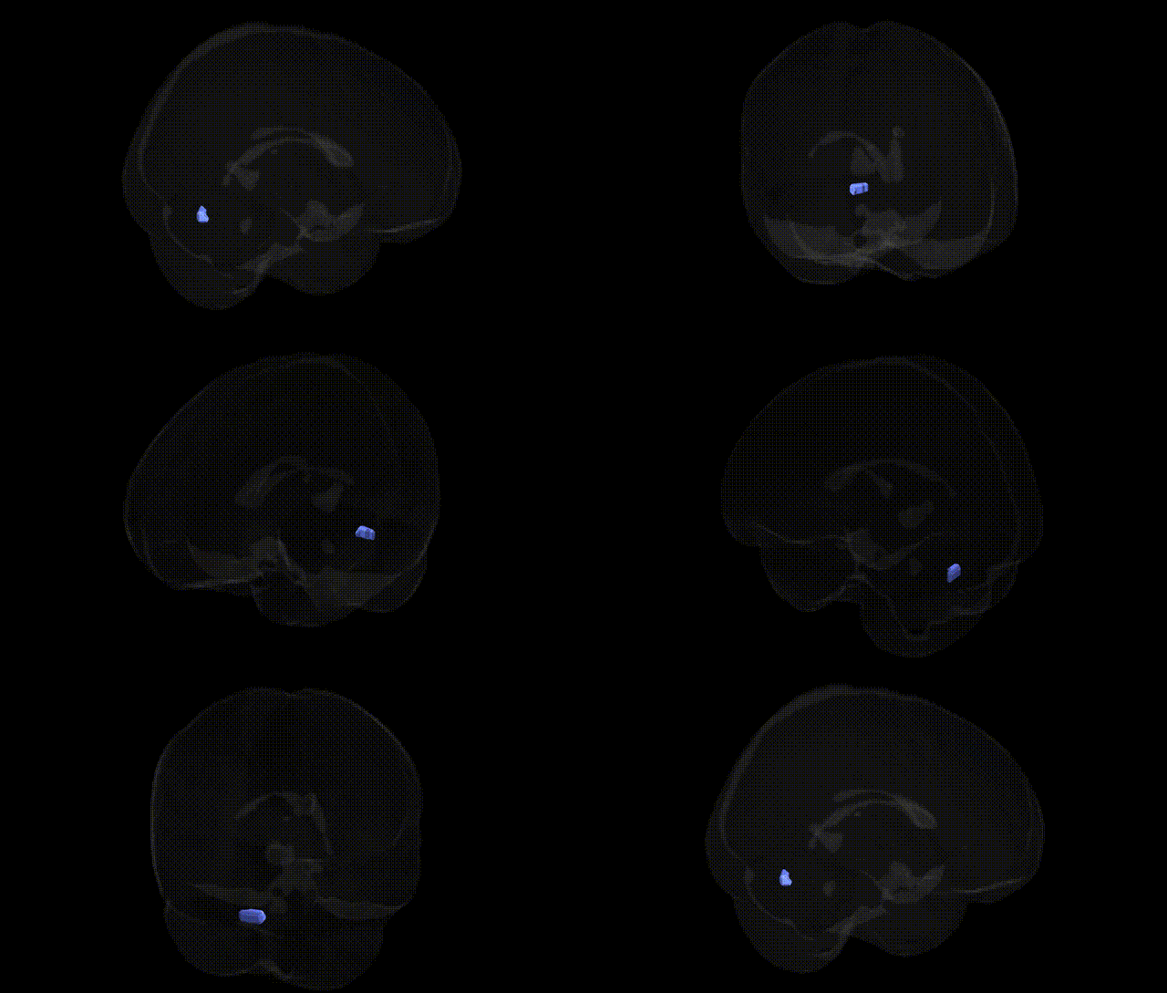
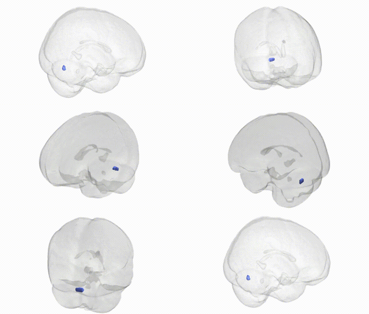
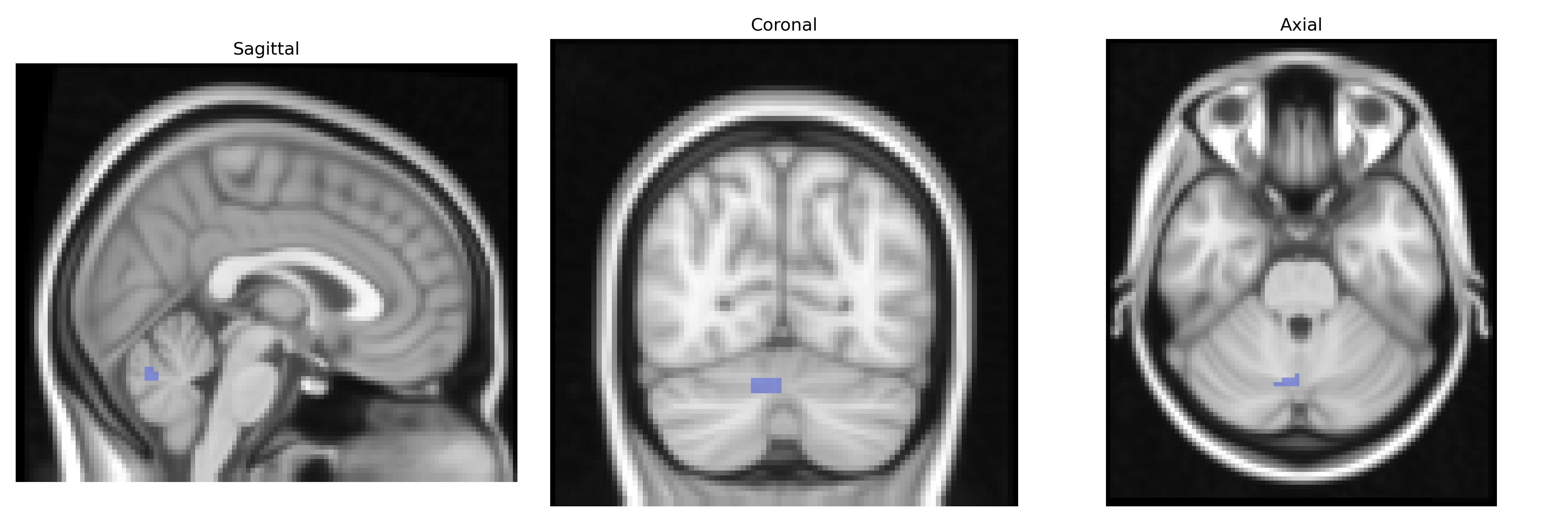
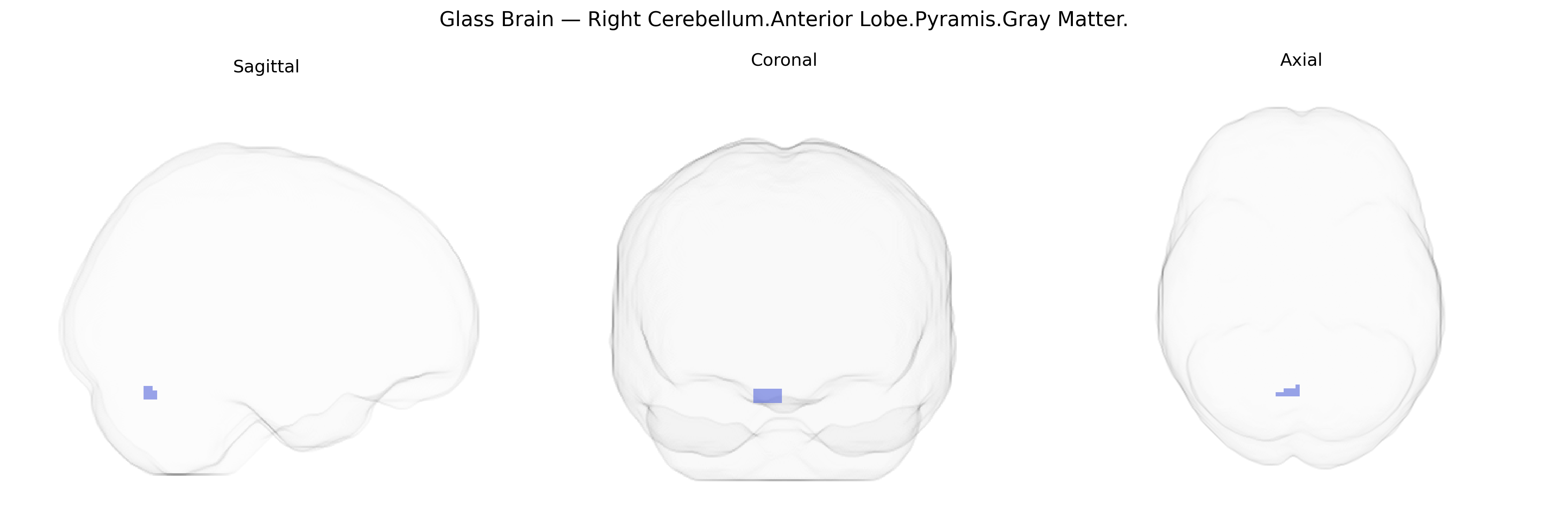
The bilateral Right Cerebellum.Anterior Lobe.Pyramis.Gray Matter corresponds to gray matter within the pyramis of the anterior lobe of the right cerebellar hemisphere, a vermal structure involved primarily in motor coordination and posture control. This region participates in the modulation of axial and proximal limb musculature, integrating proprioceptive input from the spinal cord and brainstem with cerebellar cortical processing to fine-tune ongoing movements and maintain balance. Functionally, it contributes to the regulation of muscle tone and the timing and precision of motor output, and lesions in this area can lead to truncal ataxia, gait disturbances, and impaired postural stability. There is no direct link for this specific subregion; a related structure is the Cerebellum.
The bilateral right cerebellar anterior lobe pyramis gray matter (as defined in the Talairach 2 mm atlas) corresponds to part of the anterior cerebellar vermis and adjacent hemispheric cortex, a region implicated in motor coordination, posture, and aspects of affective regulation; however, no GWAS to date has targeted this subregion specifically. Large neuroimaging genetics consortia (e.g., ENIGMA, UK Biobank) have identified numerous common variants associated with global and lobular cerebellar volume and cortical thickness—particularly in genes involved in neurodevelopment (e.g., FOXP1/FOXP2, PAX-related pathways, and general neurogenesis and axon guidance loci)—but these findings are typically reported at the level of whole cerebellum or major lobes rather than fine-grained Talairach parcels. Indirect evidence comes from GWAS of disorders with cerebellar vermis and anterior lobe abnormalities, including autism spectrum disorder, schizophrenia, bipolar disorder, and major depressive disorder, where risk loci in synaptic, calcium-channel, and immune genes (such as CACNA1C, GRM3, DRD2, and MHC-region variants) have been associated with structural and functional cerebellar alterations, though not resolved to the pyramis specifically. Similarly, genes implicated in spinocerebellar ataxias (e.g., ATXN1, ATXN2, ATXN3, CACNA1A) and other hereditary ataxias show strong effects on anterior cerebellar structures, but the anatomical mapping is usually at the level of “vermis” or “anterior lobe” rather than Talairach-defined pyramis gray matter. Overall, genetic associations for this specific bilateral right cerebellar anterior lobe pyramis parcel remain largely extrapolated from broader cerebellar and vermal imaging-genetics studies, with no robust, parcel-level GWAS currently available.
Overview generated by GPT-4o (2026).
Region ID: 117
Hemisphere: bilateral
Atlas: Talairach labels 2mm
Full Quality Version: Download MP4
Full Quality Version: Download MP4
Wali Sidiqyar*, Gaurav Rudravaram*, Elyssa M. McMaster, Trent M. Schwartz, Adam M. Saunders, Kurt G. Schilling, Bennett A. Landman "Introducing SPINS: A Shared Public Visualization Library of Neuroanatomical Structures." Medical Imaging with Deep Learning- short paper
This resource is licensed under CC0 1.0 Universal (Public Domain).