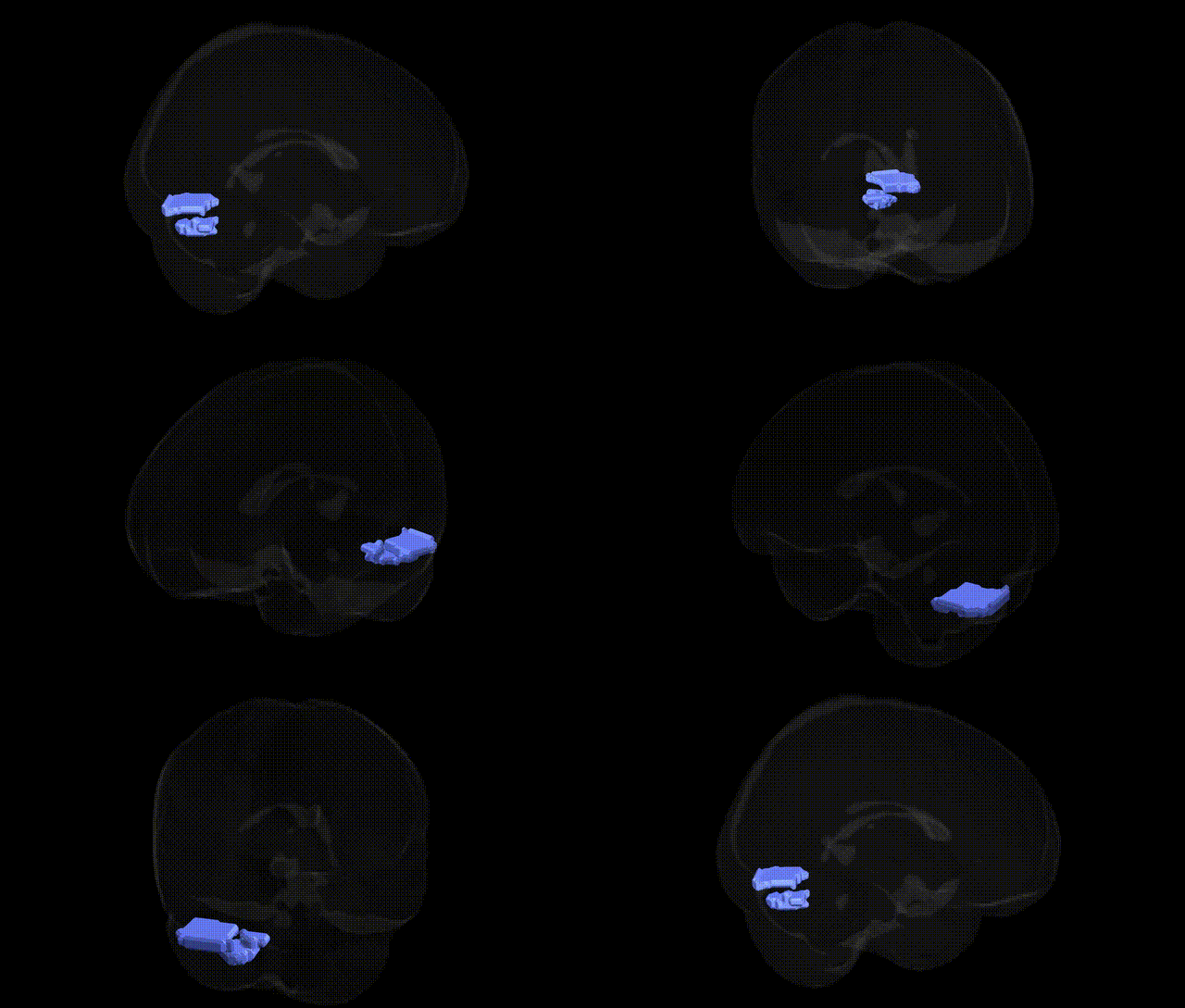
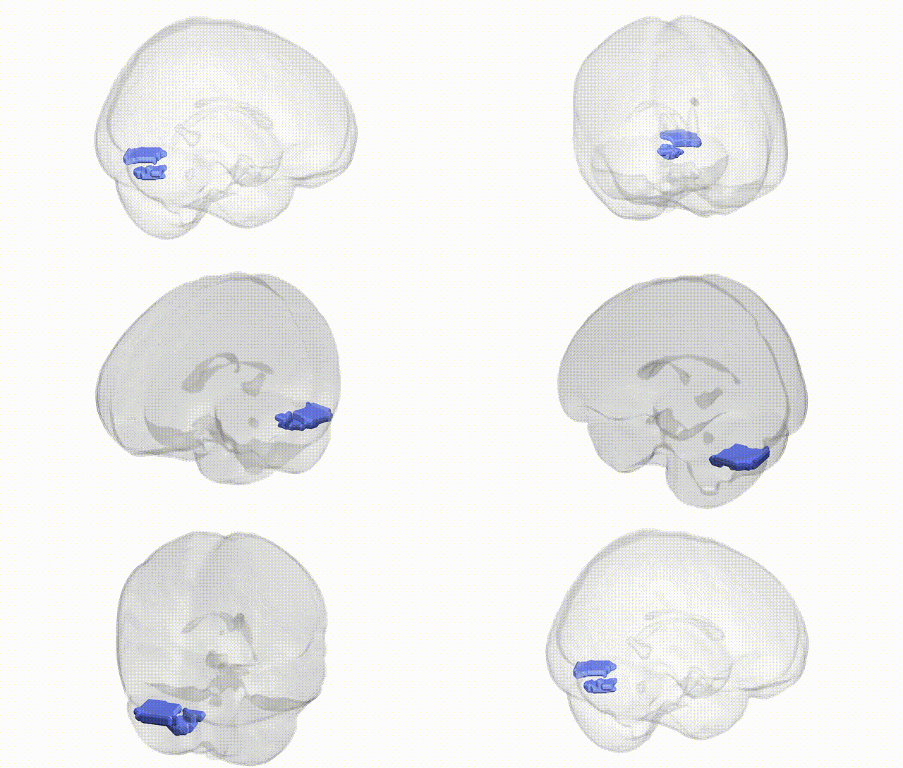
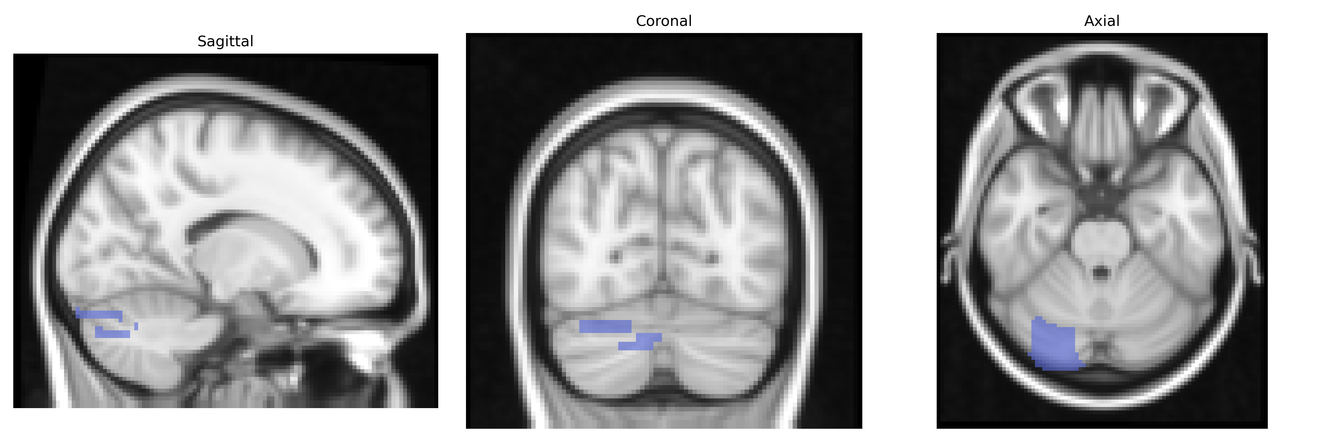
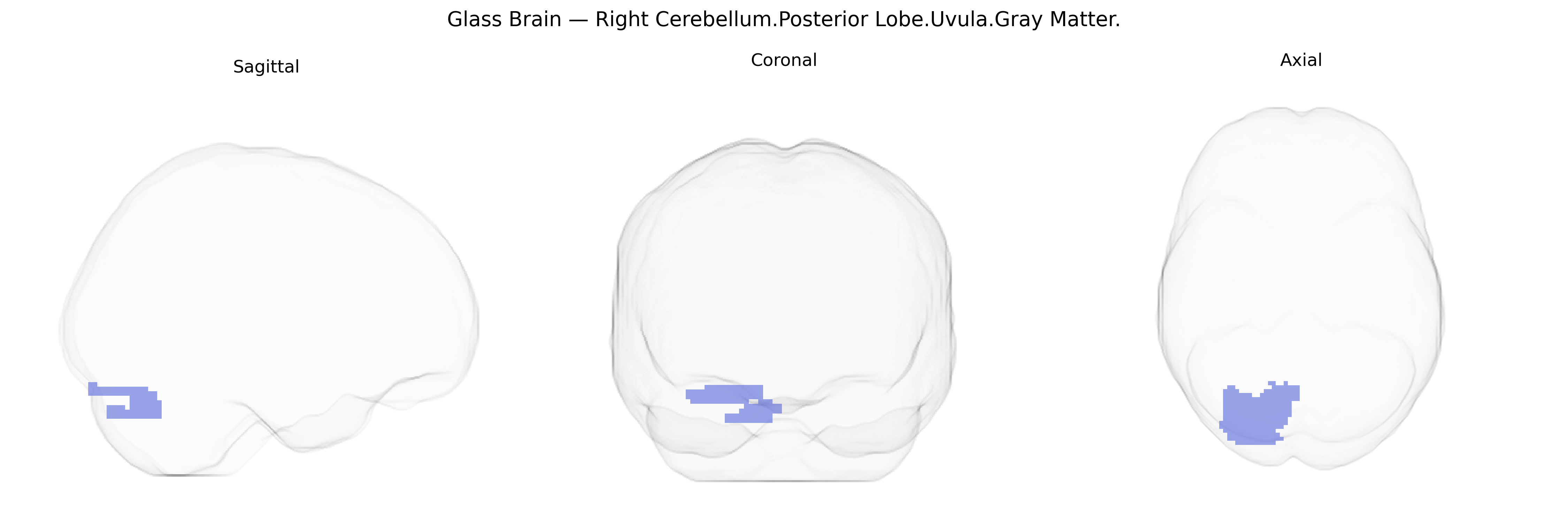
The bilateral Right Cerebellum.Posterior Lobe.Uvula.Gray Matter refers to gray matter in the uvula of the cerebellar vermis, located within the posterior lobe of the cerebellum on the right side but sampled bilaterally in the Talairach 2mm Atlas. The uvula is part of the inferior vermis and contributes to coordination of posture, balance, and eye and head movements through its connections with vestibular and reticular nuclei. Functionally, this region participates in fine-tuning motor activity and integrating sensory inputs required for equilibrium and gait, with lesions often associated with truncal ataxia and impaired postural control. There is no direct Wikipedia article for “cerebellar uvula,” but it is a subregion of the cerebellar vermis: Cerebellar vermis.
Genetic associations specifically targeting the bilateral Right Cerebellum.Posterior Lobe.Uvula.Gray Matter region, as defined in the Talairach 2 mm atlas, are not well characterized, since most imaging genetics and GWAS studies analyze larger cerebellar lobules or whole-cerebellum measures rather than fine-grained subregions like the uvula. Large brain MRI GWAS (e.g., UK Biobank–based studies) have identified multiple loci (including variants in genes such as CBLN1, GRID2, and CACNA1A, among others) associated with total cerebellar volume, lobular volumes, gray matter density, and cortical thickness, implicating pathways involved in synaptic organization, calcium signaling, and neurodevelopment, which likely influence structure and function in the posterior lobe generally but not the uvula specifically. Cerebellar gray matter variation has been genetically correlated with cognitive performance, educational attainment, and psychiatric traits, including schizophrenia and major depression, and cerebellar involvement is consistently noted in neurodevelopmental and movement disorders (e.g., autism spectrum disorder, ataxias) with known genetic underpinnings, though the evidence typically spans broader cerebellar territories. At present, no robust GWAS or candidate-gene studies have isolated the right cerebellar posterior lobe uvula gray matter as a distinct phenotype with specific genetic risk variants, so its genetic associations are inferred from studies of larger cerebellar regions rather than directly demonstrated.
Overview generated by GPT-4o (2026).
Region ID: 64
Hemisphere: bilateral
Atlas: Talairach labels 2mm
Full Quality Version: Download MP4
Full Quality Version: Download MP4
Wali Sidiqyar*, Gaurav Rudravaram*, Elyssa M. McMaster, Trent M. Schwartz, Adam M. Saunders, Kurt G. Schilling, Bennett A. Landman "Introducing SPINS: A Shared Public Visualization Library of Neuroanatomical Structures." Medical Imaging with Deep Learning- short paper
This resource is licensed under CC0 1.0 Universal (Public Domain).