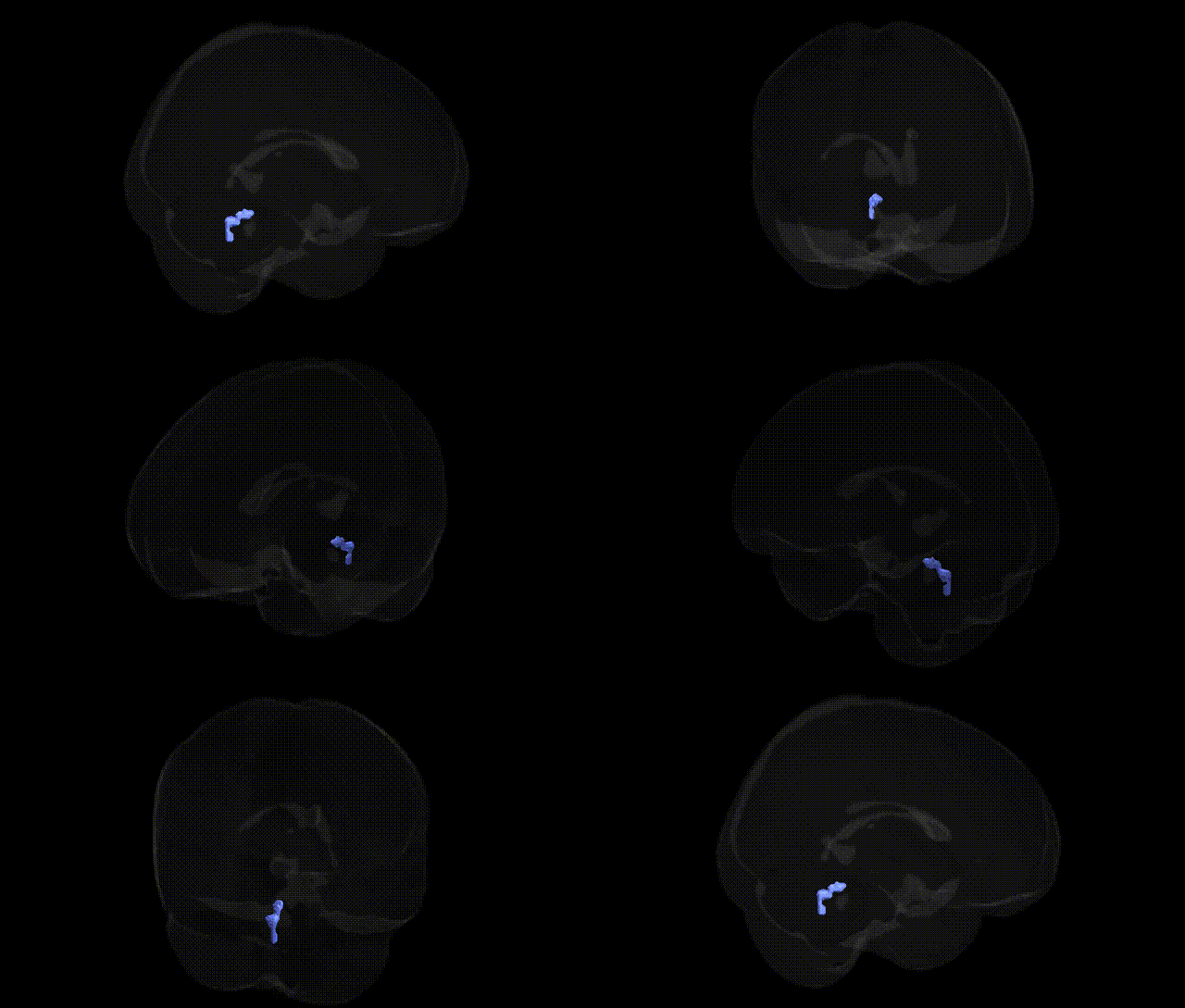
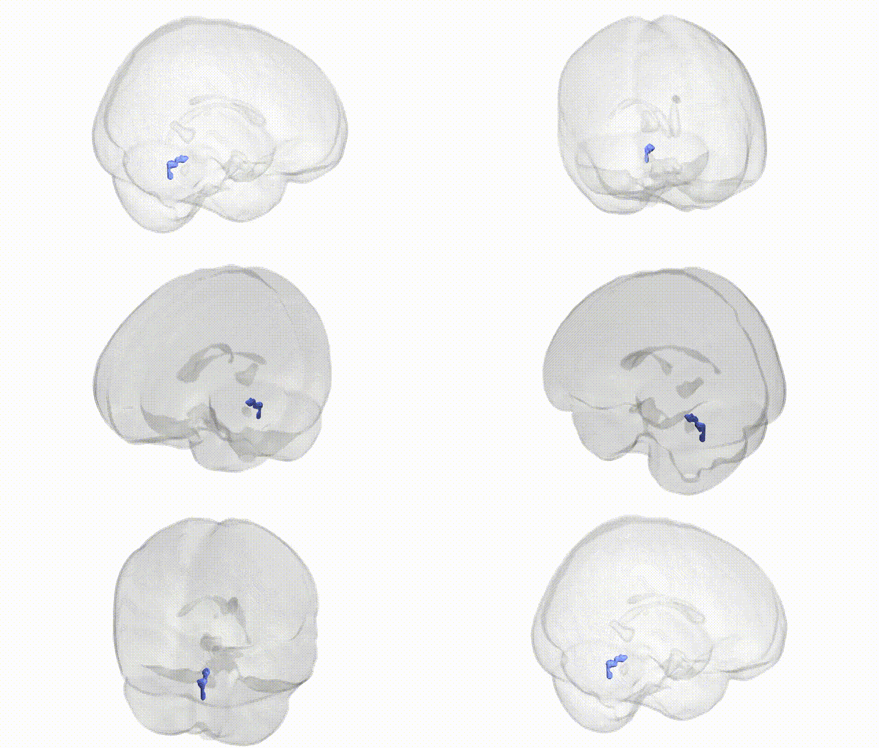
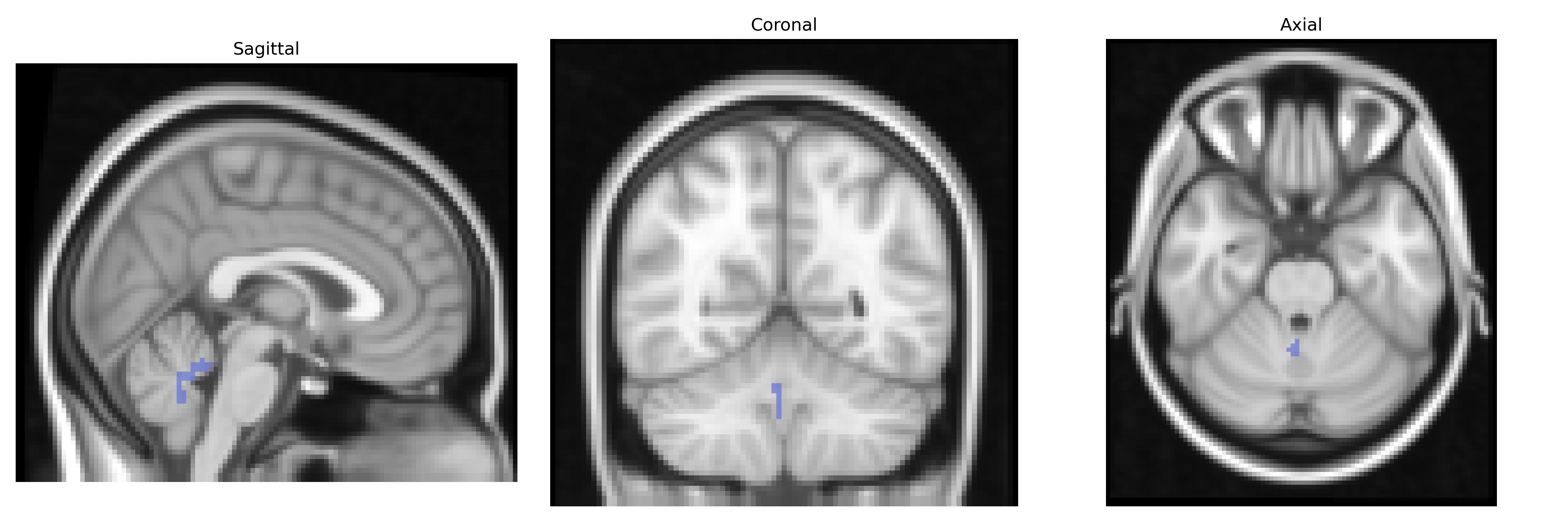
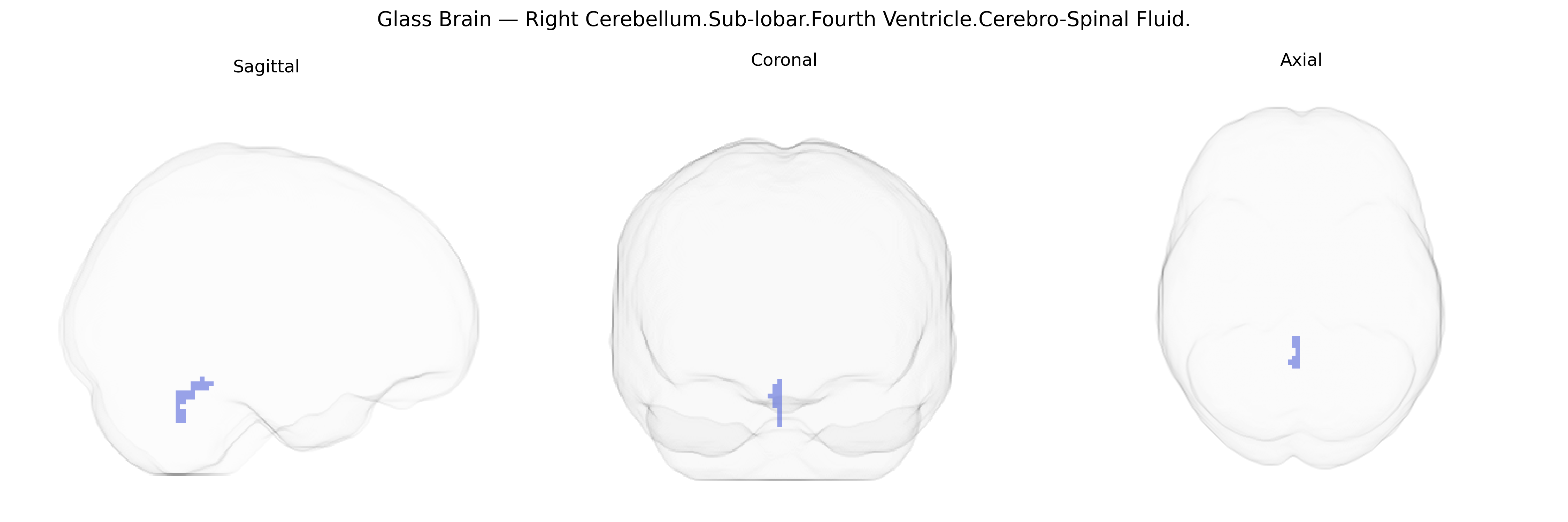
The bilateral Right Cerebellum.Sub-lobar.Fourth Ventricle.Cerebro-Spinal Fluid region, as defined in the Talairach 2 mm Atlas, corresponds to cerebrospinal fluid (CSF) occupying the fourth ventricle adjacent to the right cerebellar hemisphere. The fourth ventricle is a midline, tent-shaped cavity located between the brainstem (pons and medulla) anteriorly and the cerebellum posteriorly, forming part of the ventricular system that produces, circulates, and contains CSF. Functionally, this region is critical for CSF flow from the cerebral aqueduct into the central canal of the spinal cord and into the subarachnoid space via the median and lateral apertures, contributing to mechanical protection of neural tissue, maintenance of intracranial pressure, and homeostatic regulation of the central nervous system environment. There is no direct link for this exact Talairach label; a related structure is the Fourth ventricle.
Genetic association studies specifically targeting the bilateral Right Cerebellum.Sub-lobar.Fourth Ventricle.Cerebro-Spinal Fluid region from the Talairach 2 mm atlas are limited, and most relevant findings come from broader neuroimaging GWAS of ventricular and cerebellar volumes rather than this exact label. Large-scale GWAS of brain structure have consistently identified variants in and around genes such as FANCL, HRK, TRIOBP, PTCH1, and loci near APOE and MAPT that influence overall ventricular size and cerebrospinal fluid (CSF) spaces, traits that reflect neurodegenerative and neurodevelopmental processes affecting the fourth ventricle region indirectly. Enlarged ventricular and CSF volumes—which include the fourth ventricle—have been genetically linked to Alzheimer’s disease risk loci (e.g., APOE ε4) and other dementia-related variants, as well as to polygenic burden for schizophrenia, bipolar disorder, and general cognitive ability in imaging–genetics consortia such as ENIGMA and UK Biobank analyses. Cerebellar volume GWAS have implicated genes involved in neurodevelopment, synaptic function, and axon guidance, with polygenic overlap observed with psychiatric conditions (schizophrenia, major depression), autism spectrum traits, and motor coordination phenotypes; while these cerebellar loci do not map exclusively to the right fourth ventricle CSF space, they highlight shared genetic architecture between cerebellar morphometry and neuropsychiatric and cognitive traits. Overall, current evidence suggests that genetic influences on this Talairach-defined region are best understood as part of generalized genetic control of ventricular/CSF volumes and cerebellar structure, rather than as a distinct set of region-specific genetic associations.
Overview generated by GPT-4o (2026).
Region ID: 69
Hemisphere: bilateral
Atlas: Talairach labels 2mm
Full Quality Version: Download MP4
Full Quality Version: Download MP4
Wali Sidiqyar*, Gaurav Rudravaram*, Elyssa M. McMaster, Trent M. Schwartz, Adam M. Saunders, Kurt G. Schilling, Bennett A. Landman "Introducing SPINS: A Shared Public Visualization Library of Neuroanatomical Structures." Medical Imaging with Deep Learning- short paper
This resource is licensed under CC0 1.0 Universal (Public Domain).