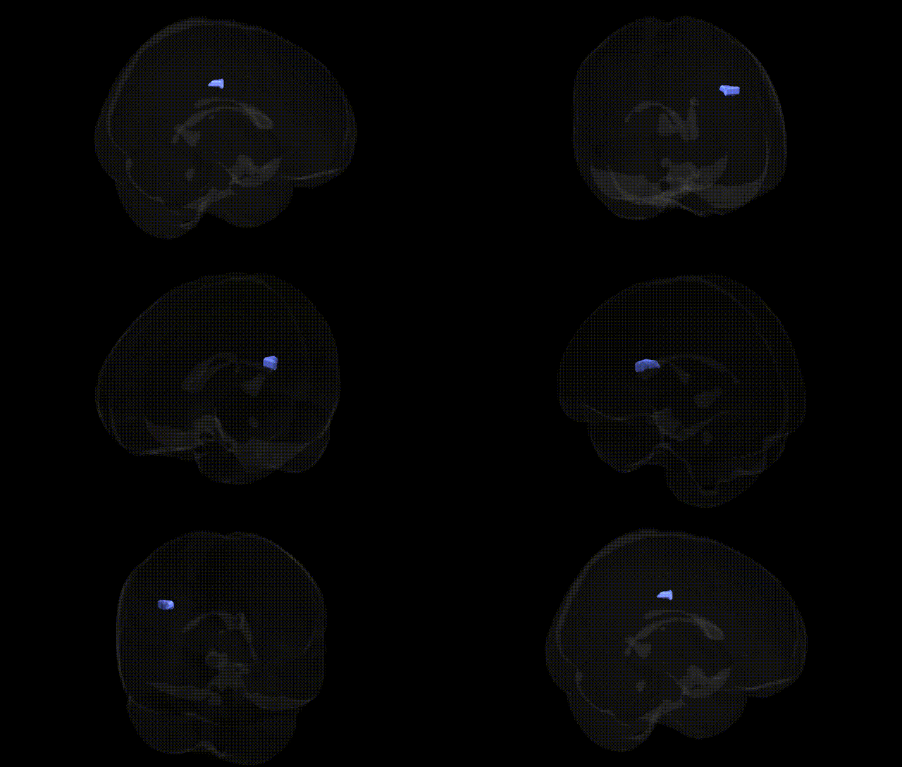
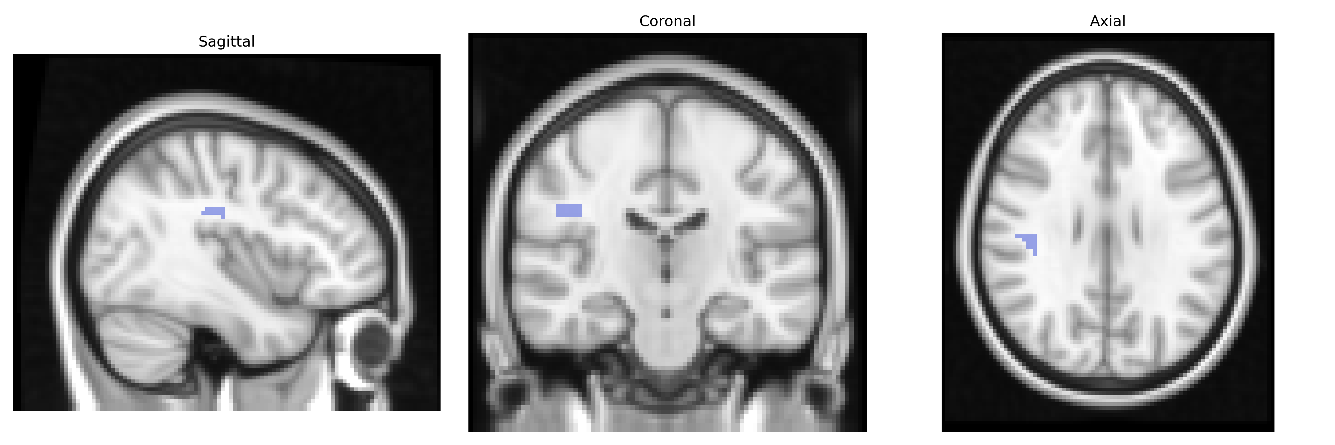
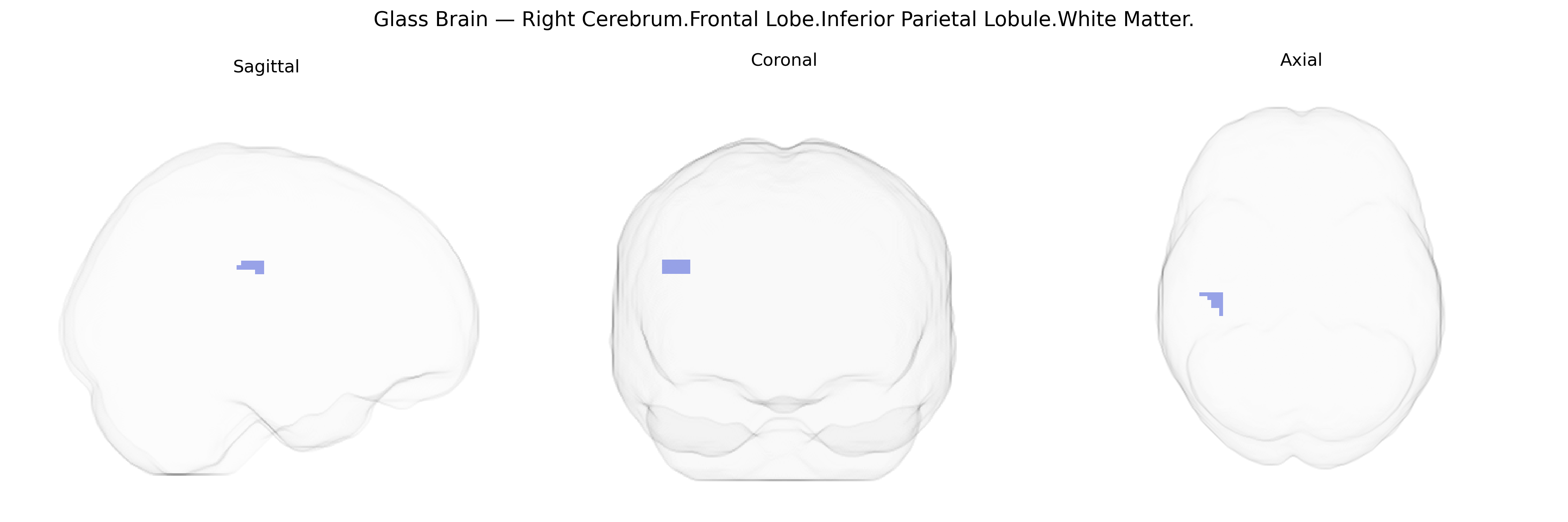
The bilateral Right Cerebrum.Frontal Lobe.Inferior Parietal Lobule.White Matter region, as defined in the Talairach 2 mm atlas, refers to subcortical white matter pathways coursing deep to the inferior parietal lobule at the junction of the frontal and parietal cortices in the right hemisphere. This white matter contains association fiber tracts that interconnect frontal executive and premotor areas with parietal regions involved in multimodal integration, spatial attention, and sensorimotor processing, forming part of frontoparietal networks critical for higher-order cognitive control and visuospatial functions. Although the atlas label is specific to white matter under the inferior parietal lobule, functionally it supports communication among cortical nodes of attention and working memory networks rather than serving as an independent processing center. There is no direct Wikipedia article for this exact white matter label; a closely related cortical structure is the Inferior parietal lobule.
The bilateral Right Cerebrum.Frontal Lobe.Inferior Parietal Lobule.White Matter, as defined in the Talairach 2 mm atlas, lies within frontoparietal association circuitry that has been implicated indirectly in numerous genetic and GWAS findings through related white-matter tracts and network-level measures rather than this exact atlas label. Large-scale imaging genetics studies, including ENIGMA and UK Biobank analyses, have identified heritable variation in frontoparietal and inferior parietal white-matter microstructure (often via diffusion metrics such as fractional anisotropy in adjacent tracts like the superior longitudinal fasciculus and frontoparietal fibers), with associated loci in genes involved in axon guidance, myelination, and neurodevelopment (for example, variants near or within genes such as CNTN4, NRG1, and multiple oligodendrocyte-related genes). GWAS of cognitive performance, educational attainment, and general intelligence frequently report associations with polygenic scores that correlate with the integrity and volume of frontoparietal and inferior parietal white-matter regions, suggesting that many small-effect variants collectively shape the structural properties of this area. Moreover, genetic risk for neuropsychiatric disorders—especially schizophrenia, bipolar disorder, major depressive disorder, and ADHD—has been linked to altered frontoparietal and inferior parietal white-matter measures, with polygenic risk scores for these conditions predicting changes in connectivity in frontoparietal control and default-mode networks that traverse this region. In neurodegenerative and vascular conditions, risk variants in genes such as APOE (e.g., APOE ε4 in Alzheimer’s disease) and loci associated with cerebral small vessel disease and white-matter hyperintensities show robust associations with abnormalities in widespread frontal and parietal white matter, including the inferior parietal lobule territory. Although no major GWAS has targeted the Talairach-defined “Right Cerebrum.Frontal Lobe.Inferior Parietal Lobule.White Matter” as a standalone phenotype, convergent imaging-genetic evidence supports a role for polygenic influences on frontoparietal white-matter microstructure and connectivity in shaping cognition, vulnerability to psychiatric and neurodevelopmental disorders, and susceptibility to age-related and vascular white-matter pathology in this region.
Overview generated by GPT-4o (2026).
Region ID: 937
Hemisphere: bilateral
Atlas: Talairach labels 2mm
Full Quality Version: Download MP4
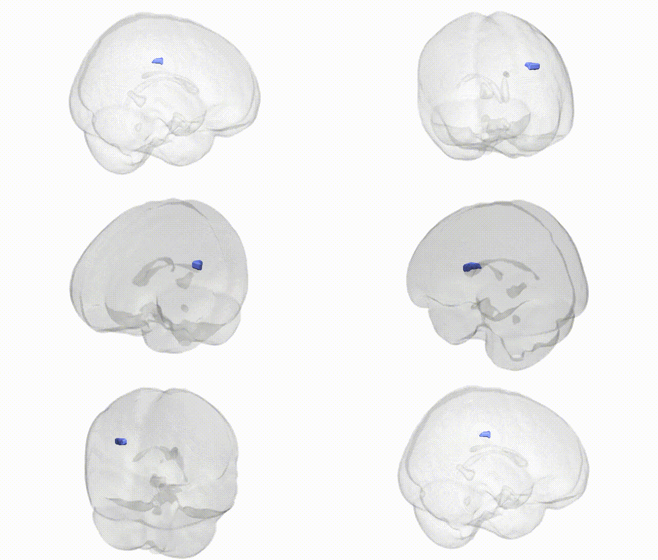
Full Quality Version: Download MP4
Wali Sidiqyar*, Gaurav Rudravaram*, Elyssa M. McMaster, Trent M. Schwartz, Adam M. Saunders, Kurt G. Schilling, Bennett A. Landman "Introducing SPINS: A Shared Public Visualization Library of Neuroanatomical Structures." Medical Imaging with Deep Learning- short paper
This resource is licensed under CC0 1.0 Universal (Public Domain).