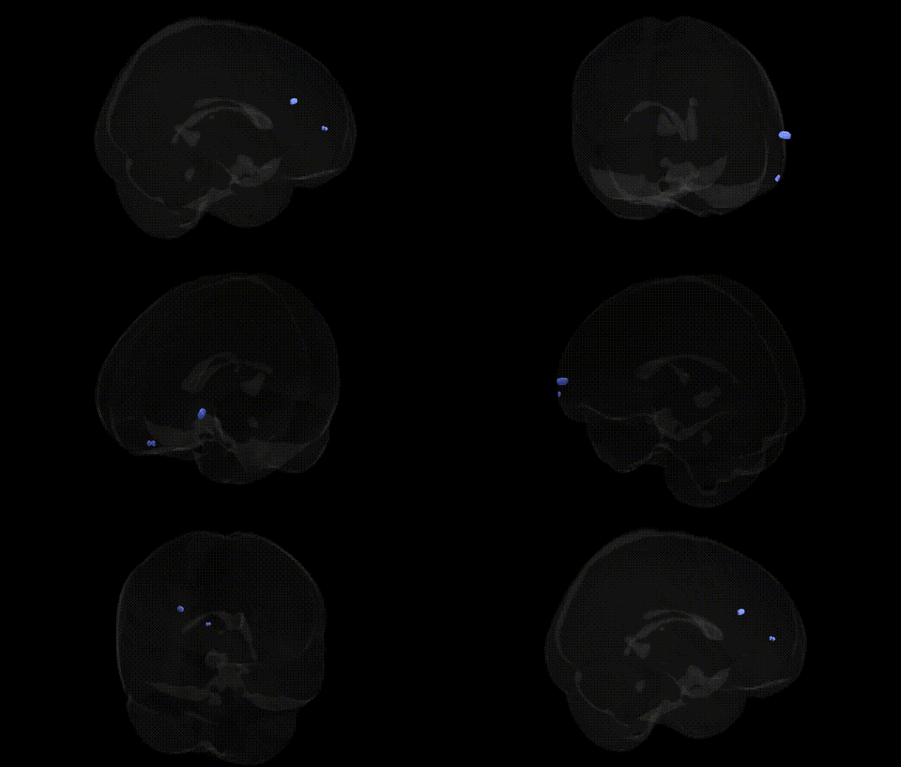
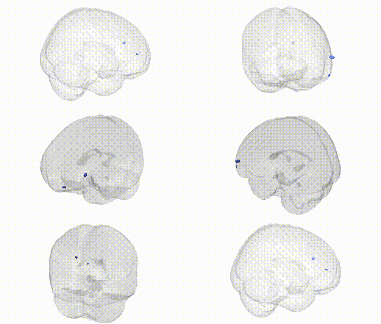
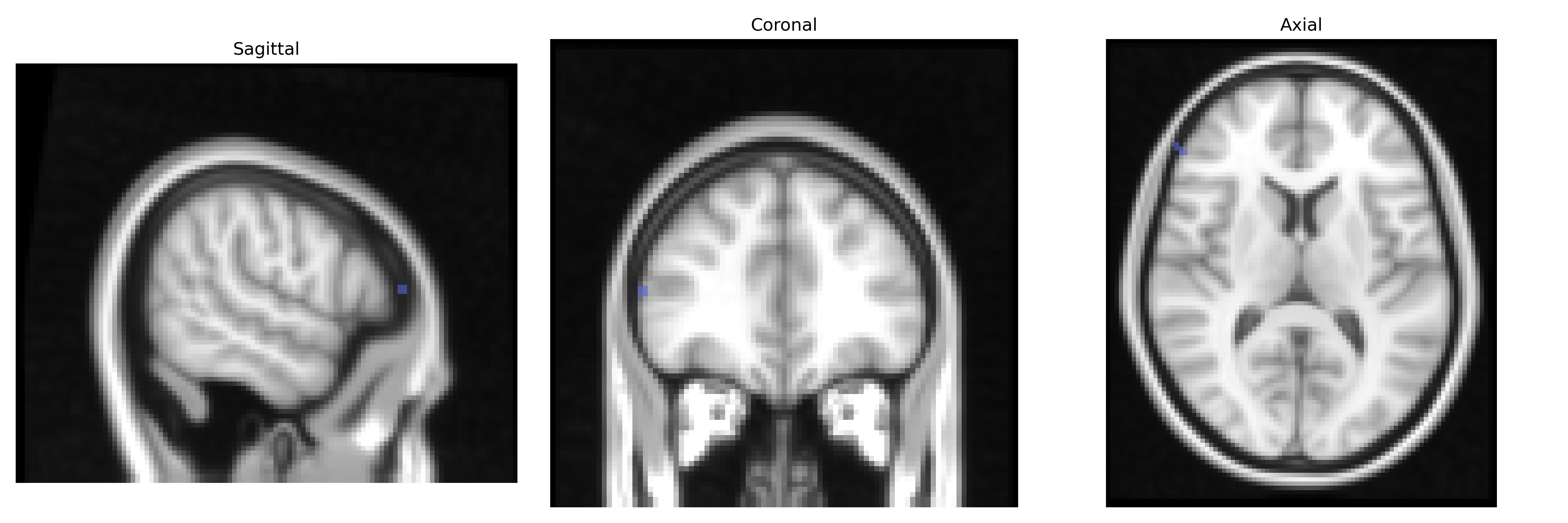
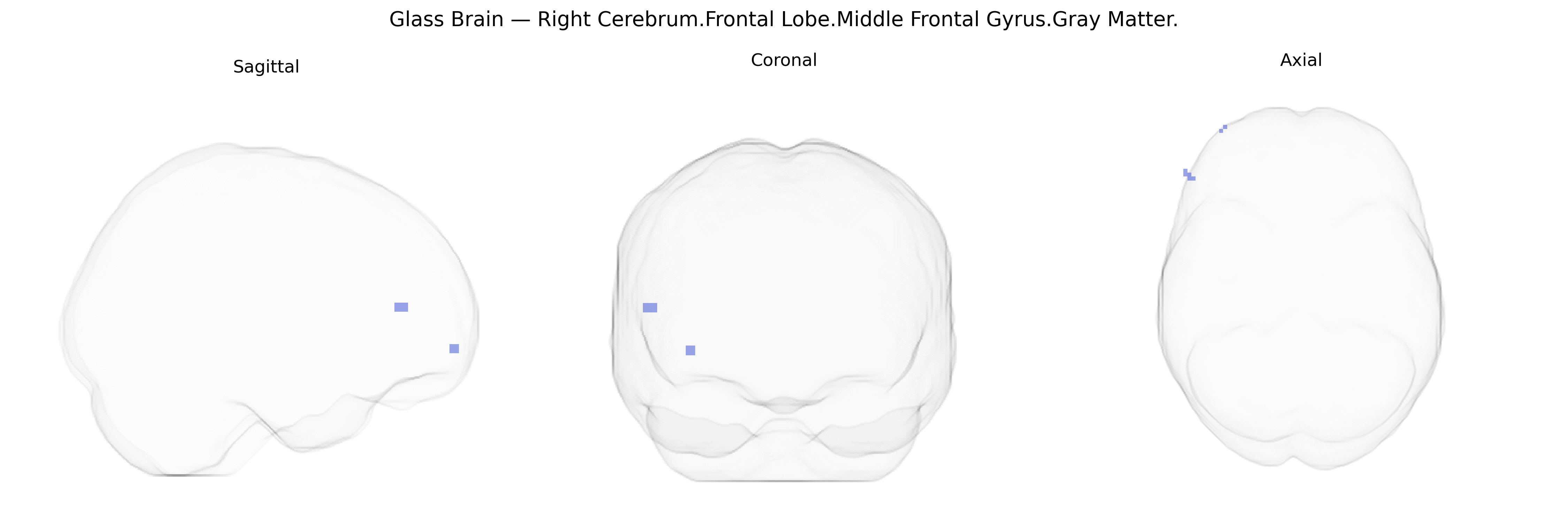
The bilateral Right Cerebrum.Frontal Lobe.Middle Frontal Gyrus.Gray Matter corresponds to cortical tissue in the dorsolateral portion of the frontal lobe, involved in higher-order executive functions such as working memory, attentional control, planning, and the regulation of goal-directed behavior. This region participates in complex cognitive processes including decision-making, task switching, and the integration of sensory and mnemonic information to guide actions, and is closely interconnected with prefrontal, parietal, and limbic structures. Neuronal populations in this area contribute to the selection and maintenance of behavioral strategies and the modulation of motor outputs based on contextual demands. There is no direct link for this exact Talairach label; a closely related structure is the Middle frontal gyrus.
The bilateral right middle frontal gyrus gray matter (Talairach 2 mm atlas: Right Cerebrum.Frontal Lobe.Middle Frontal Gyrus.Gray Matter) is a key component of the dorsolateral prefrontal cortex and has been repeatedly implicated in genetic studies of cognition, psychiatric risk, and brain structure. GWAS of cortical thickness and surface area have identified associations between common variants near genes such as PCDH family members, microtubule and synaptic genes (e.g., MAPT region, DLG4-related loci), and broad frontal lobe morphometry, with some studies reporting regionally specific signals in middle frontal gyrus measures of thickness or volume. Polygenic risk for schizophrenia, bipolar disorder, and major depression is associated with reduced gray matter volume and altered activation in this region, and risk loci in genes like CACNA1C, ZNF804A, and GRM3 have been linked to prefrontal structural and functional differences encompassing the middle frontal gyrus. GWAS of intelligence, educational attainment, and working memory show that higher polygenic scores for these traits correlate with greater right dorsolateral prefrontal (including middle frontal) volume and more efficient task-related activation, suggesting a genetically mediated contribution of this region to executive function. Additionally, large consortia (e.g., ENIGMA) have reported that variants in genes involved in neurodevelopment, synaptic plasticity, and axon guidance (such as CNTNAP2, DISC1-related pathways, and BDNF polymorphisms) are associated with prefrontal cortical measures that include the middle frontal gyrus, linking genetic architecture of neurodevelopmental and psychiatric disorders (schizophrenia, ADHD, autism spectrum conditions) to structural and functional variation in this specific frontal gray matter region.
Overview generated by GPT-4o (2026).
Region ID: 618
Hemisphere: bilateral
Atlas: Talairach labels 2mm
Full Quality Version: Download MP4
Full Quality Version: Download MP4
Wali Sidiqyar*, Gaurav Rudravaram*, Elyssa M. McMaster, Trent M. Schwartz, Adam M. Saunders, Kurt G. Schilling, Bennett A. Landman "Introducing SPINS: A Shared Public Visualization Library of Neuroanatomical Structures." Medical Imaging with Deep Learning- short paper
This resource is licensed under CC0 1.0 Universal (Public Domain).