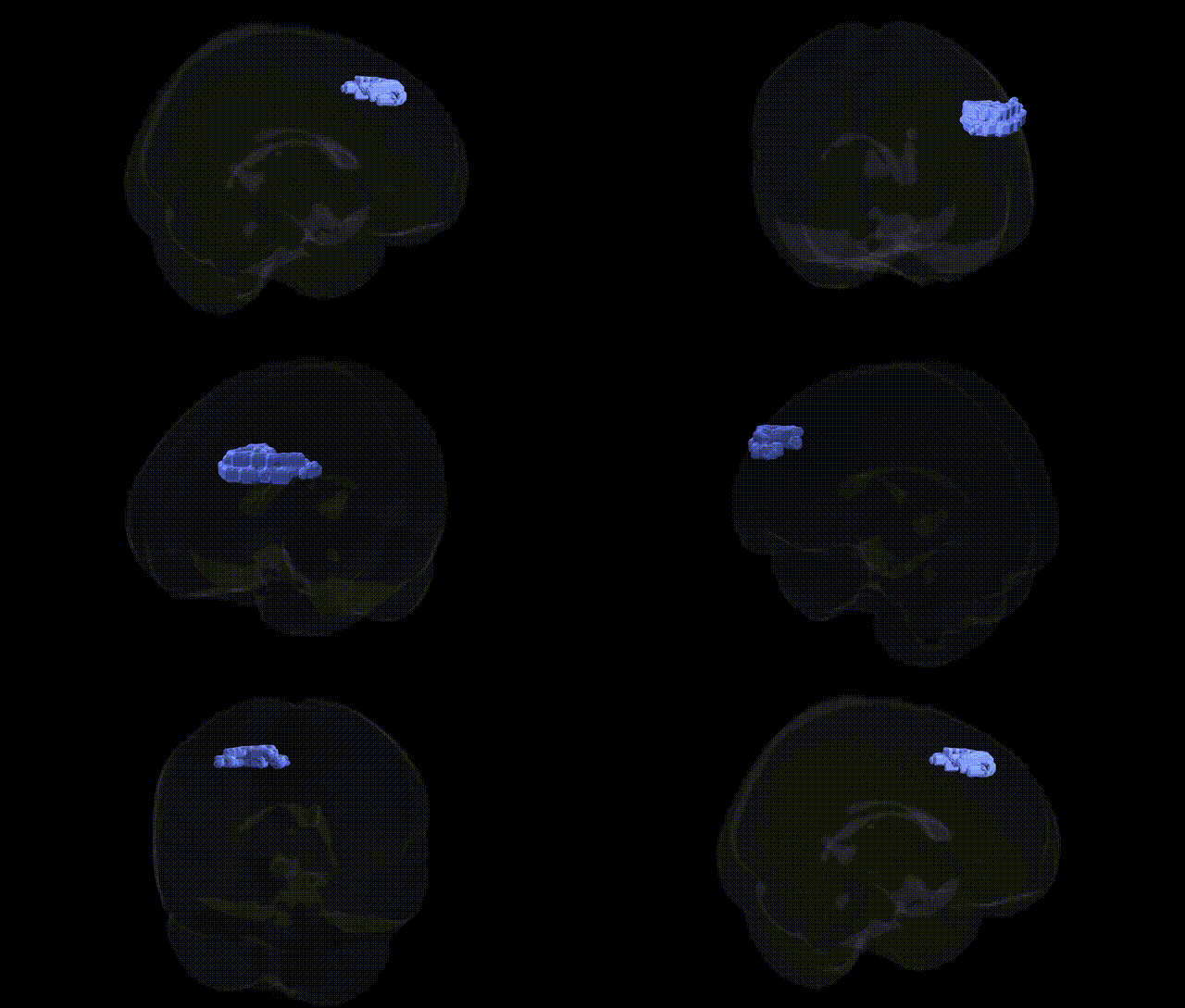
Bilateral Right Cerebrum.Frontal Lobe.Middle Frontal Gyrus.Gray Matter.Brodmann area 8 corresponds to a dorsolateral frontal cortical region implicated in higher-order motor control, including the planning, selection, and voluntary initiation of saccadic eye movements, as well as aspects of executive functioning such as attention, working memory, and response inhibition. Cytoarchitectonically defined by Brodmann as area 8, this territory lies anterior to the primary motor and premotor cortices and contributes to the frontal eye fields and associated oculomotor networks that integrate visual, spatial, and goal-directed signals for orienting behavior. Through extensive reciprocal connections with parietal, temporal, and subcortical structures, Brodmann area 8 participates in top-down control of behavior, flexible adaptation to changing task demands, and the coordination of complex action sequences. There is no direct link for “Brodmann area 8” as a standalone article; see the related structure Brodmann area.
The bilateral right middle frontal gyrus gray matter in Brodmann area 8 (Talairach 2 mm atlas) corresponds to a key dorsal prefrontal/superior frontal region implicated in executive control, working memory, and oculomotor functions, and genetic associations are typically inferred from imaging‑genetics and GWAS of brain structure and function rather than region‑specific candidate gene studies. Large neuroimaging GWAS consortia (e.g., ENIGMA, UK Biobank) have identified common variants influencing cortical thickness and surface area in nearby superior/middle frontal territories—often overlapping BA8—including loci near genes involved in neurodevelopment and synaptic function such as HMGA2, FOXO3, RSPO3, and microtubule or axon guidance genes, with polygenic scores for general cognitive ability and educational attainment showing robust associations with BA8‑adjacent frontal morphology and activation patterns during working memory or cognitive control tasks. Structural and functional abnormalities in this region have been repeatedly observed in psychiatric GWAS contexts: risk variants for schizophrenia, bipolar disorder, and major depressive disorder (e.g., within CACNA1C, GRM3, ZNF804A, and other synaptic/ion‑channel genes) correlate with altered activation and connectivity in right middle frontal/superior frontal areas during executive and emotional processing, and polygenic risk scores for these disorders predict reduced cortical thickness or altered resting‑state connectivity in BA8‑proximal regions. In ADHD and autism spectrum disorder, GWAS‑derived polygenic risk is associated with differences in frontal cortical development, including right middle frontal gyrus, consistent with this region’s role in attention and cognitive flexibility, while GWAS of neuroticism, anxiety, and impulsivity link frontal control networks incorporating BA8 with genetic variants affecting fronto‑striatal and fronto‑limbic circuitry. Additionally, GWAS of brain functional networks show heritable variation in the dorsal frontoparietal/attention network that includes BA8, indicating that numerous small‑effect variants collectively shape structure and function of the right middle frontal gyrus rather than any single gene exerting a specific, isolated effect on this Talairach‑defined region.
Overview generated by GPT-4o (2026).
Region ID: 1026
Hemisphere: bilateral
Atlas: Talairach labels 2mm
Full Quality Version: Download MP4
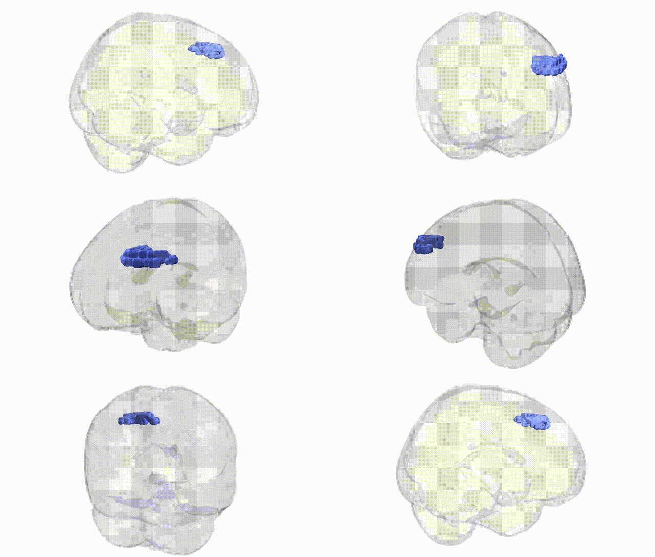
Full Quality Version: Download MP4
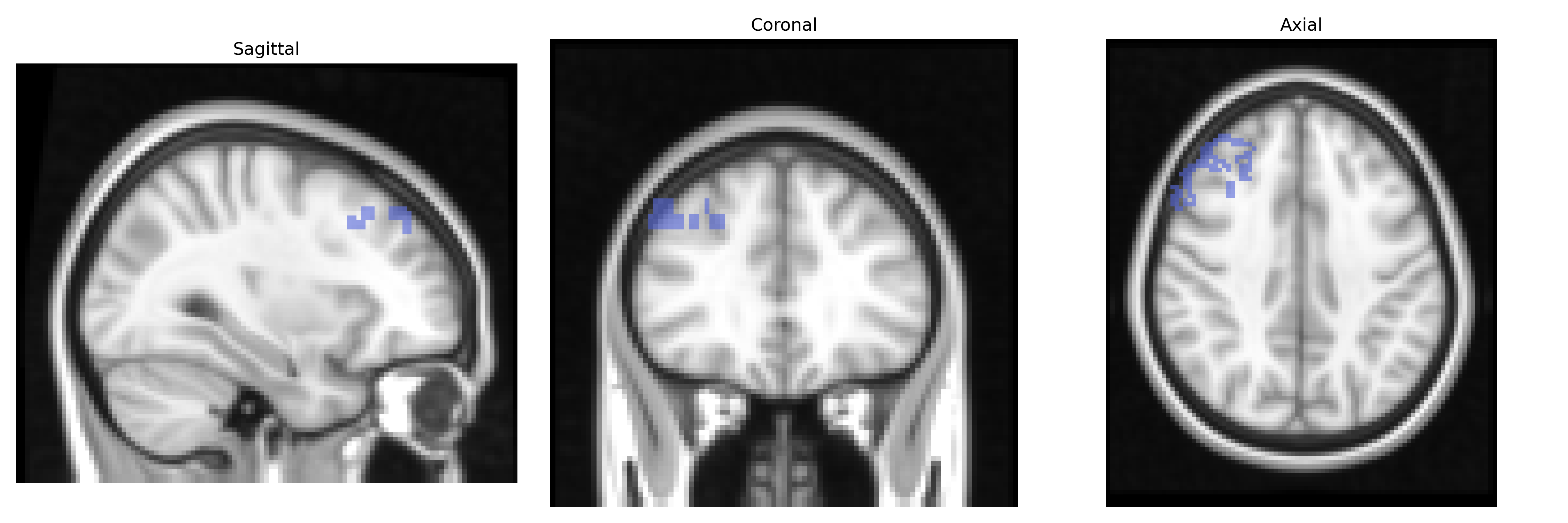
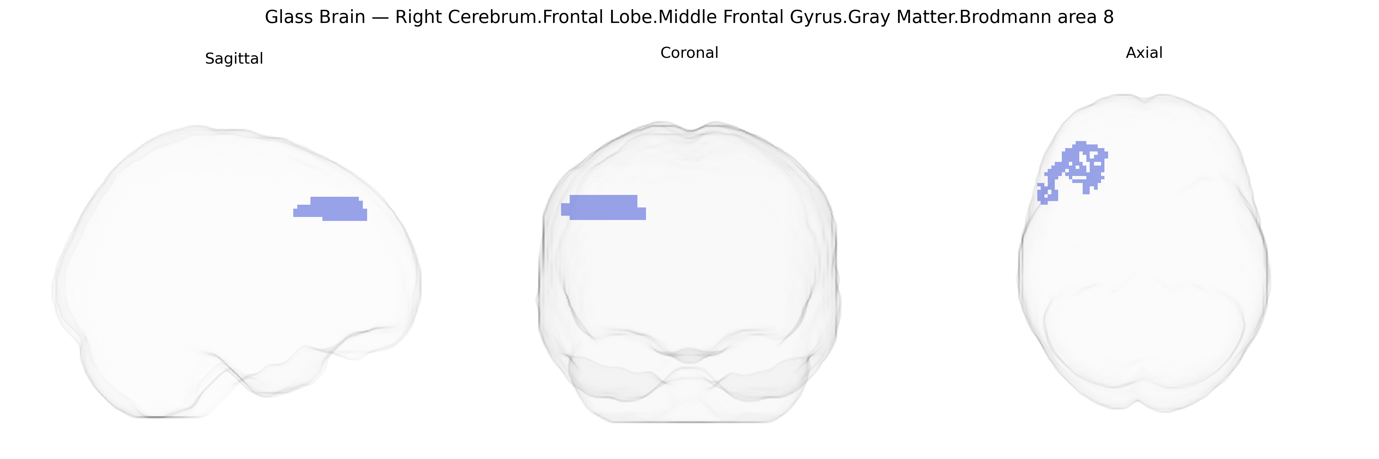
Wali Sidiqyar*, Gaurav Rudravaram*, Elyssa M. McMaster, Trent M. Schwartz, Adam M. Saunders, Kurt G. Schilling, Bennett A. Landman "Introducing SPINS: A Shared Public Visualization Library of Neuroanatomical Structures." Medical Imaging with Deep Learning- short paper
This resource is licensed under CC0 1.0 Universal (Public Domain).