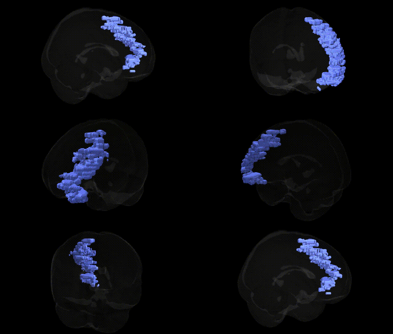
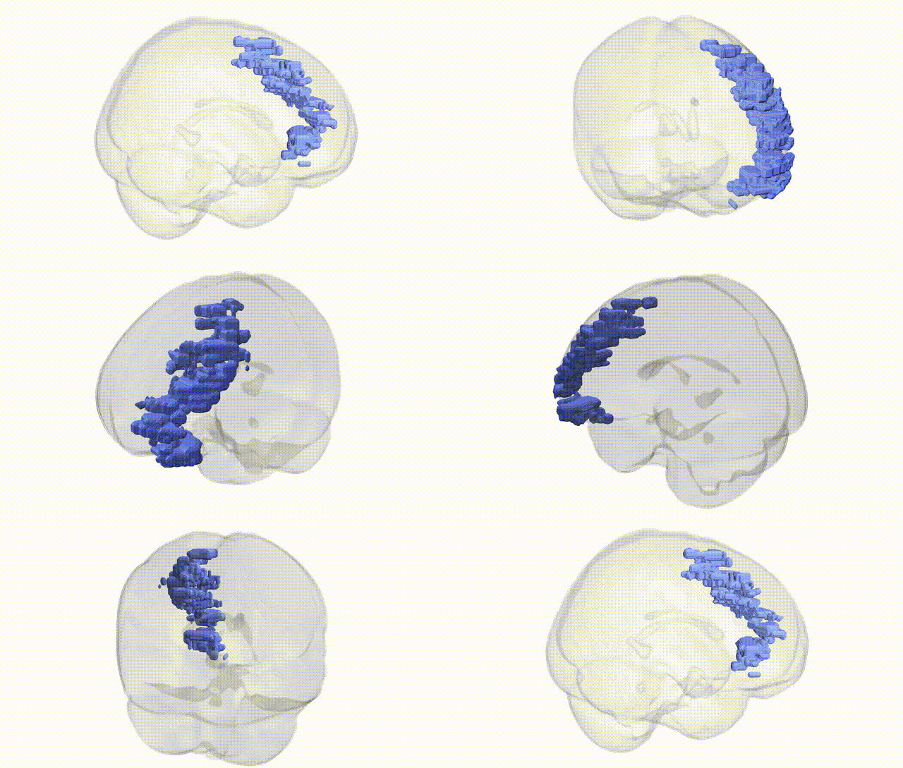
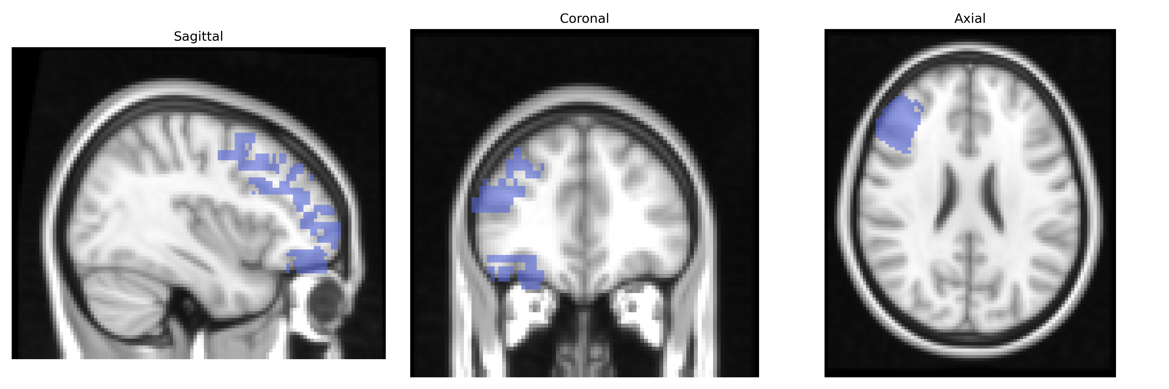
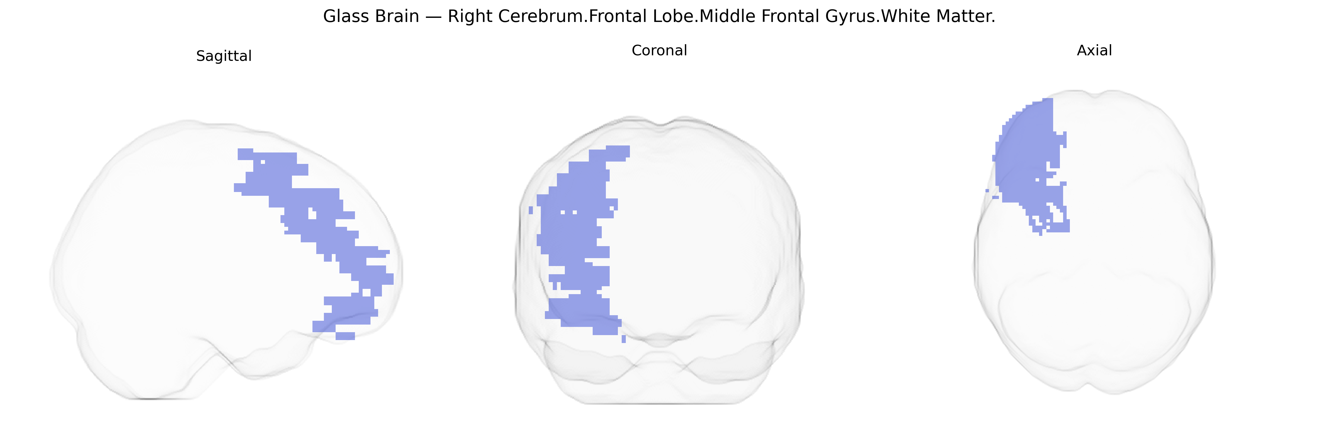
The bilateral Right Cerebrum.Frontal Lobe.Middle Frontal Gyrus.White Matter corresponds to the subcortical fiber pathways underlying the middle frontal gyrus in the right frontal lobe. This region contains association fibers connecting dorsolateral prefrontal cortical areas with other frontal, parietal, and temporal regions, supporting higher-order executive functions such as working memory, cognitive control, planning, and attentional modulation. Its white matter tracts contribute to large-scale networks involved in goal-directed behavior and integration of sensory and mnemonic information. Although there is no direct Wikipedia article for this specific white matter subdivision, it is structurally related to the Middle frontal gyrus.
Genetic associations involving the middle frontal gyrus white matter of the right frontal lobe (as defined in the Talairach 2 mm atlas) typically arise from imaging genetics and MRI-based GWAS of white matter microstructure and frontal lobe connectivity rather than region-specific candidate gene studies. Large GWAS of diffusion tensor imaging (DTI) metrics have linked common variants near genes involved in axonal guidance, myelination, and synaptic development—such as CNTNAP2, NRG1, MAG, and genes within the major histocompatibility complex (MHC) region—to variation in fractional anisotropy and other microstructural measures in frontal association tracts that traverse or connect the middle frontal gyrus (e.g., superior longitudinal fasciculus, anterior corona radiata). Polygenic risk scores for schizophrenia, bipolar disorder, major depressive disorder, ADHD, and autism spectrum disorder have been repeatedly associated with altered frontal white matter integrity, including middle frontal regions, in large cohorts like UK Biobank; specific loci in CACNA1C, ZNF804A, DRD2, and GRM3 have been implicated in modulating prefrontal connectivity and working-memory–related circuits. Frontal white matter serving the middle frontal gyrus also shows genetic correlations with cognitive traits—particularly general intelligence, executive function, and educational attainment—where GWAS hits in genes related to neuronal development (e.g., KIBRA/WFS1, SHANK3, and others) track with prefrontal microstructure. Additionally, frontally distributed white matter abnormalities influenced by APOE, CLU, and other Alzheimer’s disease–related variants have been observed in aging and neurodegeneration studies, suggesting that genetic risk for dementia affects middle frontal gyrus white matter pathways. Overall, while no single gene is uniquely or exclusively tied to this Talairach-defined region, convergent GWAS and imaging-genetic evidence indicate that common variants affecting neurodevelopmental, myelination, and synaptic pathways contribute to structural and functional variation in the right middle frontal gyrus white matter and to its involvement in psychiatric disorders, cognitive traits, and neurodegenerative risk.
Overview generated by GPT-4o (2026).
Region ID: 196
Hemisphere: bilateral
Atlas: Talairach labels 2mm
Full Quality Version: Download MP4
Full Quality Version: Download MP4
Wali Sidiqyar*, Gaurav Rudravaram*, Elyssa M. McMaster, Trent M. Schwartz, Adam M. Saunders, Kurt G. Schilling, Bennett A. Landman "Introducing SPINS: A Shared Public Visualization Library of Neuroanatomical Structures." Medical Imaging with Deep Learning- short paper
This resource is licensed under CC0 1.0 Universal (Public Domain).