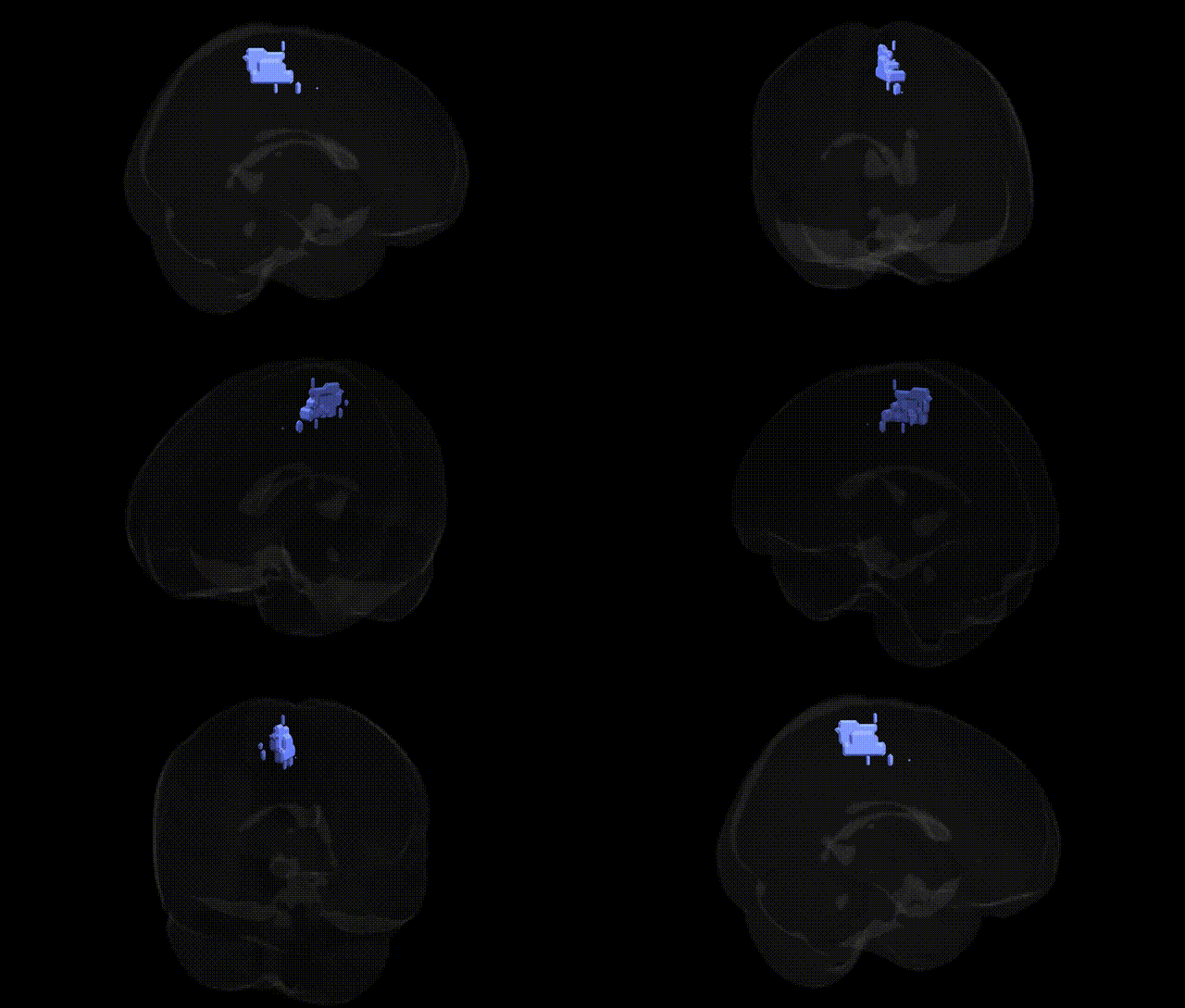
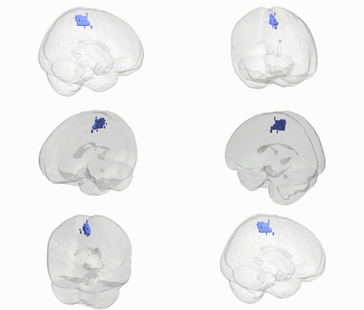
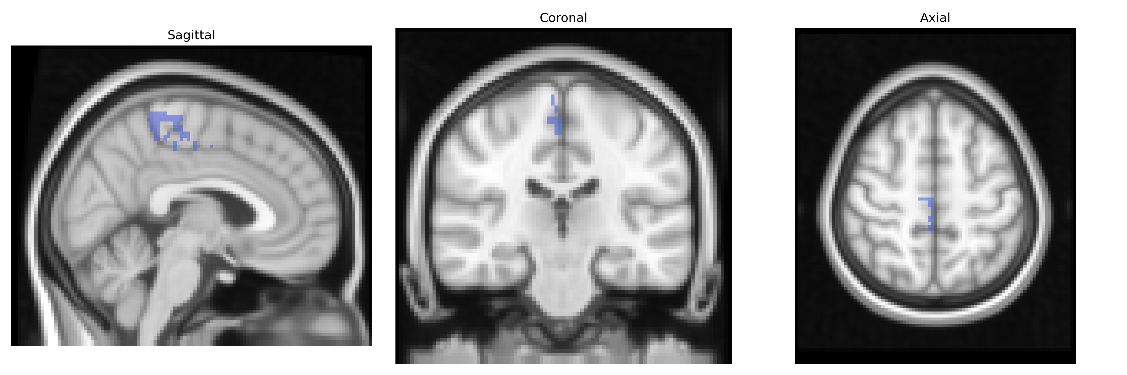
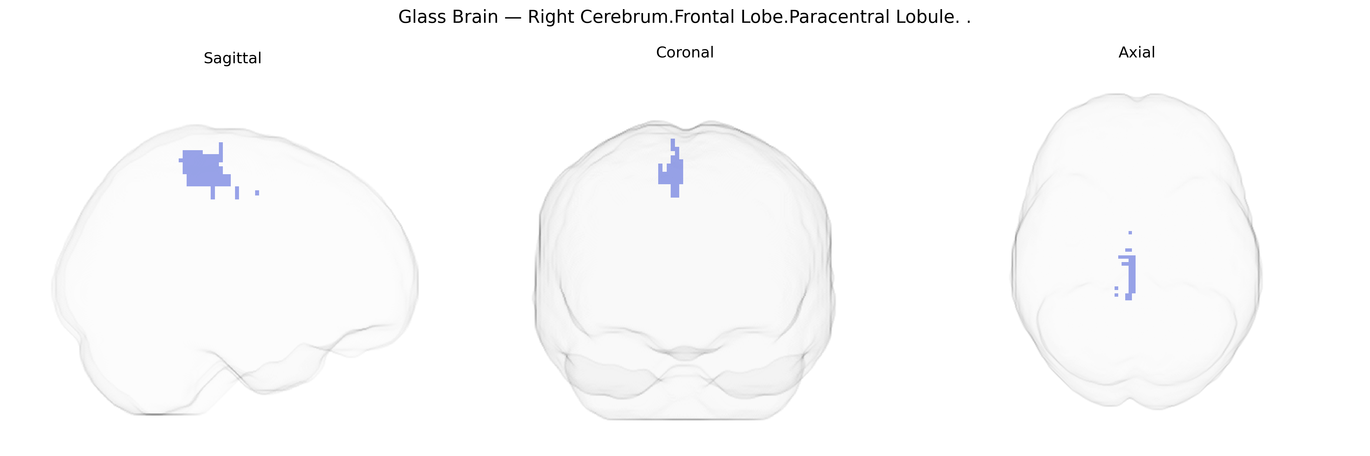
The bilateral Right Cerebrum.Frontal Lobe.Paracentral Lobule corresponds to the right paracentral lobule, a medial frontal cortical region bordering the central sulcus and spanning portions of the primary motor (precentral gyrus) and primary somatosensory (postcentral gyrus) cortices for the contralateral lower limb and perineal areas. Functionally, it is crucial for voluntary motor control of the leg and foot, integration of somatosensory input from these regions, and participation in sensorimotor aspects of gait and posture. The paracentral lobule lies on the medial surface of the hemisphere within the frontal lobe’s extension into the interhemispheric fissure and is closely connected with supplementary motor areas and other sensorimotor networks. There is no direct link for “Paracentral lobule,” but it is a subdivision of the Frontal lobe.
The bilateral right paracentral lobule (medial frontal lobe, encompassing primary motor and somatosensory regions for lower limbs and aspects of motor control) has been implicated in several genetic and GWAS-based associations, though often under broader labels such as “medial frontal,” “supplementary motor area,” or “sensorimotor cortex.” Imaging genetics studies from large cohorts (e.g., ENIGMA, UK Biobank) have reported SNPs and loci affecting cortical thickness, surface area, and volume in medial frontal/paracentral regions, including variants near genes involved in neurodevelopment and synaptic function (such as MECP2, MAPT region 17q21, and multiple glutamatergic and axon-guidance pathways), with heritability estimates in the moderate range. GWAS of motor phenotypes and gait parameters, as well as lower-extremity strength and coordination, have indirectly implicated genetic influences on paracentral lobule structure and function, consistent with its role in leg and foot representation. In disease-focused genetics, this region shows altered activation and structural changes associated with risk alleles in multiple sclerosis (e.g., HLA-DRB1*15:01 and other immune-related loci) and hereditary spastic paraplegia genes affecting corticospinal tract integrity, aligning with motor symptoms involving the lower limbs. Genetic risk for neurodegenerative disorders such as amyotrophic lateral sclerosis and Parkinson’s disease has also been tied to sensorimotor cortical changes that include paracentral lobule involvement, although specific locus-to-region mappings are typically indirect. In psychiatric genetics, common risk variants for attention-deficit/hyperactivity disorder and schizophrenia have been associated with altered medial frontal and paracentral activation during motor inhibition and cognitive control tasks, and polygenic risk scores for these disorders correlate with subtle structural variation in adjacent medial frontal areas in some imaging genetics analyses, suggesting that the paracentral lobule participates in broader genetic networks influencing motor, cognitive, and sensorimotor integration.
Overview generated by GPT-4o (2026).
Region ID: 1048
Hemisphere: bilateral
Atlas: Talairach labels 2mm
Full Quality Version: Download MP4
Full Quality Version: Download MP4
Wali Sidiqyar*, Gaurav Rudravaram*, Elyssa M. McMaster, Trent M. Schwartz, Adam M. Saunders, Kurt G. Schilling, Bennett A. Landman "Introducing SPINS: A Shared Public Visualization Library of Neuroanatomical Structures." Medical Imaging with Deep Learning- short paper
This resource is licensed under CC0 1.0 Universal (Public Domain).