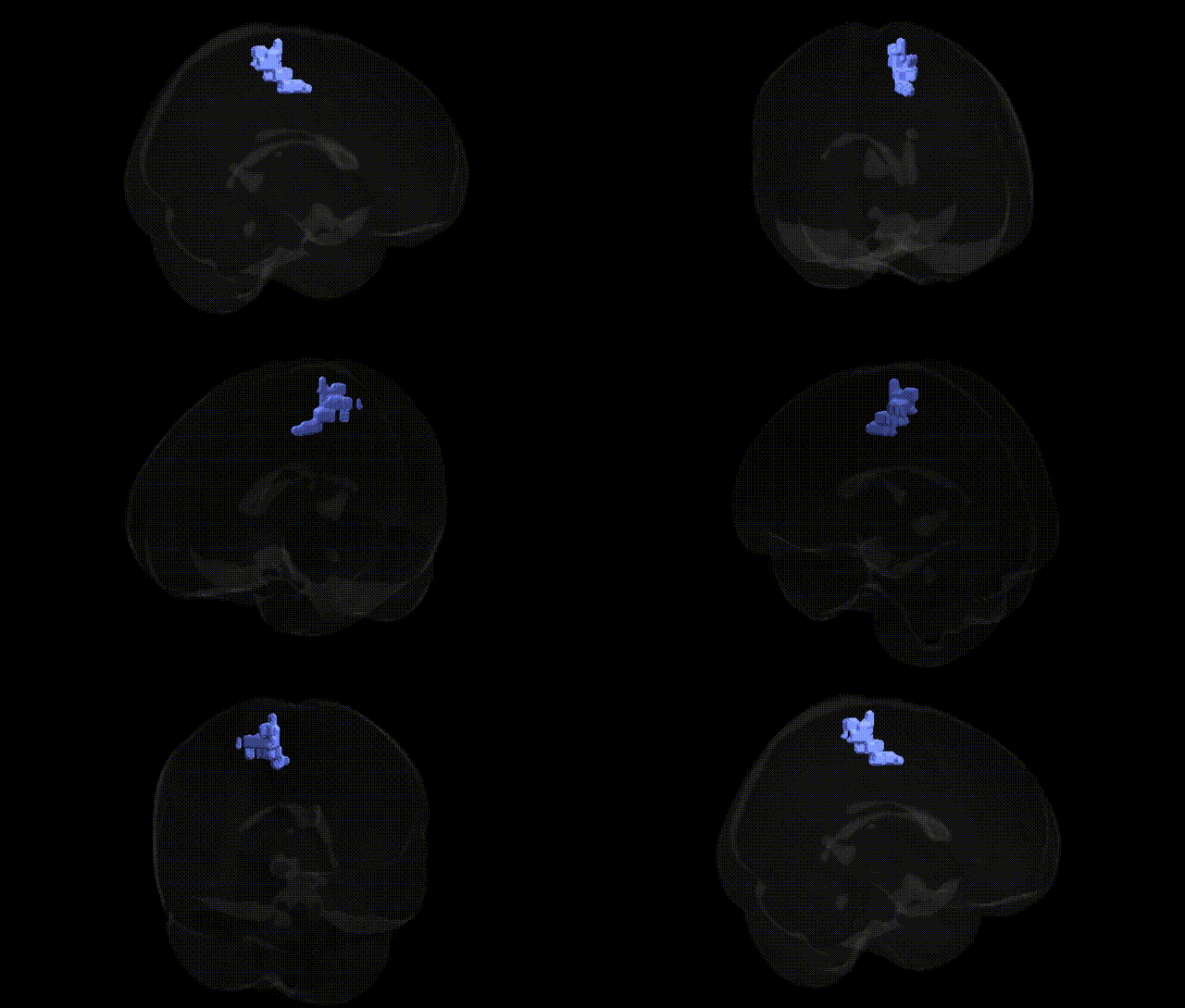
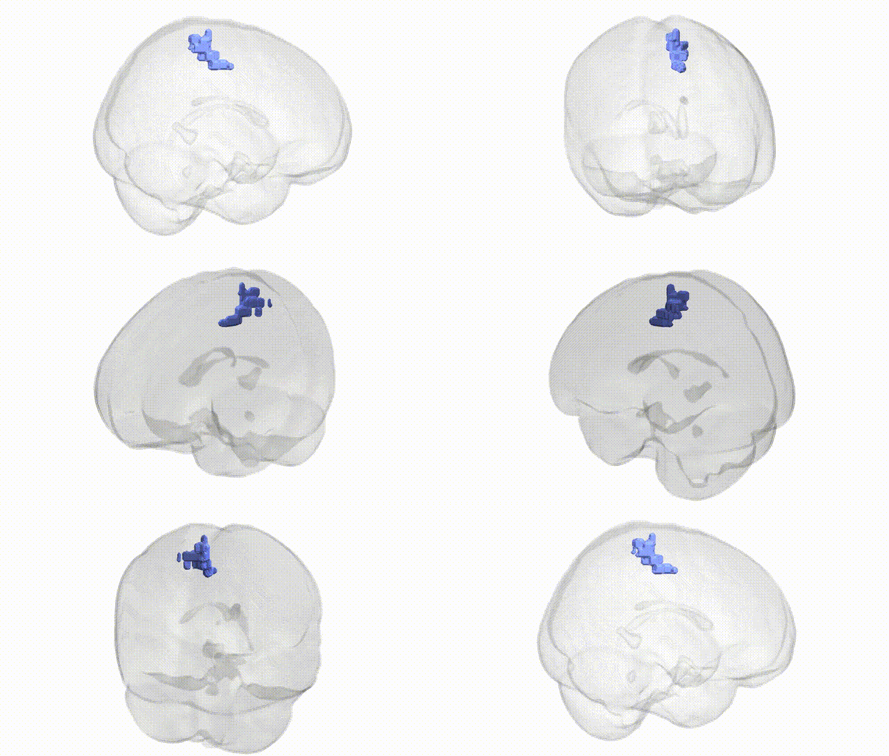
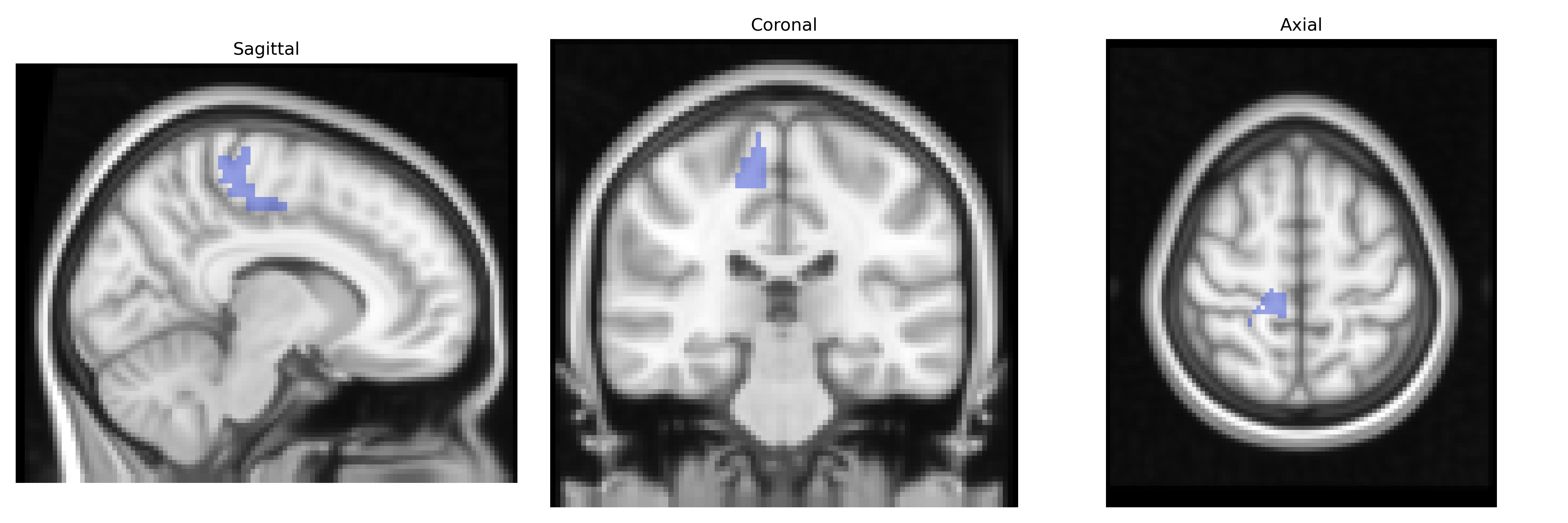
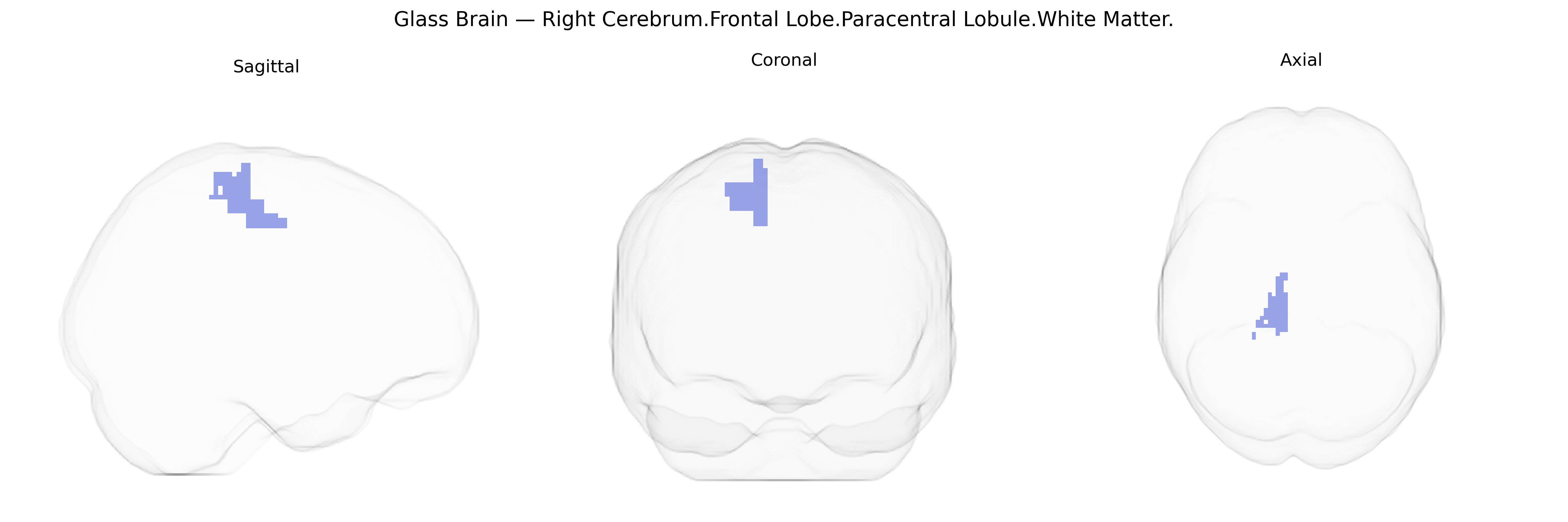
The bilateral Right Cerebrum.Frontal Lobe.Paracentral Lobule.White Matter corresponds to the subcortical myelinated fiber tracts underlying the paracentral lobule of the frontal lobe, located along the medial surface of the hemisphere adjacent to the central sulcus. This white matter region contains association and projection fibers that interconnect the primary motor cortex (particularly the representations of the lower limb and supplementary motor areas) with other frontal, parietal, and subcortical structures, supporting motor planning, execution, and sensorimotor integration. Functionally, it contributes to the coordination and modulation of voluntary movements and integration of somatosensory feedback, especially for lower extremity control, via its role in corticospinal and intra-frontal connectivity. There is no direct link for this exact white matter subdivision; a related cortical structure is the Paracentral lobule.
The bilateral white matter of the right paracentral lobule—part of the medial frontal lobe near the primary motor and somatosensory cortices and represented in the Talairach 2 mm atlas—has been implicated in multiple genetic and GWAS-based neuroimaging findings, although most studies do not isolate this tract by name and instead examine adjacent sensorimotor and medial frontal white matter. Polygenic influences on cortical thickness, surface area, and white matter microstructure in this region have been identified in large imaging-genetics datasets (e.g., ENIGMA, UK Biobank), with common variants near genes involved in axon guidance, myelination, and synaptic development (such as CNTNAP2, NRG1, MAG, and multiple loci in the major histocompatibility complex) contributing to regional structure. GWAS of motor-related phenotypes (e.g., fine motor skills, gait, and muscle strength) and somatosensory traits show associations with structural variation in paracentral and adjacent sensorimotor white matter, reflecting genetic effects on corticospinal and sensorimotor pathways. In psychiatric and neurodevelopmental disorders, schizophrenia, bipolar disorder, autism spectrum disorder, and ADHD have all demonstrated heritable alterations in medial frontal and paracentral white matter integrity—often measured via fractional anisotropy or tract-based spatial statistics—with risk variants in genes like CACNA1C, GRIN2A, and common polygenic risk scores for these conditions correlating with microstructural changes in this area. Neurodegenerative disorders, particularly multiple sclerosis and, to a lesser extent, ALS and Parkinson’s disease, show lesion burden or microstructural abnormalities in paracentral white matter that correlate with risk alleles in immune, myelin, and neuroinflammatory genes (e.g., HLA-DRB1 for MS). Although highly specific GWAS hits for “Right Cerebrum.Frontal Lobe.Paracentral Lobule.White Matter” are not typically reported as a standalone region, convergent evidence from imaging-genetics, polygenic risk analyses, and disorder-focused studies supports a role for this tract as a genetically influenced node in motor, somatosensory, and frontal networks affected in both normal variation and multiple brain disorders.
Overview generated by GPT-4o (2026).
Region ID: 1052
Hemisphere: bilateral
Atlas: Talairach labels 2mm
Full Quality Version: Download MP4
Full Quality Version: Download MP4
Wali Sidiqyar*, Gaurav Rudravaram*, Elyssa M. McMaster, Trent M. Schwartz, Adam M. Saunders, Kurt G. Schilling, Bennett A. Landman "Introducing SPINS: A Shared Public Visualization Library of Neuroanatomical Structures." Medical Imaging with Deep Learning- short paper
This resource is licensed under CC0 1.0 Universal (Public Domain).