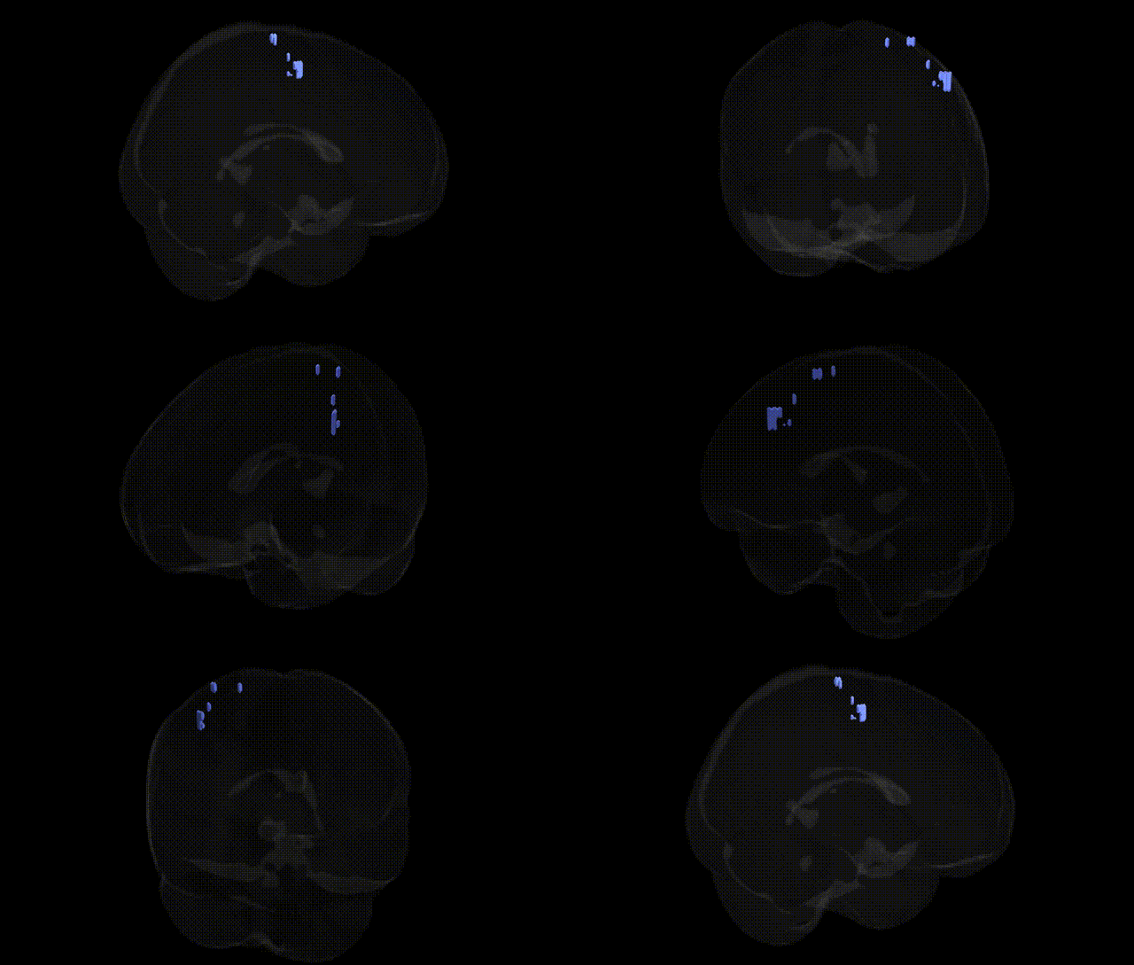
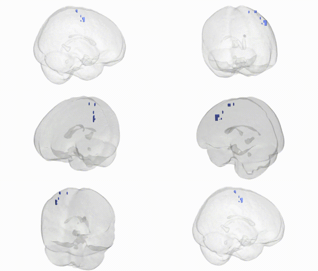
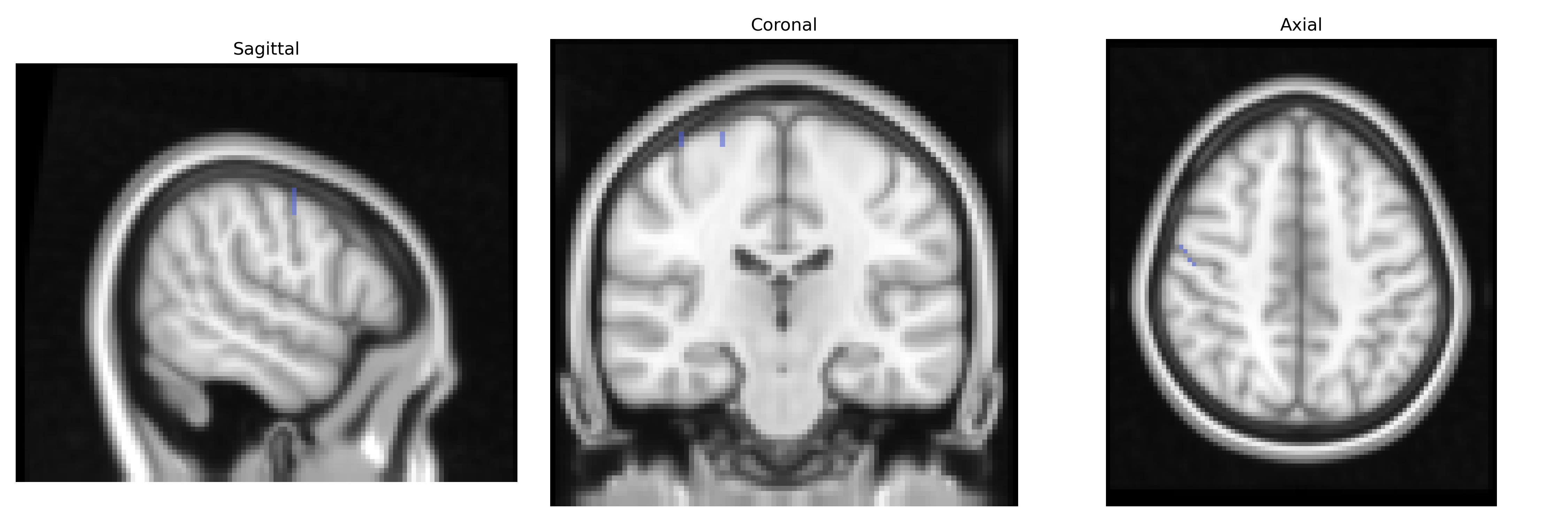
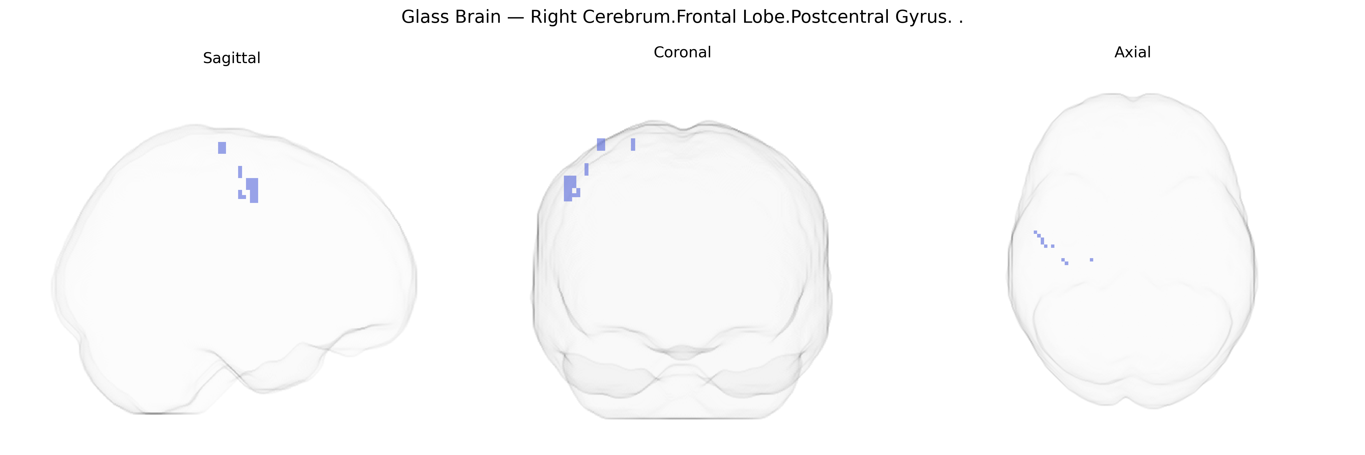
The bilateral Right Cerebrum.Frontal Lobe.Postcentral Gyrus label in the Talairach 2 mm atlas appears to reflect a misclassification, as the postcentral gyrus is anatomically part of the parietal lobe rather than the frontal lobe. The postcentral gyrus is the primary somatosensory cortex (Brodmann areas 3, 1, and 2), receiving and processing afferent input from the body via thalamic relays, and is crucial for conscious perception of tactile stimuli, proprioception, nociception, and temperature. It displays somatotopic organization (sensory homunculus), with different body regions mapped along its mediolateral axis, and it integrates sensory information for fine discrimination of texture, shape, and localization of stimuli. Functionally, activity in this region contributes to sensorimotor integration, influences motor planning via projections to frontal motor areas, and participates in higher-order processes such as tactile attention and body representation. There is no direct Wikipedia article for “Right Cerebrum.Frontal Lobe.Postcentral Gyrus”; a closely related and more accurate structure is the Postcentral gyrus.
The bilateral right postcentral gyrus (Talairach: Right Cerebrum.Frontal Lobe.Postcentral Gyrus) corresponds primarily to primary somatosensory cortex, and genetic associations involving this region largely emerge from imaging genetics and GWAS of brain structure and function rather than region-specific candidate gene work. Large-scale neuroimaging GWAS (e.g., ENIGMA and UK Biobank) have shown that cortical thickness and surface area in somatosensory regions, including the postcentral gyrus, are highly heritable and influenced by variants in genes related to neurodevelopment, synaptic function, and neuronal migration, such as those in Wnt signaling and cytoskeletal pathways, though typically reported at the level of parietal or global cortical measures rather than this narrow Talairach-defined label. Variants near genes implicated in neurodevelopmental and psychiatric conditions—such as CNTNAP2, CACNA1C, and others identified in schizophrenia, autism spectrum disorder, and ADHD GWAS—have been associated with altered somatosensory or sensorimotor cortex morphology and activation, and postcentral gyrus abnormalities have been reported in these disorders, but the evidence generally links gene variants to broader network-level changes rather than uniquely to the right postcentral gyrus. Pain-related and somatosensory traits (e.g., chronic pain, tactile sensitivity) show associations with cortical measures and activation patterns in postcentral gyrus, with GWAS identifying multiple loci (including in genes related to nociception, immune function, and neuroinflammation such as OPRM1 and HLA-region variants) that may indirectly influence structure or responsivity of this region, although spatial resolution in most genetic imaging studies does not isolate the bilateral right postcentral gyrus specifically. Overall, genetic influences on this Talairach-defined region are well supported in terms of heritability of somatosensory cortical anatomy and function, but current GWAS and imaging-genetic findings tend to implicate broader somatosensory and parietal circuits rather than providing highly specific, uniquely validated gene associations for this single right-hemisphere label.
Overview generated by GPT-4o (2026).
Region ID: 980
Hemisphere: bilateral
Atlas: Talairach labels 2mm
Full Quality Version: Download MP4
Full Quality Version: Download MP4
Wali Sidiqyar*, Gaurav Rudravaram*, Elyssa M. McMaster, Trent M. Schwartz, Adam M. Saunders, Kurt G. Schilling, Bennett A. Landman "Introducing SPINS: A Shared Public Visualization Library of Neuroanatomical Structures." Medical Imaging with Deep Learning- short paper
This resource is licensed under CC0 1.0 Universal (Public Domain).