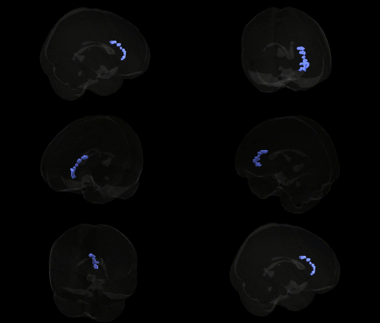
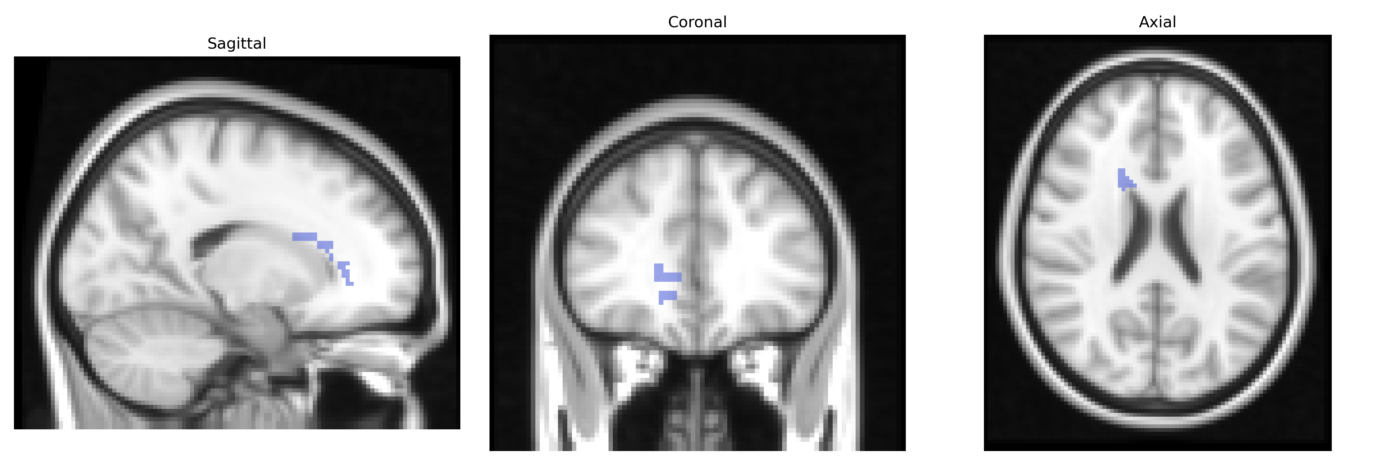
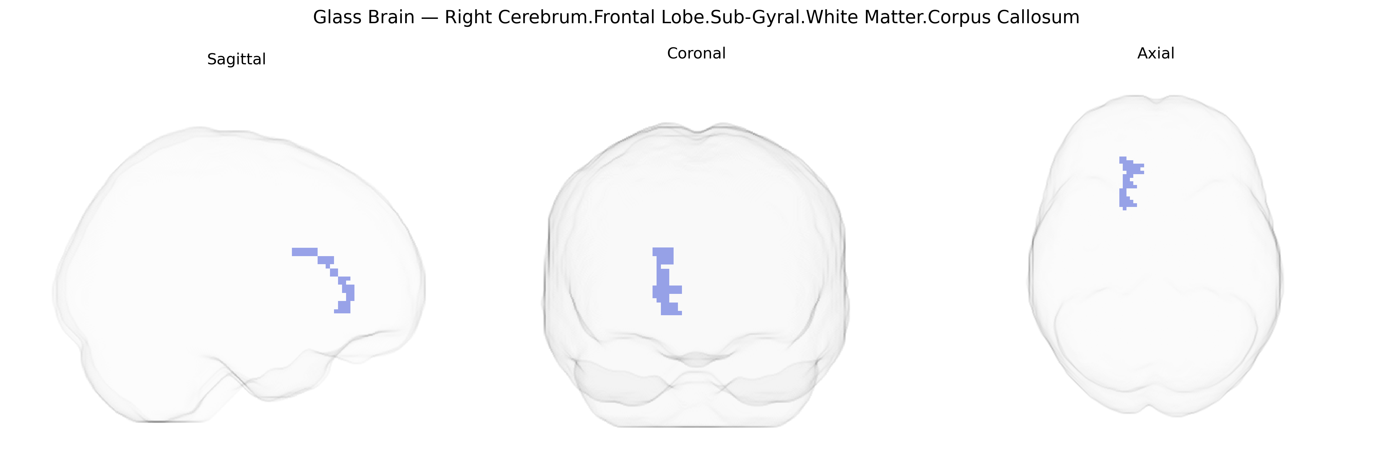
The bilateral Right Cerebrum.Frontal Lobe.Sub-Gyral.White Matter.Corpus Callosum region refers to commissural white matter fibers situated beneath the frontal cortical gyri, within the frontal lobe, that traverse the corpus callosum to interconnect homologous and associated regions of the left and right frontal cortex. These fibers support interhemispheric integration of executive functions, motor planning, working memory, and higher-order cognitive and behavioral regulation by facilitating rapid communication between frontal networks in both hemispheres. Structurally, this region consists primarily of heavily myelinated axons organized within callosal segments projecting to frontal areas, and it plays a crucial role in coordinated bilateral motor activity and complex cognitive processing dependent on frontal lobe circuitry. There is no direct link for this exact composite label; a closely related and encompassing structure is the Corpus callosum.
The bilateral Right Cerebrum.Frontal Lobe.Sub-Gyral.White Matter.Corpus Callosum region, as delineated in the Talairach 2 mm atlas, corresponds primarily to interhemispheric frontal white matter tracts that are frequently implicated in genetic studies of brain structure and connectivity. Large-scale GWAS and imaging-genetics consortia (e.g., ENIGMA and UK Biobank) have identified multiple loci associated with corpus callosum microstructure (such as fractional anisotropy and mean diffusivity) and callosal volume, including variants near or within genes involved in axon guidance and myelination (e.g., CNTNAP2, MAG, NRG1, and various oligodendrocyte-related genes), though findings are generally distributed across the corpus callosum rather than restricted to a specific frontal segment. Callosal and frontal white matter phenotypes in this region have shown genetic overlap with neurodevelopmental and psychiatric disorders, including schizophrenia, bipolar disorder, autism spectrum disorder, and ADHD, as well as with traits like general cognitive ability and processing speed, where polygenic risk scores and GWAS hits (for example in GRIN2A, CACNA1C, and other synaptic genes) correlate with altered frontal interhemispheric connectivity. Additionally, rare and common variants affecting midline development (such as mutations in L1CAM and other axon guidance molecules) can produce callosal dysgenesis involving frontal portions of the corpus callosum, linking this region to congenital disorders of connectivity. Overall, genetic findings indicate that variation in genes regulating neuronal migration, axon outgrowth, synaptic function, and myelin formation contributes to differences in the structure and integrity of this frontal callosal white matter region and its involvement in neurodevelopmental, psychiatric, and cognitive phenotypes.
Overview generated by GPT-4o (2026).
Region ID: 515
Hemisphere: bilateral
Atlas: Talairach labels 2mm
Full Quality Version: Download MP4
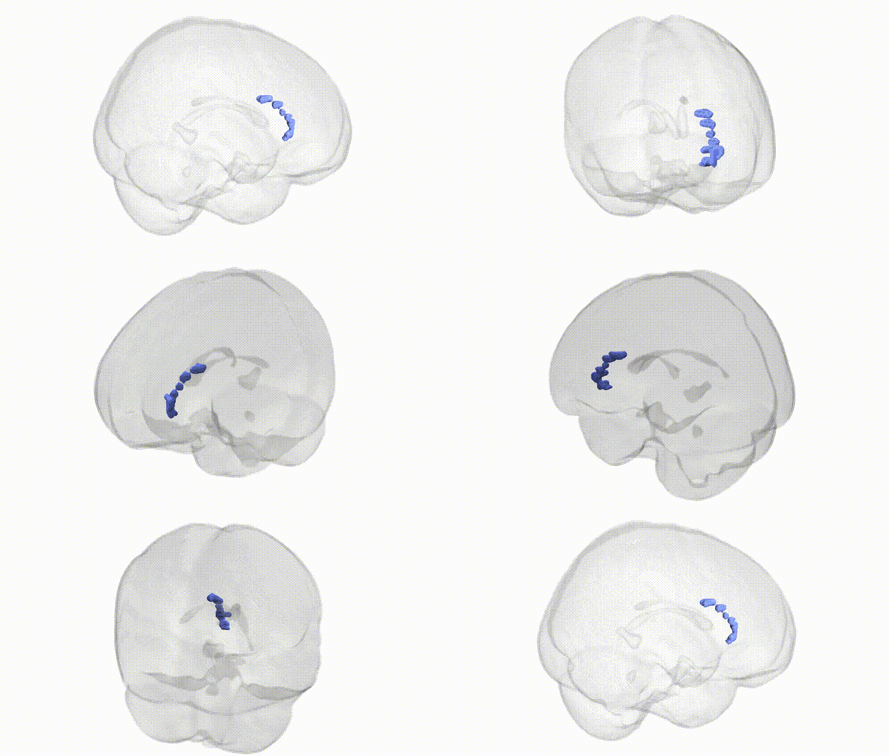
Full Quality Version: Download MP4
Wali Sidiqyar*, Gaurav Rudravaram*, Elyssa M. McMaster, Trent M. Schwartz, Adam M. Saunders, Kurt G. Schilling, Bennett A. Landman "Introducing SPINS: A Shared Public Visualization Library of Neuroanatomical Structures." Medical Imaging with Deep Learning- short paper
This resource is licensed under CC0 1.0 Universal (Public Domain).