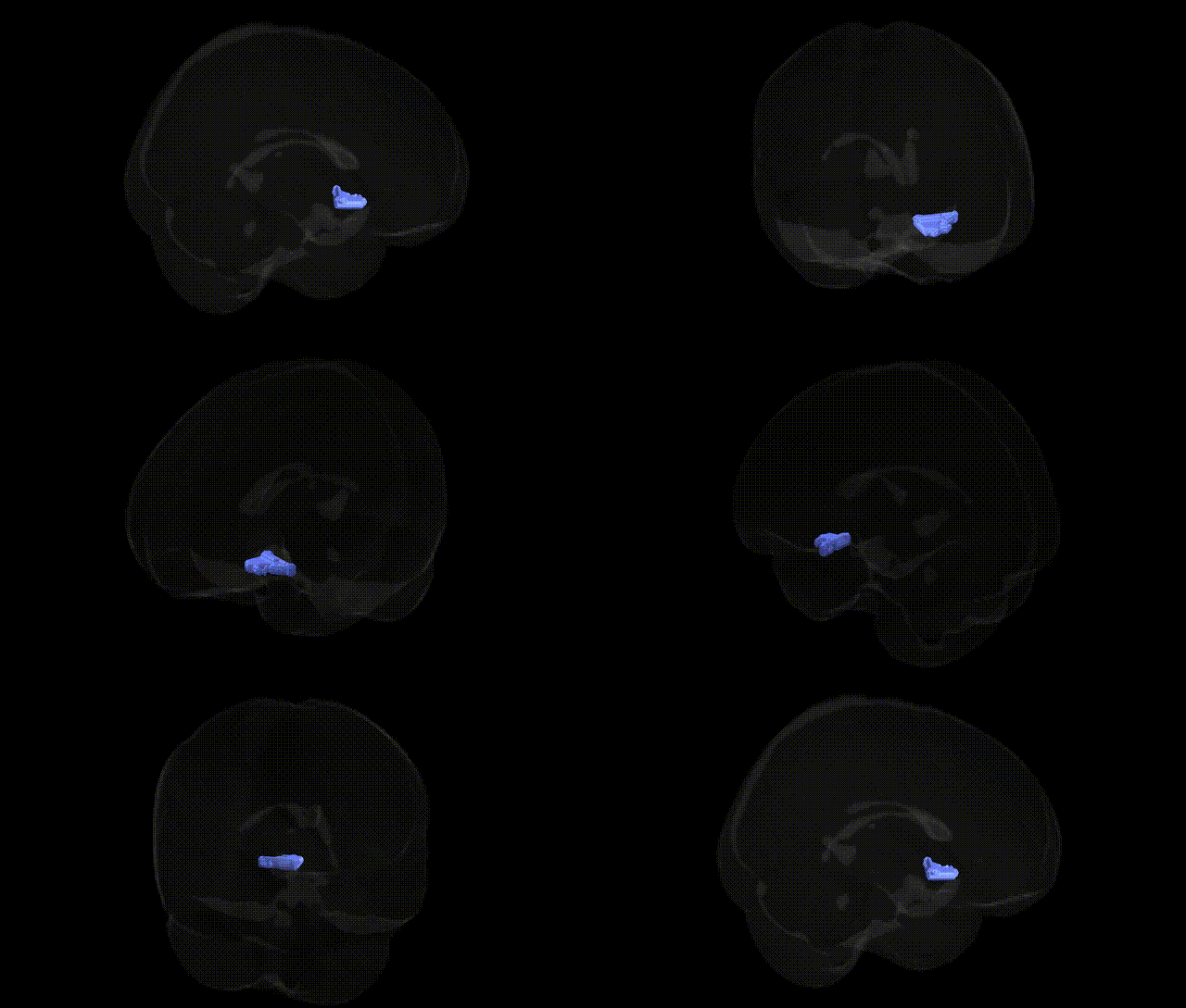
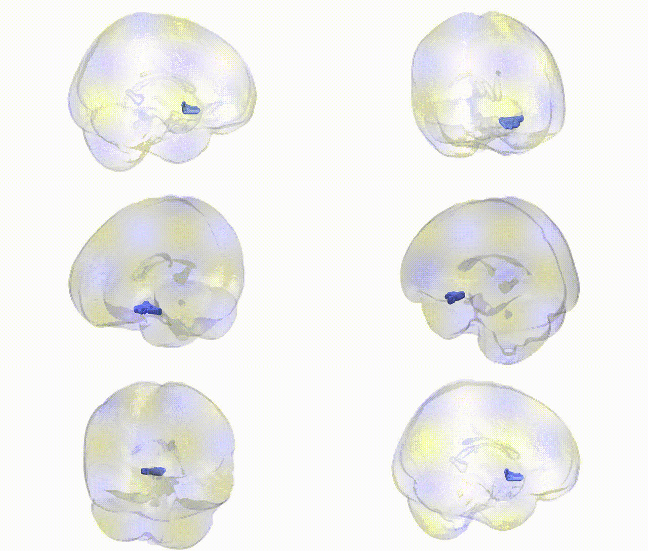
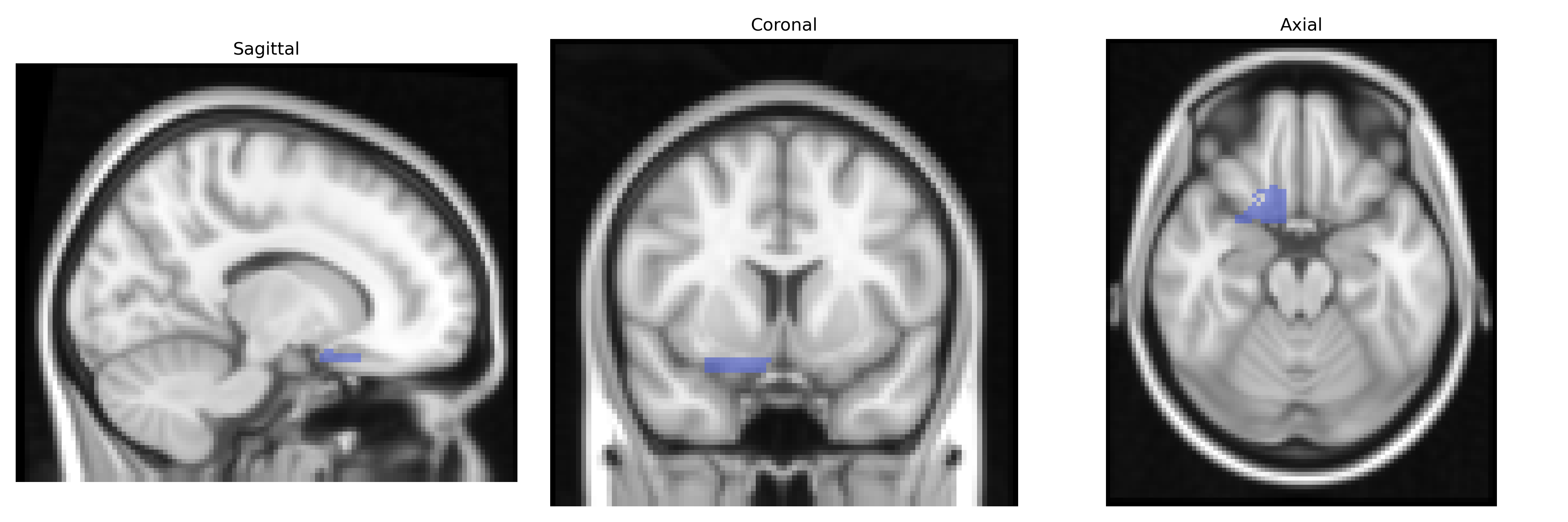
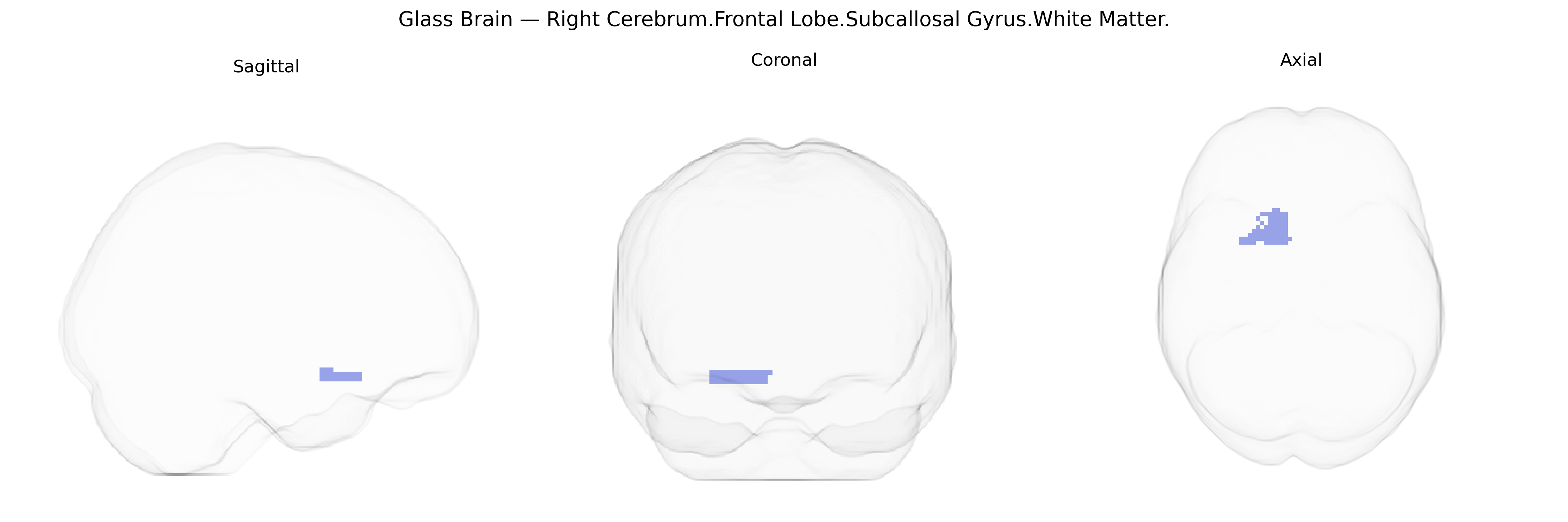
The bilateral Right Cerebrum.Frontal Lobe.Subcallosal Gyrus.White Matter corresponds to the deep white matter underlying the subcallosal area, a ventromedial frontal region located inferior to the genu of the corpus callosum and adjacent to the septal and limbic structures. This white matter contains association and projection fibers interconnecting the orbitofrontal and medial prefrontal cortices with limbic regions such as the hypothalamus, septal nuclei, and anterior cingulate cortex, thereby contributing to circuits involved in affect regulation, autonomic control, and reward-related processing. Functionally, this region is part of fronto-limbic networks implicated in mood and emotional behavior, and it is of particular interest in neuropsychiatric research, including depression and anxiety. There is no direct Wikipedia article for the subcallosal gyrus white matter; a closely related structure is the subcallosal gyrus itself: Subcallosal area.
The subcallosal gyrus white matter in the right frontal lobe, as defined in the Talairach 2mm atlas, overlaps with anterior cingulate/ventromedial prefrontal circuitry that has been repeatedly implicated in genetic studies of mood, cognition, and structural brain variation. GWAS of MRI-derived phenotypes (e.g., ENIGMA, UK Biobank) have identified common variants in genes involved in axonal guidance, myelination, and synaptic plasticity (such as those in the neuregulin–ERBB, cell-adhesion, and Wnt signaling pathways) that influence regional white matter volume, microstructure (e.g., fractional anisotropy), and cortical connectivity in frontal midline regions including the subcallosal area. Genetic risk for major depressive disorder, bipolar disorder, and schizophrenia—captured both by single loci (e.g., CACNA1C, ZNF804A, GRM3, and other neurodevelopmental genes) and polygenic risk scores—has been associated with alterations of subcallosal/ventromedial prefrontal white matter integrity and connectivity to limbic regions such as the amygdala and hippocampus, linking this region to affect regulation and treatment response (e.g., structural predictors of response to antidepressants and deep brain stimulation targeting the subcallosal cingulate). Variants associated with neuroticism, anxiety, and stress reactivity also show imaging–genetics correlations with white matter characteristics in adjacent cingulo-frontal tracts, while GWAS of cognitive traits and educational attainment report effects on frontal white matter organization that include fibers traversing or bordering the subcallosal gyrus. Although relatively few studies isolate the bilateral right subcallosal gyrus white matter as a standalone ROI, convergent evidence from imaging genetics, psychiatric GWAS, and endophenotype analyses supports this region as a genetically modulated hub in fronto-limbic circuits underlying mood disorders, emotional processing, and higher-order cognitive control.
Overview generated by GPT-4o (2026).
Region ID: 291
Hemisphere: bilateral
Atlas: Talairach labels 2mm
Full Quality Version: Download MP4
Full Quality Version: Download MP4
Wali Sidiqyar*, Gaurav Rudravaram*, Elyssa M. McMaster, Trent M. Schwartz, Adam M. Saunders, Kurt G. Schilling, Bennett A. Landman "Introducing SPINS: A Shared Public Visualization Library of Neuroanatomical Structures." Medical Imaging with Deep Learning- short paper
This resource is licensed under CC0 1.0 Universal (Public Domain).