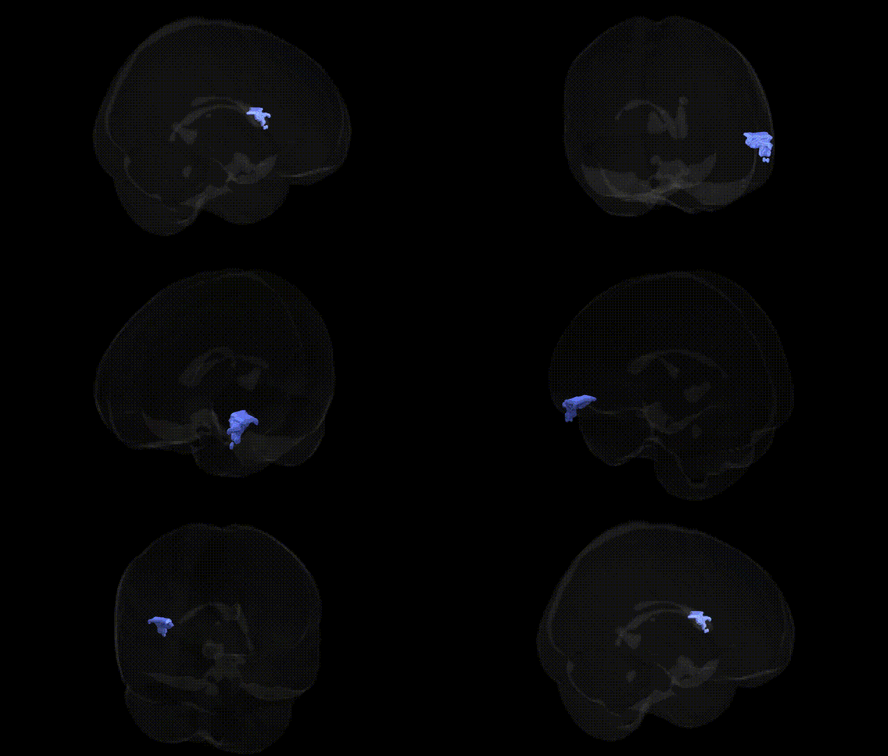
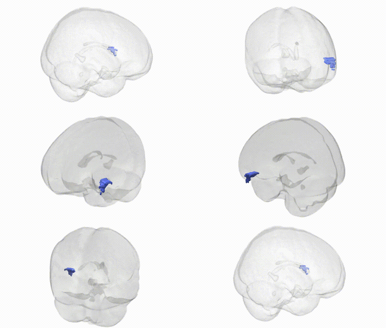
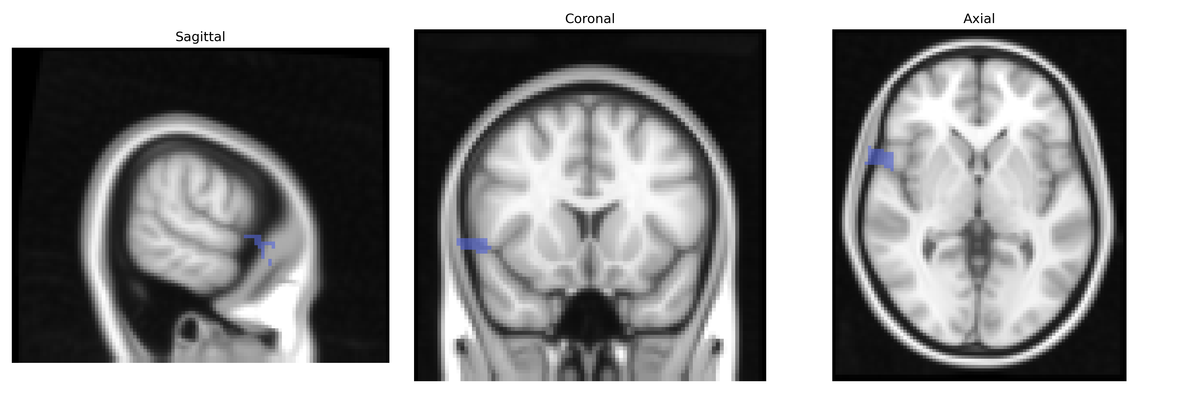
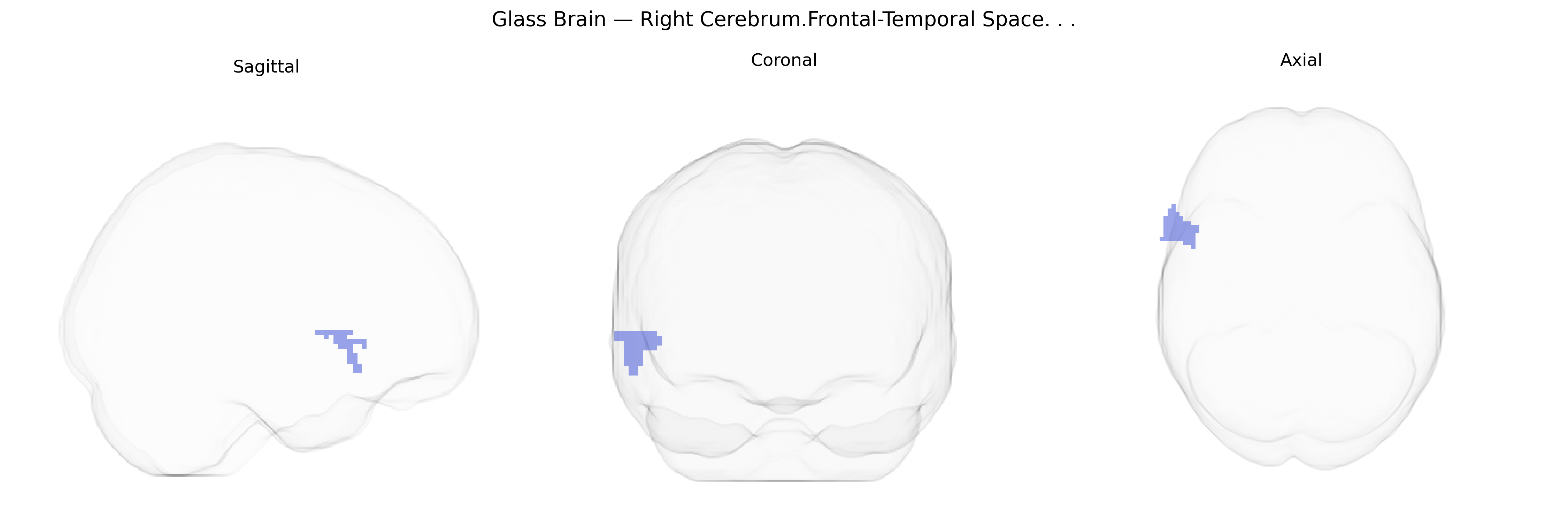
The bilateral Right Cerebrum.Frontal-Temporal space in the 2 mm Talairach atlas refers to a distributed set of voxels spanning portions of the right frontal and right temporal lobes, encompassing association cortices involved in higher-order cognition, language, executive control, and socio-emotional processing. Functionally, frontal components contribute to planning, decision-making, working memory, inhibitory control, and aspects of speech production, while temporal components subserve auditory perception, lexical-semantic processing, memory encoding and retrieval, and interpretation of complex social and emotional cues. This composite region thus integrates multimodal sensory input with executive and language networks, supporting the coordination of behavior, communication, and memory within the right cerebral hemisphere. There is no direct Wikipedia article for this composite Talairach label; a closely related structure is the Cerebral cortex.
Genetic associations specific to the bilateral Right Cerebrum.Frontal-Temporal Space as labeled in the Talairach 2 mm atlas are not typically reported at the fine-grained “Talairach label” level, but convergent GWAS and imaging-genetics work implicate overlapping frontal and temporal circuits in several traits and disorders. Large-scale studies from consortia such as ENIGMA and UK Biobank have identified common variants influencing frontal and temporal cortical thickness, surface area, and gyrification (e.g., in genes like HMGA2, PAX6, KIAA0586, and multiple loci in neurodevelopmental and synaptic pathways), and these structural measures in right frontal–temporal regions are frequently associated with cognitive performance, general intelligence, educational attainment, and social cognition. GWAS of psychiatric disorders—particularly schizophrenia, bipolar disorder, major depression, ADHD, and autism spectrum disorder—have linked polygenic risk scores to altered volume, connectivity, and activation within right frontal and temporal areas (including inferior frontal and superior temporal regions involved in language and social perception), suggesting that many risk loci act through neurodevelopmental and synaptic mechanisms affecting these circuits. Variants in genes related to glutamatergic neurotransmission (e.g., GRIN2A/GRIN2B), calcium signaling (e.g., CACNA1C), and neuroplasticity (e.g., BDNF) have been repeatedly associated with both psychiatric risk and structural or functional differences in frontal–temporal networks, while APOE and other Alzheimer’s disease loci show associations with atrophy and hypometabolism in temporal components of this space. Overall, genetic influences on the frontal–temporal region are highly polygenic, reflecting broad neurodevelopmental, synaptic, and cognitive pathways rather than a set of variants uniquely tied to the specific Talairach-labeled bilateral right frontal–temporal space.
Overview generated by GPT-4o (2026).
Region ID: 397
Hemisphere: bilateral
Atlas: Talairach labels 2mm
Full Quality Version: Download MP4
Full Quality Version: Download MP4
Wali Sidiqyar*, Gaurav Rudravaram*, Elyssa M. McMaster, Trent M. Schwartz, Adam M. Saunders, Kurt G. Schilling, Bennett A. Landman "Introducing SPINS: A Shared Public Visualization Library of Neuroanatomical Structures." Medical Imaging with Deep Learning- short paper
This resource is licensed under CC0 1.0 Universal (Public Domain).