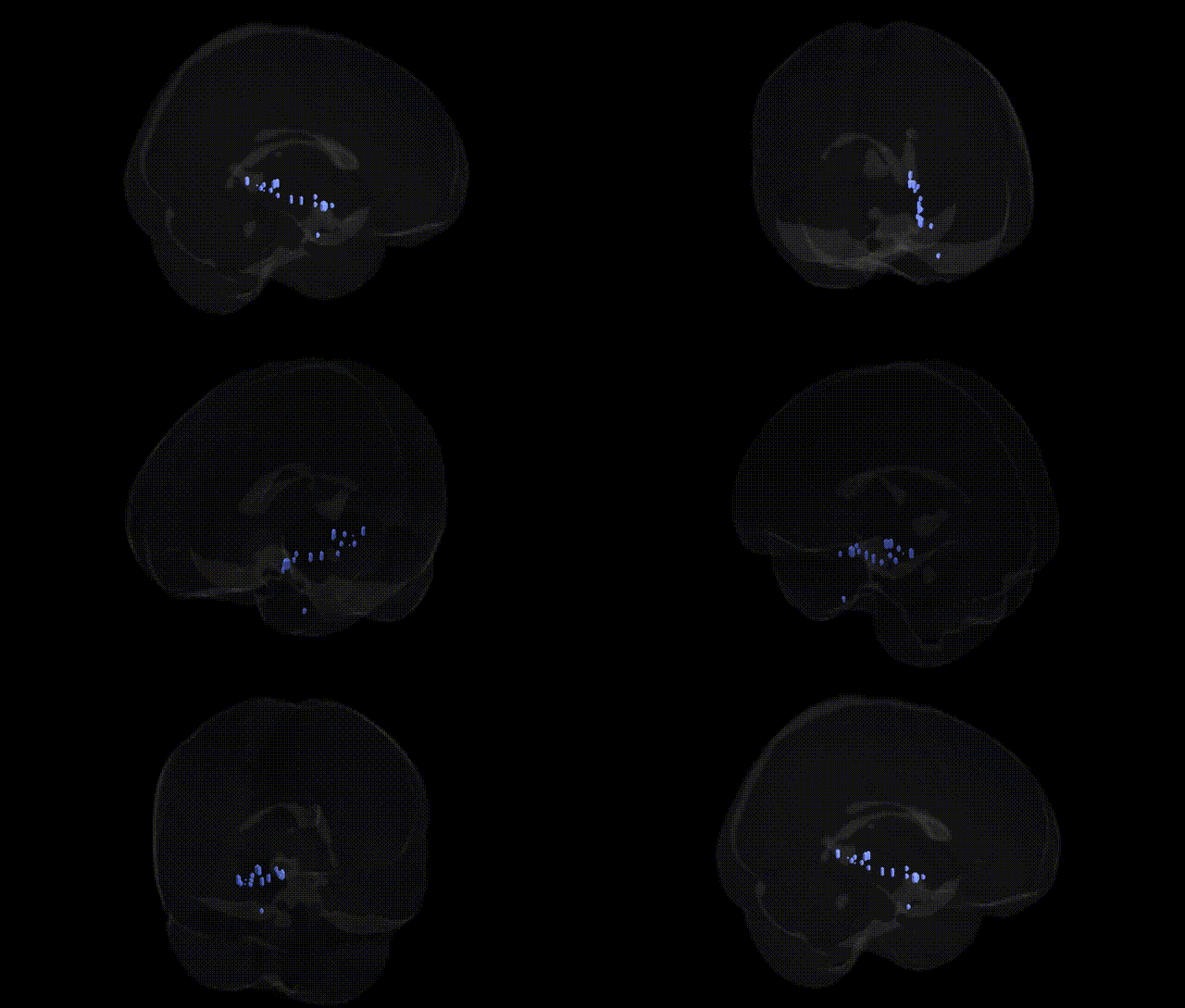
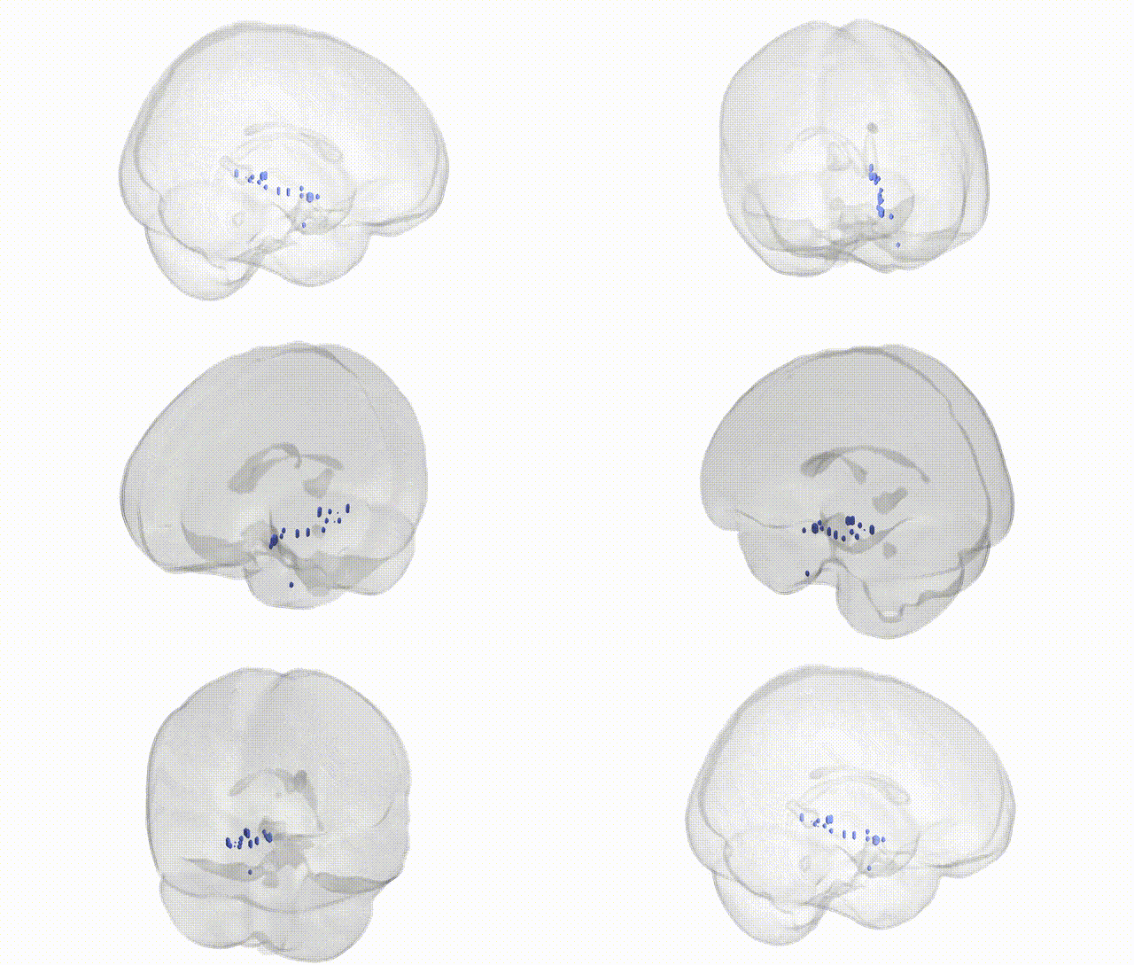
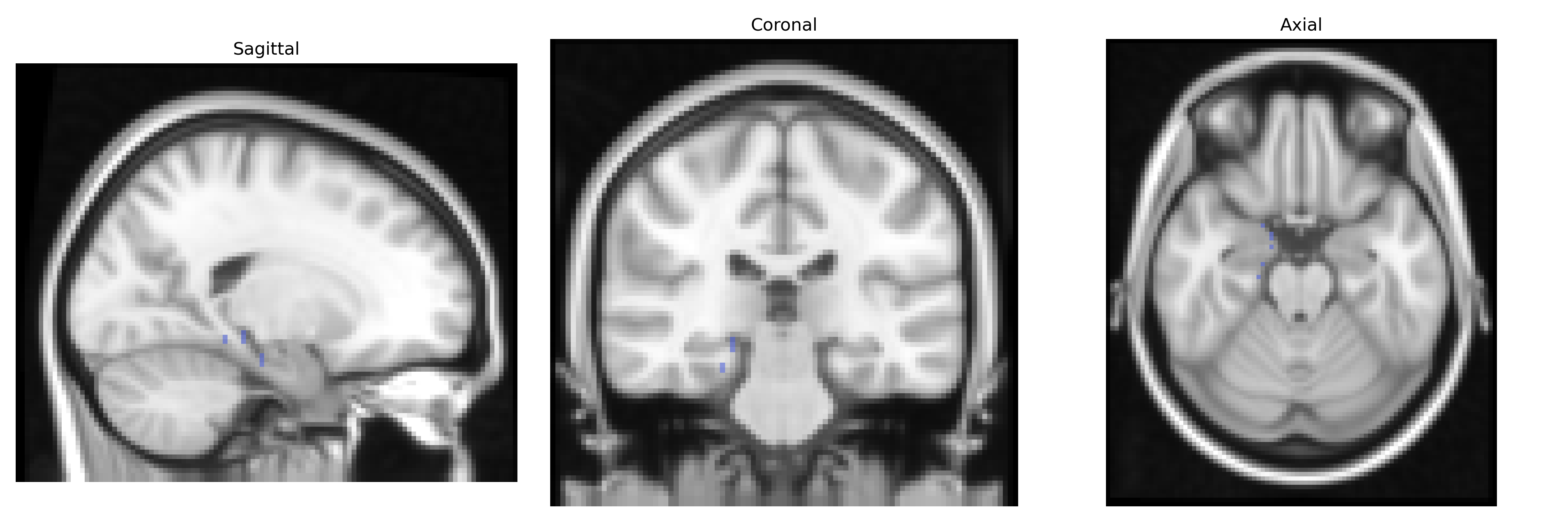
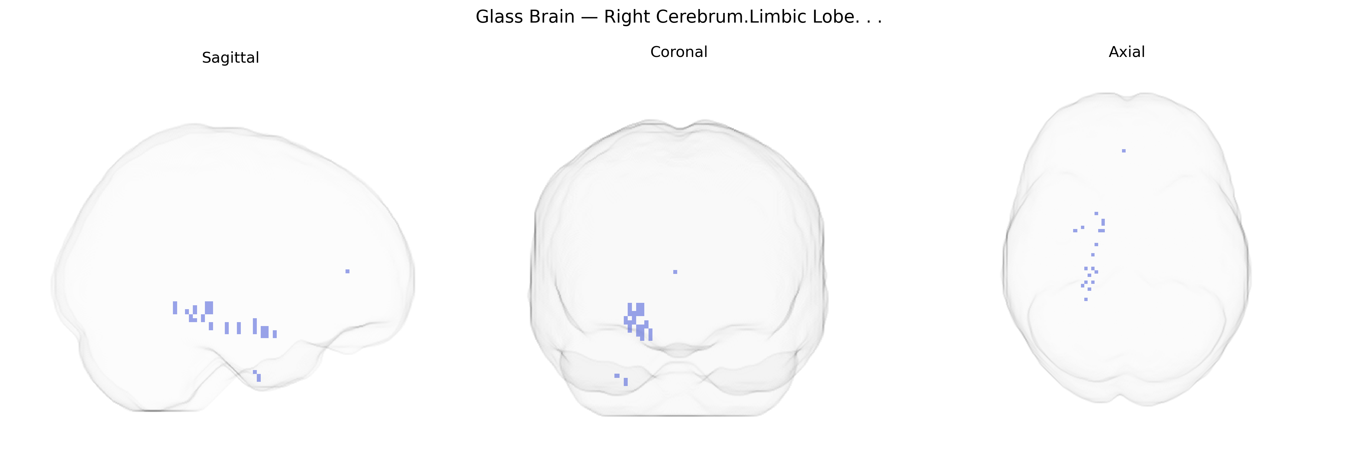
The bilateral Right Cerebrum Limbic Lobe, as defined in the Talairach 2mm Atlas, encompasses cortical and subcortical structures along the medial aspect of the right cerebral hemisphere that are central to emotional processing, memory, and autonomic regulation. This lobe includes key components such as the cingulate gyrus, parahippocampal gyrus, and associated limbic cortices, which form part of the broader limbic system interconnected with the hippocampus, amygdala, hypothalamus, and prefrontal regions. Functionally, the right limbic lobe is implicated in affective evaluation, visceromotor integration, contextual and episodic memory, and modulation of neuroendocrine and autonomic responses, contributing to the integration of internal physiological states with motivational and emotional behavior. There is no direct Wikipedia article for “Right Cerebrum Limbic Lobe”; see instead the related article: Limbic system.
The bilateral right cerebrum limbic lobe (encompassing structures such as the hippocampus, parahippocampal gyrus, amygdala, cingulate cortex, and adjacent medial temporal areas as defined in Talairach 2 mm atlases) is strongly implicated in genetic studies of psychiatric, cognitive, and neurodegenerative traits, though GWAS typically reference these component structures rather than the composite label. Large-scale GWAS and imaging-genetics studies have repeatedly associated common variants in genes such as APOE (especially ε4), BIN1, CLU, and CR1 with limbic-related atrophy and connectivity changes in Alzheimer’s disease, while variants near MAPT and GRN influence limbic and medial temporal degeneration in frontotemporal dementia. Schizophrenia and bipolar disorder GWAS meta-analyses (e.g., PGC) have identified risk loci (CACNA1C, ZNF804A, DRD2, among many others) whose burden correlates with structural and functional alterations in limbic circuits, particularly hippocampal volume and amygdala–prefrontal connectivity. Limbic structures defined within this region show robust heritability in twin and family studies, and imaging GWAS (e.g., ENIGMA, UK Biobank) link variants in genes like HMGA2, KIBRA/WDR45B, and others to hippocampal and amygdala volumes, cingulate cortical thickness, and resting-state networks. Genetic risk for major depression, anxiety, and PTSD has been connected to limbic activation patterns and structural measures, with loci in genes such as SLC6A4, FKBP5, and CRHR1 often highlighted in candidate and polygenic risk studies. Additional GWAS findings relate this limbic territory to personality traits (e.g., neuroticism), memory performance, emotional processing, and stress reactivity, underscoring the region’s role as a genetically modulated hub for affective and mnemonic functions, even though “bilateral Right Cerebrum.Limbic Lobe” itself is not typically targeted as a standalone label in genetic literature.
Overview generated by GPT-4o (2026).
Region ID: 22
Hemisphere: bilateral
Atlas: Talairach labels 2mm
Full Quality Version: Download MP4
Full Quality Version: Download MP4
Wali Sidiqyar*, Gaurav Rudravaram*, Elyssa M. McMaster, Trent M. Schwartz, Adam M. Saunders, Kurt G. Schilling, Bennett A. Landman "Introducing SPINS: A Shared Public Visualization Library of Neuroanatomical Structures." Medical Imaging with Deep Learning- short paper
This resource is licensed under CC0 1.0 Universal (Public Domain).