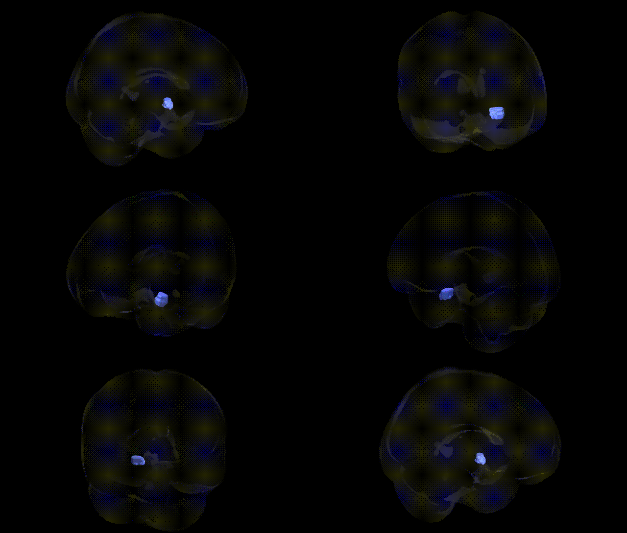
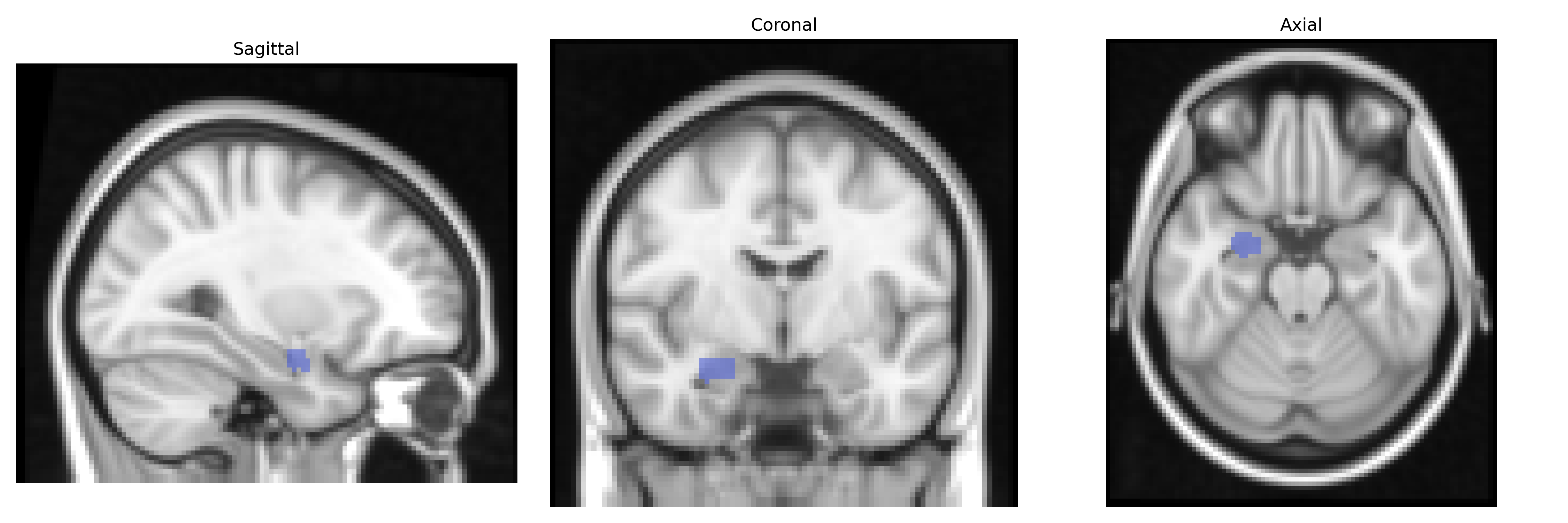
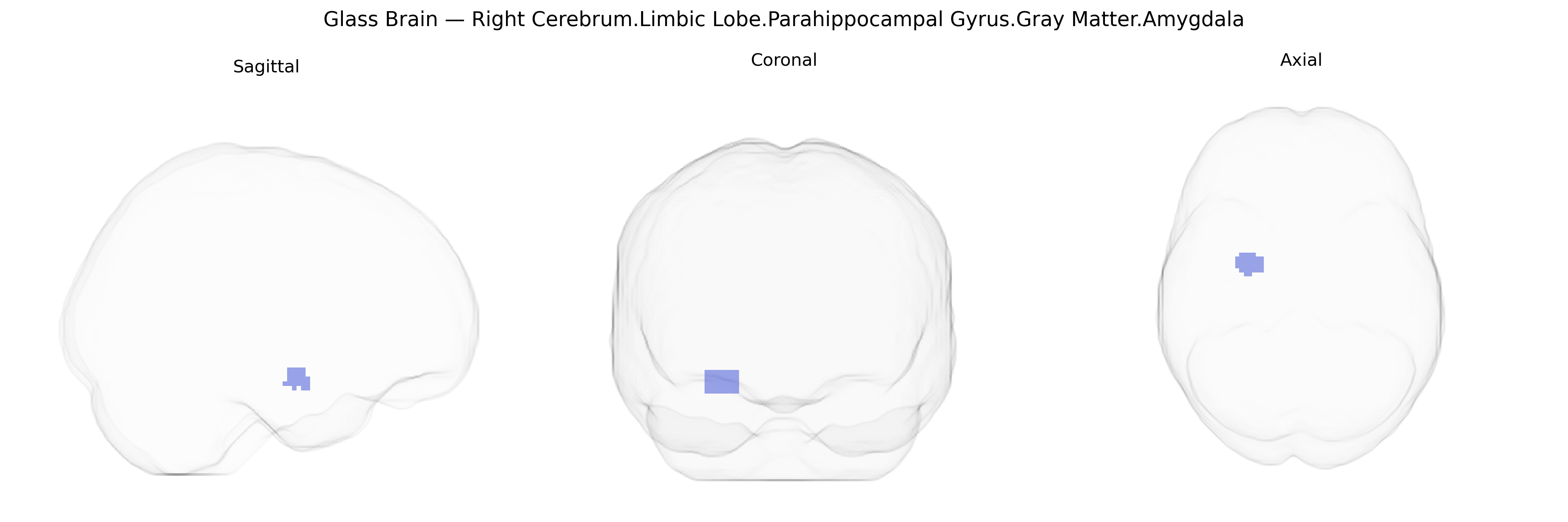
The bilateral Right Cerebrum.Limbic Lobe.Parahippocampal Gyrus.Gray Matter.Amygdala corresponds to the gray matter nuclei of the amygdala embedded within the anterior portion of the parahippocampal region of the limbic lobe in the right cerebral hemisphere, as defined in the Talairach 2 mm Atlas. The amygdala is a complex of nuclei (including basolateral, centromedial, and cortical groups) that receives multimodal sensory inputs from cortical and subcortical structures and projects to hypothalamic, brainstem, and prefrontal regions to modulate autonomic, endocrine, and behavioral responses. Functionally, this region is central to emotional processing (particularly fear and threat detection), affective learning, consolidation of emotional memories, and the integration of visceral states with higher-order cognitive and social behavior. It plays a key role in stress reactivity and is frequently implicated in psychiatric and neurological conditions involving dysregulated emotion and anxiety.
Amygdala
The bilateral right parahippocampal–amygdala region (Talairach Right Cerebrum.Limbic Lobe.Parahippocampal Gyrus.Gray Matter.Amygdala) has been repeatedly implicated in genetic studies of emotion, threat processing, and memory-related phenotypes, with GWAS and candidate-gene work linking its structure and function to multiple traits and disorders. Common variants in genes affecting monoaminergic signaling—especially serotonin transporter (SLC6A4, including 5-HTTLPR), serotonin receptors (e.g., HTR1A, HTR2A), and catechol-O-methyltransferase (COMT)—have been associated with individual differences in amygdala reactivity to emotional stimuli and, in some studies, with gray matter volume in amygdala/parahippocampal regions. Large neuroimaging GWAS (e.g., ENIGMA, UK Biobank) have identified polygenic influences on amygdala volume and limbic cortical thickness, involving loci in and around genes such as DCC, MEF2C, and others related to neurodevelopment, synaptic plasticity, and axonal guidance, which overlap partially with risk loci for major depressive disorder, schizophrenia, bipolar disorder, and anxiety traits. The amygdala-parahippocampal complex shows heritable variation in both structure and functional connectivity, and twin and GWAS studies link this heritability to genetic liability for PTSD, social anxiety, neuroticism, and broader internalizing psychopathology, with several risk alleles (e.g., in FKBP5, BDNF Val66Met, and CRHR1) associated with altered fear conditioning, stress responsivity, and volume or activation patterns in this region. Additionally, Alzheimer’s disease risk genes such as APOE and CLU, as well as loci identified in memory and cognitive GWAS, have been tied to atrophy and functional alterations in the medial temporal lobe including the parahippocampal gyrus and amygdala, supporting a genetically influenced role of this limbic structure in episodic memory, emotional learning, and vulnerability to affective and neurodegenerative disorders.
Overview generated by GPT-4o (2026).
Region ID: 183
Hemisphere: bilateral
Atlas: Talairach labels 2mm
Full Quality Version: Download MP4
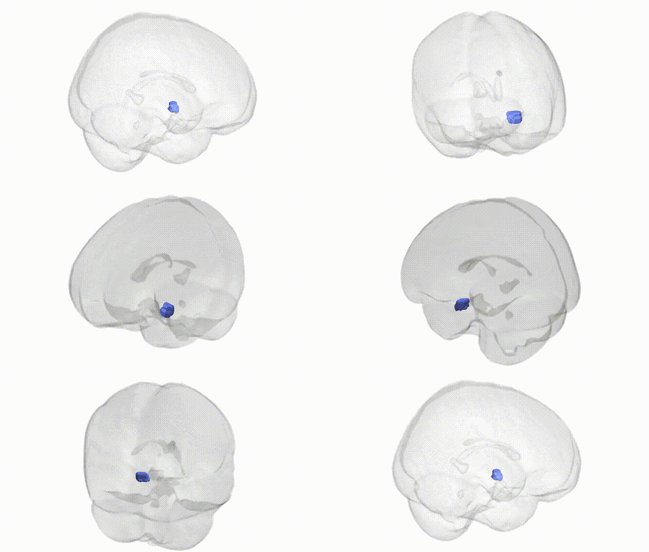
Full Quality Version: Download MP4
Wali Sidiqyar*, Gaurav Rudravaram*, Elyssa M. McMaster, Trent M. Schwartz, Adam M. Saunders, Kurt G. Schilling, Bennett A. Landman "Introducing SPINS: A Shared Public Visualization Library of Neuroanatomical Structures." Medical Imaging with Deep Learning- short paper
This resource is licensed under CC0 1.0 Universal (Public Domain).