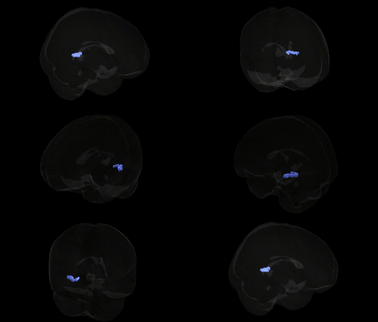
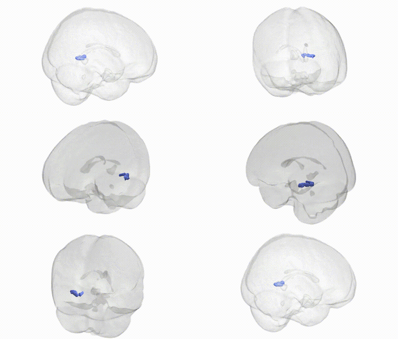
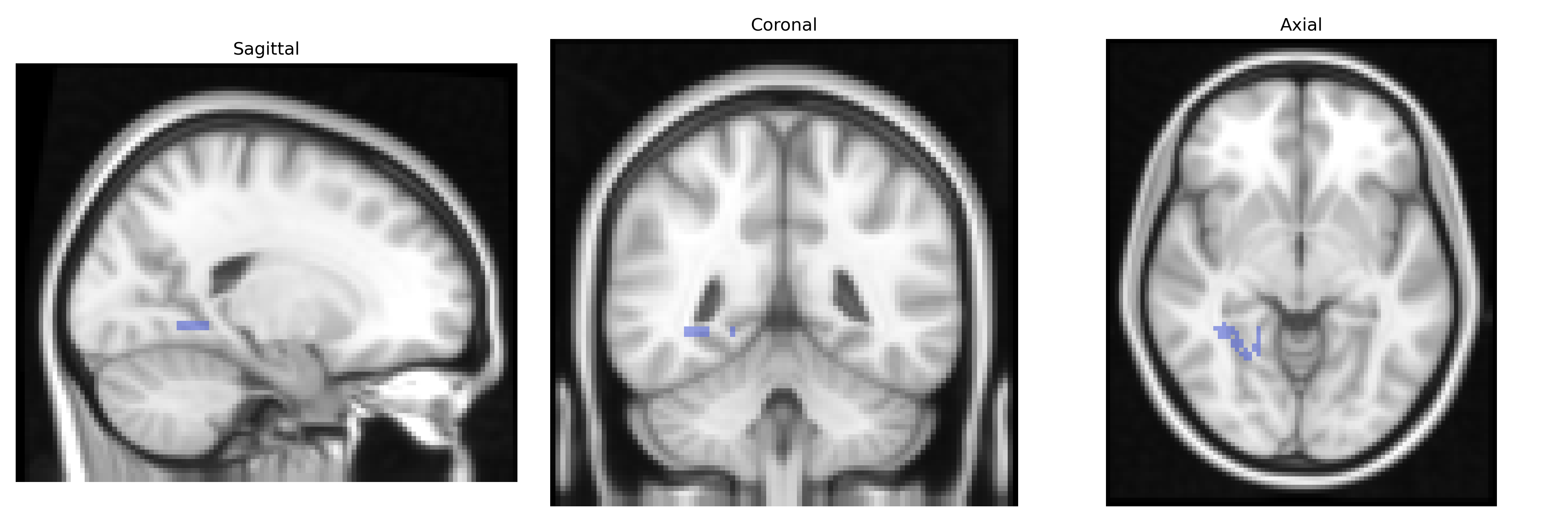
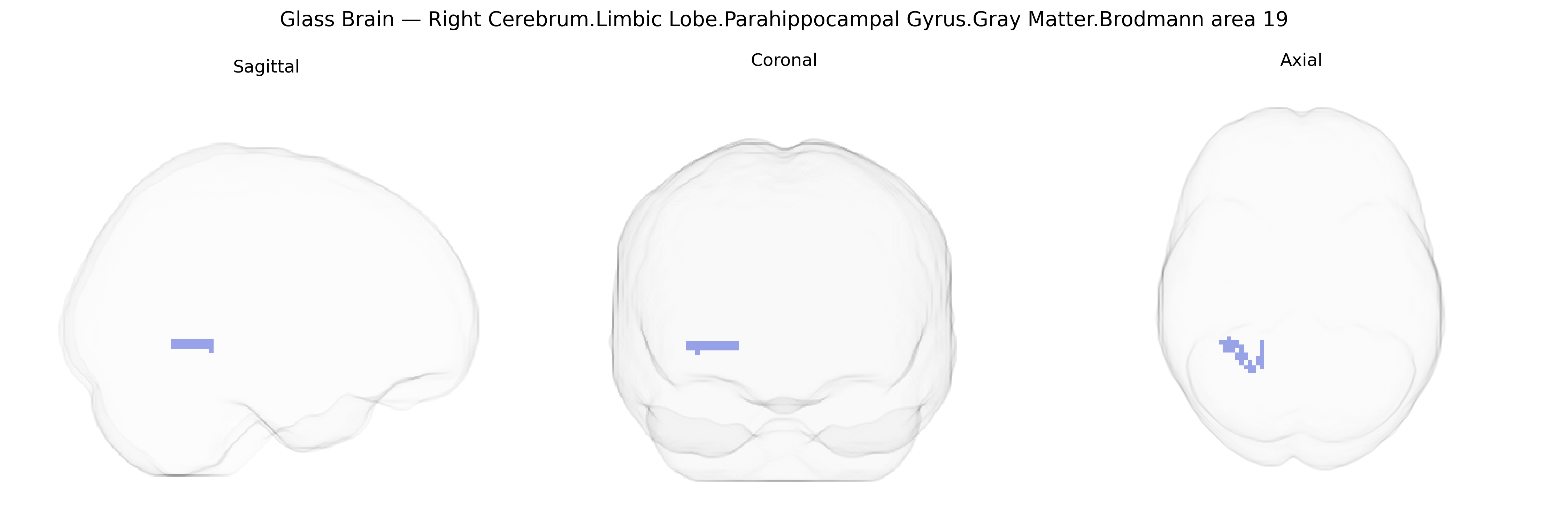
The bilateral Right Cerebrum.Limbic Lobe.Parahippocampal Gyrus.Gray Matter.Brodmann area 19 corresponds to cortical tissue located at the intersection of limbic and visual association territories, where the parahippocampal gyrus abuts the occipitotemporal region classified cytoarchitectonically as Brodmann area 19. Functionally, this zone participates in higher-order visual processing, including complex scene analysis and integration of visual context with mnemonic and spatial information relayed through limbic circuits. Its gray matter contains pyramidal neurons that contribute to associative visual networks, interfacing with the hippocampal formation and adjacent temporal and occipital cortices to support visuospatial memory, environmental navigation, and the contextualization of visual stimuli. There is no direct Wikipedia article for this exact composite region; a closely related structure is the Parahippocampal gyrus.
The bilateral right parahippocampal gyrus gray matter region, as localized in the Talairach 2 mm Atlas and overlapping with visual-associative territory including Brodmann area 19, has been implicated in multiple genetic and GWAS findings, largely through imaging genetics and neuropsychiatric studies rather than region-specific GWAS alone. Variants in genes involved in synaptic plasticity, neurodevelopment, and glutamatergic signaling (for example BDNF, DISC1, and CACNA1C) have shown associations with parahippocampal and adjacent medial temporal lobe volume or activation patterns, often in the context of schizophrenia, bipolar disorder, and major depressive disorder. Large-scale GWAS of brain structure (e.g., ENIGMA and UK Biobank-based studies) have identified common variants near genes such as KIAA0586, DPP4, and others that affect medial temporal lobe and hippocampal/parahippocampal cortical thickness or surface area, which in turn relate to genetic risk for Alzheimer’s disease, cognitive performance, and educational attainment. In Alzheimer’s disease and other dementias, risk loci including APOE and CLU are associated with accelerated atrophy and altered connectivity in parahippocampal and adjacent posterior temporal regions that contribute to episodic memory deficits. Additional imaging-genetic work has linked polygenic risk scores for schizophrenia, depression, and neuroticism to structural and functional changes in limbic and parahippocampal networks, including right-sided regions, although these effects are generally modest and distributed across cortex rather than uniquely confined to Brodmann area 19.
Overview generated by GPT-4o (2026).
Region ID: 426
Hemisphere: bilateral
Atlas: Talairach labels 2mm
Full Quality Version: Download MP4
Full Quality Version: Download MP4
Wali Sidiqyar*, Gaurav Rudravaram*, Elyssa M. McMaster, Trent M. Schwartz, Adam M. Saunders, Kurt G. Schilling, Bennett A. Landman "Introducing SPINS: A Shared Public Visualization Library of Neuroanatomical Structures." Medical Imaging with Deep Learning- short paper
This resource is licensed under CC0 1.0 Universal (Public Domain).