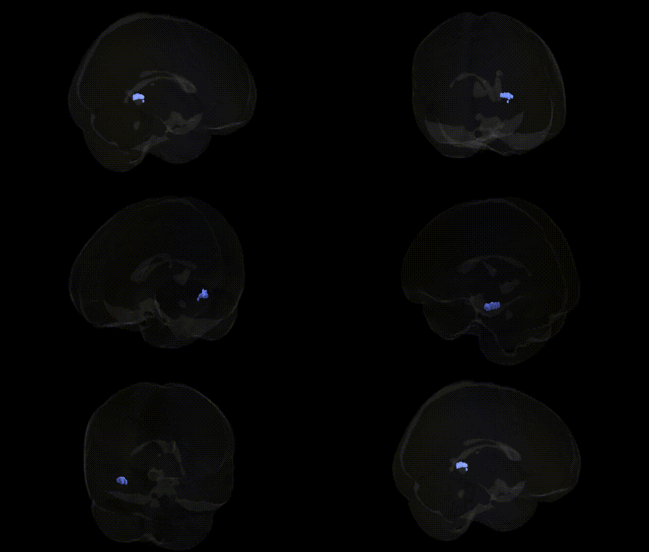
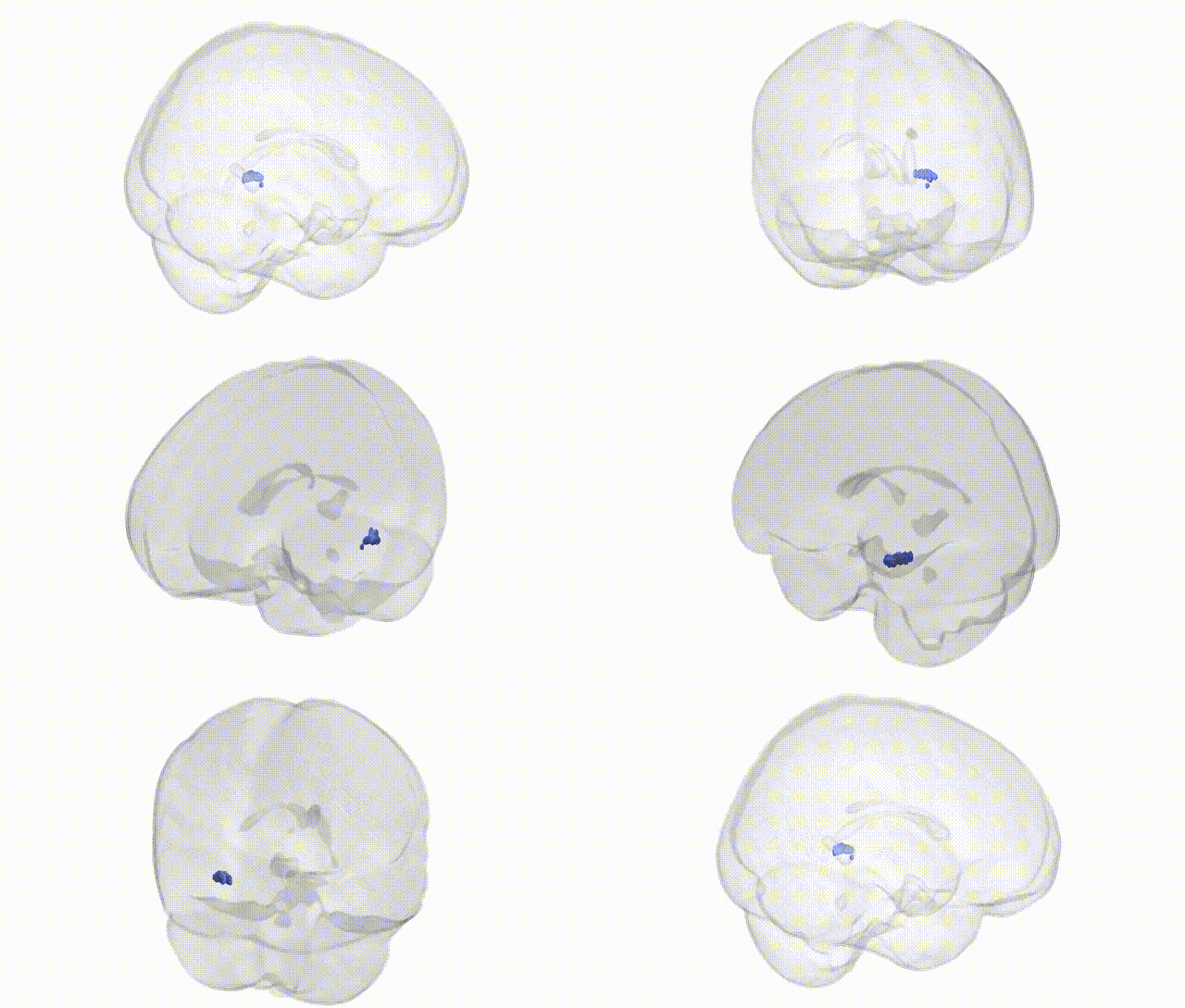
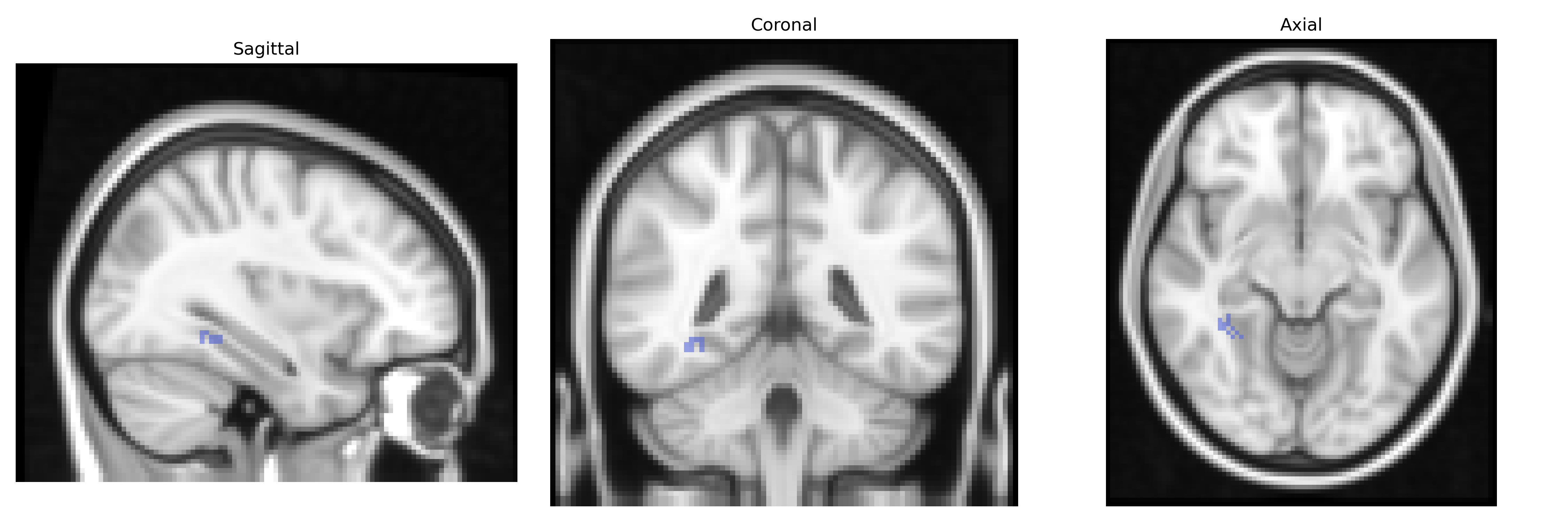
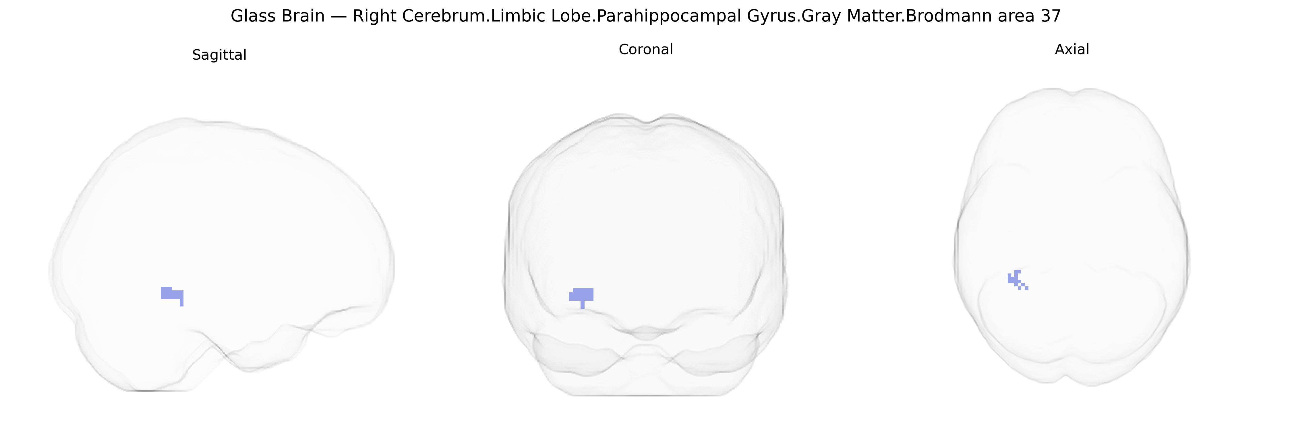
The bilateral Right Cerebrum.Limbic Lobe.Parahippocampal Gyrus.Gray Matter.Brodmann area 37 corresponds to cortical tissue in the posterior portion of the parahippocampal gyrus that functionally and cytoarchitectonically aligns with Brodmann area 37, a region traditionally associated with the fusiform and adjacent inferior temporal cortex. This area participates in high-level visual processing, including object and face recognition, and interfaces with limbic structures to support the integration of visual information with memory and emotional context. Its location within the parahippocampal gyrus places it near medial temporal lobe memory circuits, suggesting a role in binding complex visual percepts to stored representations and contextual information. There is no direct Wikipedia article for this exact Talairach-defined region; however, a closely related structure is Brodmann area 37.
The bilateral right parahippocampal gyrus gray matter in Brodmann area 37, an occipito-temporal limbic-associated region implicated in memory, scene processing, and higher-order visual recognition, has been repeatedly highlighted in imaging genetics and GWAS of brain structure and function. Large-scale studies such as ENIGMA and UK Biobank have identified heritable variation in parahippocampal and fusiform volumes and cortical thickness, with associated loci including genes involved in neurodevelopment, synaptic plasticity, and axonal guidance (for example, variants near DAAM2, WNT3, KCNK2, and other neuronal signaling genes), although most findings are regionally distributed and not specific to a single Brodmann area. GWAS of cognitive traits (general intelligence, memory performance, reading and language abilities) and face/object recognition have implicated BA37 and adjacent fusiform/parahippocampal cortex as mediating effects of polygenic risk scores on behavior, particularly through pathways regulating cortical surface area and microstructure. In psychiatric and neurologic genetics, parahippocampal and BA37 alterations have been linked to risk variants for schizophrenia (e.g., in complement and synaptic genes such as C4A and CACNA1C), autism spectrum disorder (genes affecting synaptic development and social cognition networks), and major depression and anxiety (genes related to stress response and hippocampal-limbic circuitry), typically showing reduced volume or altered activation in carriers of higher polygenic risk. Alzheimer’s disease and other dementias show strong genetic influences (e.g., APOE, BIN1, CLU, CR1) on medial temporal atrophy that extend into parahippocampal regions, including BA37, with GWAS revealing overlapping loci for hippocampal/parahippocampal integrity and dementia risk. Overall, genetic associations for this Talairach-defined region arise primarily from polygenic effects on temporal-limbic morphology and connectivity, rather than single-region-specific loci, and converge on pathways governing neurodevelopment, synaptic function, and neurodegeneration that influence susceptibility to memory disorders, language and visual recognition deficits, and major psychiatric illness.
Overview generated by GPT-4o (2026).
Region ID: 271
Hemisphere: bilateral
Atlas: Talairach labels 2mm
Full Quality Version: Download MP4
Full Quality Version: Download MP4
Wali Sidiqyar*, Gaurav Rudravaram*, Elyssa M. McMaster, Trent M. Schwartz, Adam M. Saunders, Kurt G. Schilling, Bennett A. Landman "Introducing SPINS: A Shared Public Visualization Library of Neuroanatomical Structures." Medical Imaging with Deep Learning- short paper
This resource is licensed under CC0 1.0 Universal (Public Domain).