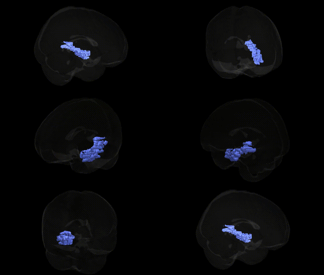
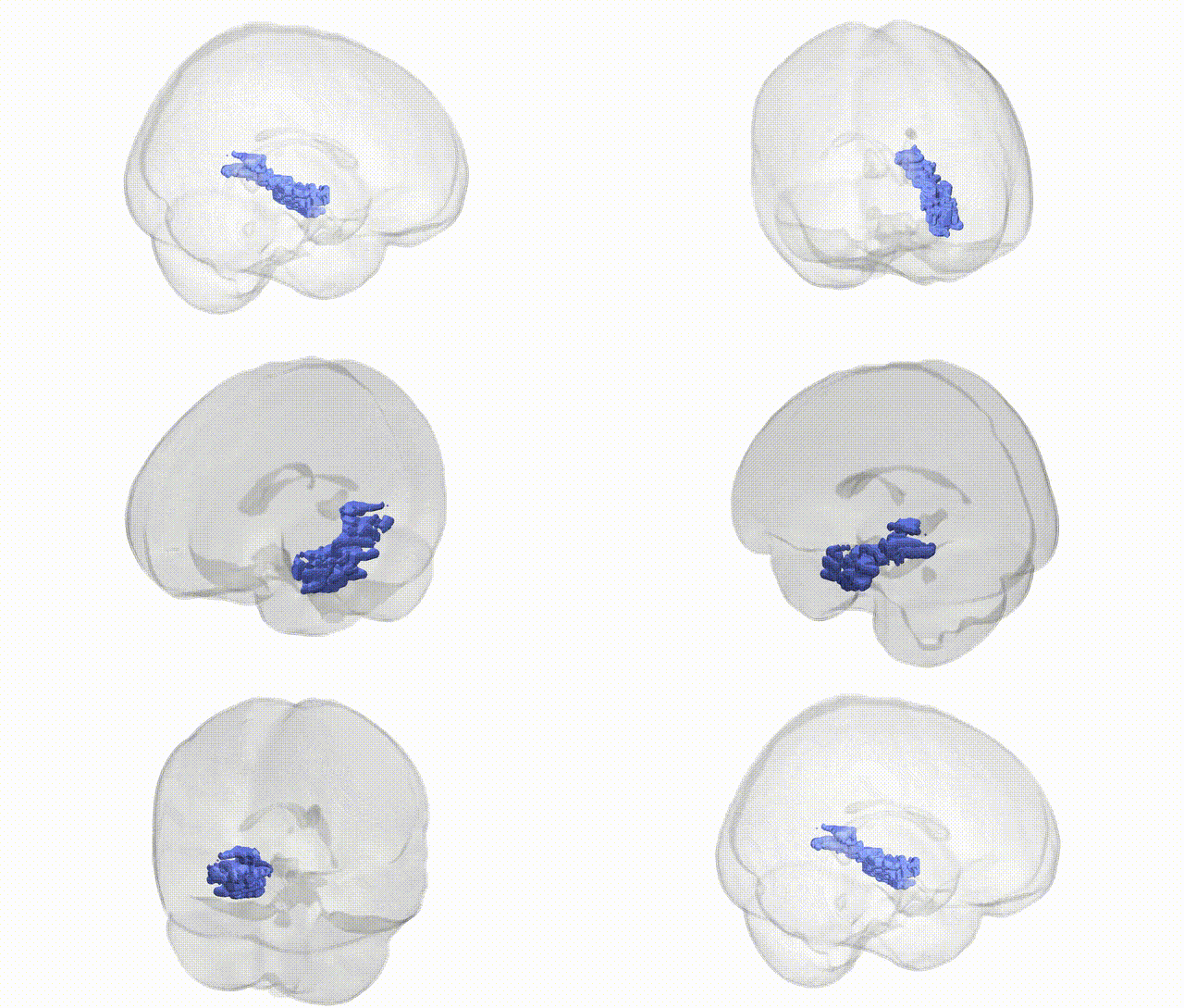
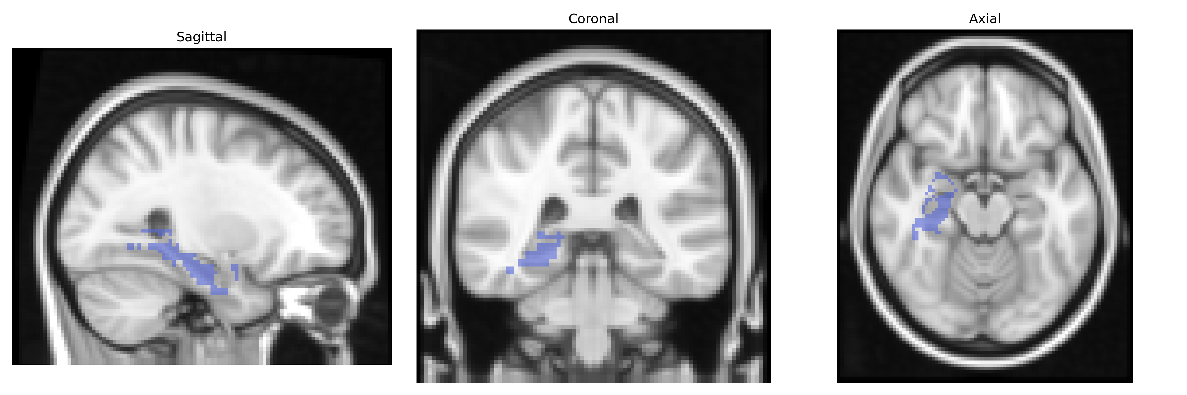
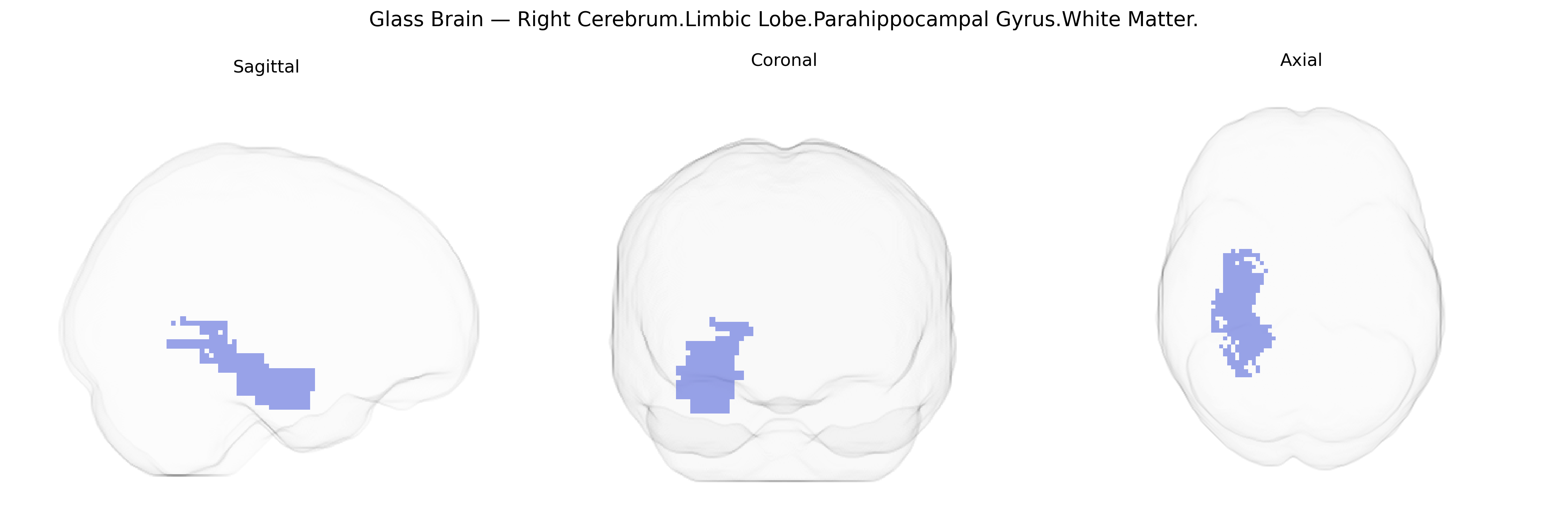
The bilateral Right Cerebrum Limbic Lobe Parahippocampal Gyrus white matter comprises the subcortical fiber pathways underlying the parahippocampal gyrus in the medial temporal lobe, forming part of the limbic system’s connectivity infrastructure. These white matter tracts include associative and projection fibers that interconnect parahippocampal cortex with the hippocampal formation, entorhinal cortex, and other limbic and neocortical regions, supporting functions such as episodic memory encoding, spatial navigation, and contextual processing of environmental cues. The right-sided parahippocampal white matter is particularly implicated in visuospatial memory and scene recognition, and its integrity is critical for the transmission of information between cortical inputs and the hippocampus. There is no direct Wikipedia article for this specific white matter subdivision; a closely related structure is the Parahippocampal gyrus.
The bilateral right parahippocampal gyrus white matter, as defined in the Talairach 2 mm atlas, is a key limbic tract region connecting medial temporal lobe structures involved in episodic memory, contextual processing, and aspects of emotional regulation, and its microstructure has been repeatedly implicated in genetic studies of brain imaging and neuropsychiatric traits. Large-scale GWAS of diffusion MRI metrics have identified numerous loci influencing white matter integrity (e.g., fractional anisotropy, mean diffusivity) in parahippocampal and adjacent medial temporal tracts, with notable contributions from genes involved in axonal guidance, myelination, and synaptic plasticity—such as variants in genes like CNTNAP2, NRG1, and BDNF—although findings are typically distributed across networks rather than uniquely specific to this tract. Imaging genetics work in schizophrenia, bipolar disorder, and major depressive disorder has linked risk alleles in loci including CACNA1C, ZNF804A, and DISC1 to altered connectivity and microstructure in medial temporal white matter pathways encompassing the parahippocampal region, consistent with these genes’ roles in neurodevelopment and synaptic signaling. In Alzheimer’s disease and related dementias, APOE ε4 and other GWAS-identified risk variants (e.g., in CLU, PICALM, BIN1) have been associated with reduced integrity of parahippocampal and perforant-pathway white matter, aligning with the early involvement of medial temporal structures in amyloid and tau pathology and memory decline. GWAS of cognitive traits, including episodic memory performance and general cognitive ability, have shown that polygenic scores for cognition and education correlate with structural and microstructural features of medial temporal white matter, including the parahippocampal region, suggesting that many small-effect variants collectively shape the connectivity of this limbic pathway. Additionally, genetic studies of anxiety, PTSD, and stress-related traits indicate that polymorphisms in stress-response and serotonergic genes (e.g., FKBP5, 5-HTTLPR/SLC6A4) can modulate white matter integrity in parahippocampal and hippocampal circuits, potentially influencing vulnerability to intrusive memories and altered contextual fear processing, although these effects are modest and embedded within broader limbic–fronto-parietal networks. Overall, current GWAS and imaging genetics evidence supports a polygenic, network-level influence on the parahippocampal gyrus white matter, with risk and trait-associated variants shaping its development and integrity in conjunction with other medial temporal and limbic pathways rather than acting in isolation on this single Talairach-defined region.
Overview generated by GPT-4o (2026).
Region ID: 134
Hemisphere: bilateral
Atlas: Talairach labels 2mm
Full Quality Version: Download MP4
Full Quality Version: Download MP4
Wali Sidiqyar*, Gaurav Rudravaram*, Elyssa M. McMaster, Trent M. Schwartz, Adam M. Saunders, Kurt G. Schilling, Bennett A. Landman "Introducing SPINS: A Shared Public Visualization Library of Neuroanatomical Structures." Medical Imaging with Deep Learning- short paper
This resource is licensed under CC0 1.0 Universal (Public Domain).