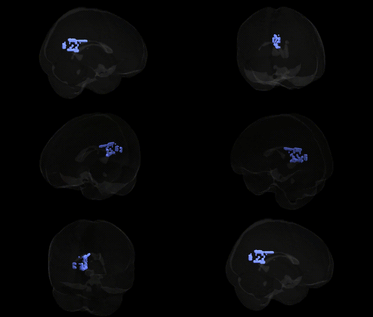
The bilateral Right Cerebrum.Limbic Lobe.Posterior Cingulate corresponds to the posterior portion of the cingulate gyrus, a key hub of the limbic system situated on the medial surface of the cerebral hemisphere, just dorsal to the corpus callosum. This region is heavily interconnected with precuneus, medial parietal cortex, hippocampal formation, and medial prefrontal areas, and is a central node of the default mode network. Functionally, the posterior cingulate is implicated in internally directed cognition, autobiographical and episodic memory retrieval, visuospatial orientation, and evaluation of emotional and motivational significance of stimuli. It shows robust metabolic and hemodynamic activity at rest, is sensitive to disruptions in consciousness and attention, and exhibits characteristic structural and functional alterations in neurodegenerative and psychiatric conditions. Posterior cingulate cortex
Genetic associations involving the bilateral right cerebrum limbic lobe posterior cingulate (as defined in the Talairach 2 mm atlas) largely emerge from imaging genetics and GWAS of brain structure, function, and related neuropsychiatric traits rather than direct region-specific GWAS hits. Variants in APOE (especially ε4) and nearby TOMM40 are consistently associated with altered posterior cingulate volume, metabolism, and functional connectivity, particularly in Alzheimer’s disease and preclinical cognitive decline. Large neuroimaging GWAS consortia (e.g., ENIGMA, UK Biobank) have identified multiple loci linked to cortical thickness, surface area, and functional connectivity in cingulate/precuneus–default mode network regions, including variants near genes such as MEF2C, BIN1, CLU, CR1, and PICALM that are also established Alzheimer’s risk loci. Polygenic risk scores for Alzheimer’s disease, schizophrenia, major depression, and autism spectrum disorder have been associated with posterior cingulate structural and functional changes, suggesting that distributed genetic risk for these disorders modulates this hub of the default mode network. Additionally, GWAS of cognitive traits (general intelligence, memory, and attentional control) and personality dimensions (e.g., neuroticism) show overlapping polygenic architectures with posterior cingulate morphology and connectivity measures, indicating that this region’s genetics are intertwined with broader neurodevelopmental, neurodegenerative, and cognitive-emotional pathways rather than being driven by a single, highly specific locus.
Overview generated by GPT-4o (2026).
Region ID: 668
Hemisphere: bilateral
Atlas: Talairach labels 2mm
Full Quality Version: Download MP4
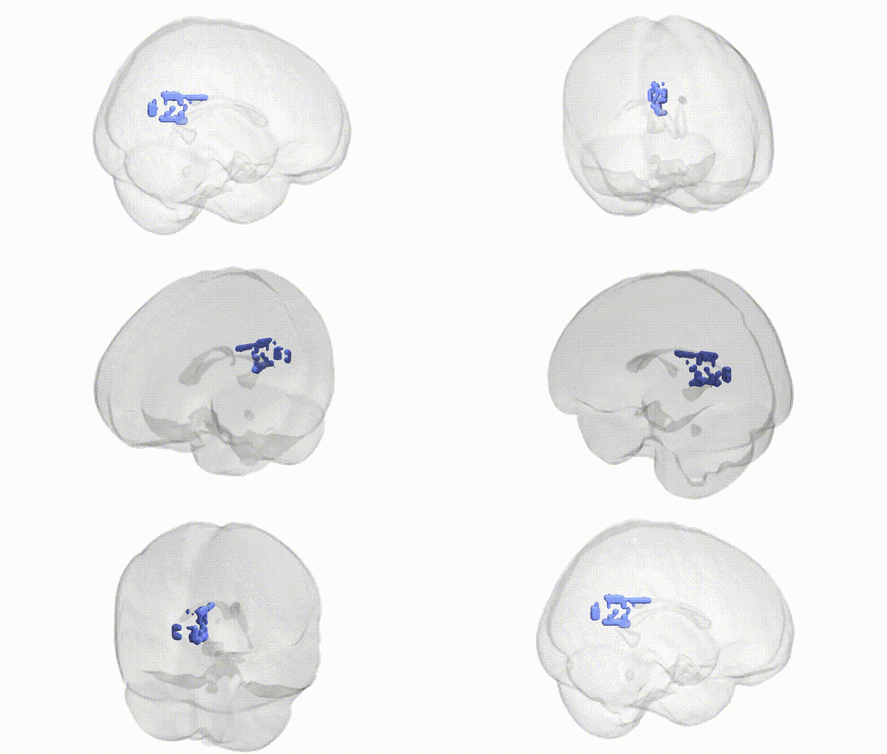
Full Quality Version: Download MP4
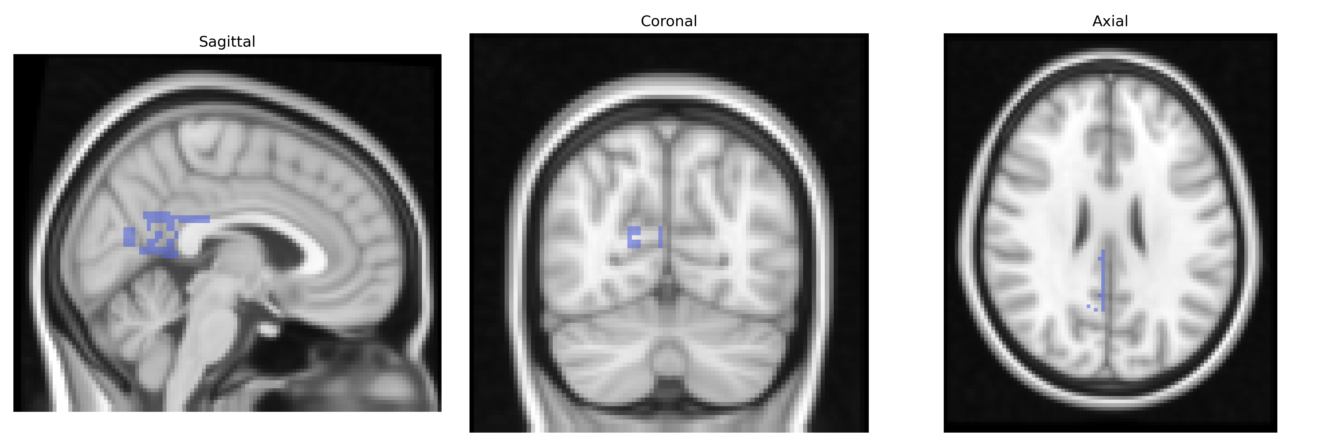
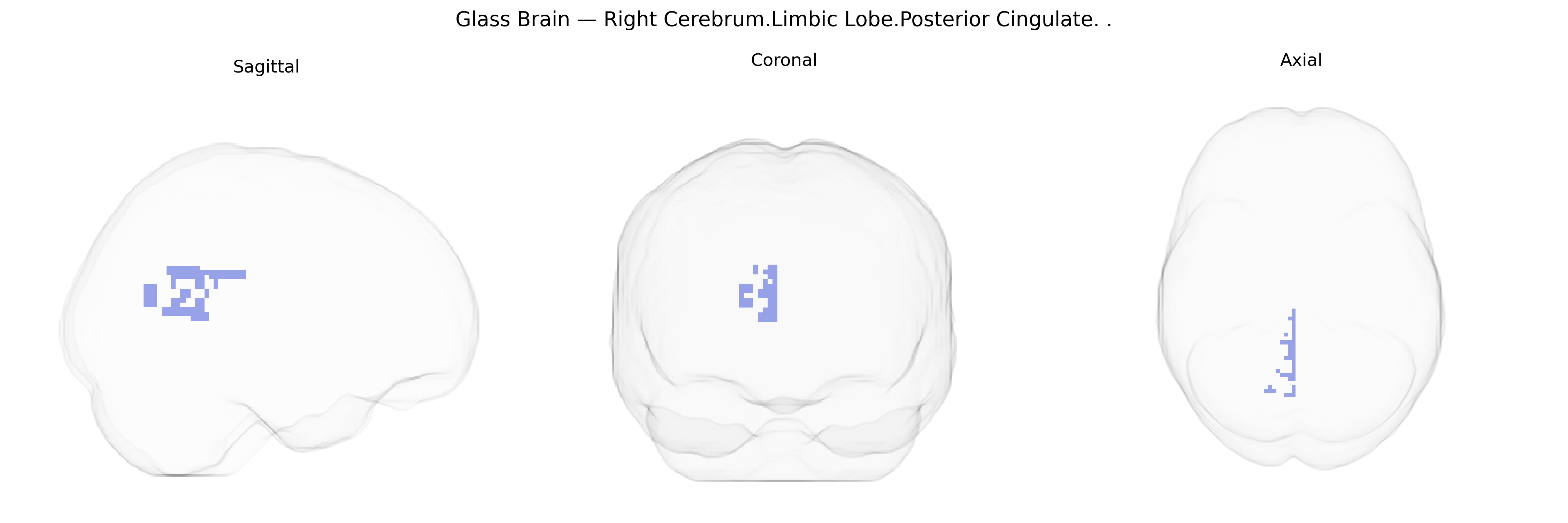
Wali Sidiqyar*, Gaurav Rudravaram*, Elyssa M. McMaster, Trent M. Schwartz, Adam M. Saunders, Kurt G. Schilling, Bennett A. Landman "Introducing SPINS: A Shared Public Visualization Library of Neuroanatomical Structures." Medical Imaging with Deep Learning- short paper
This resource is licensed under CC0 1.0 Universal (Public Domain).