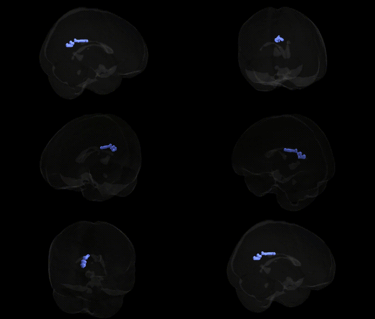
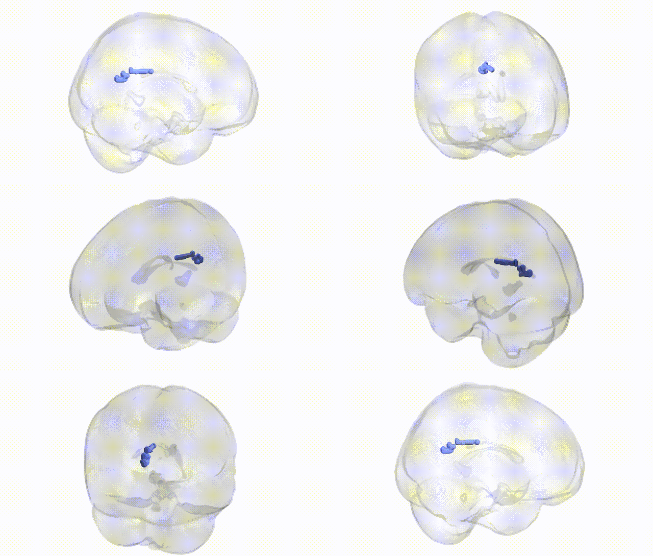
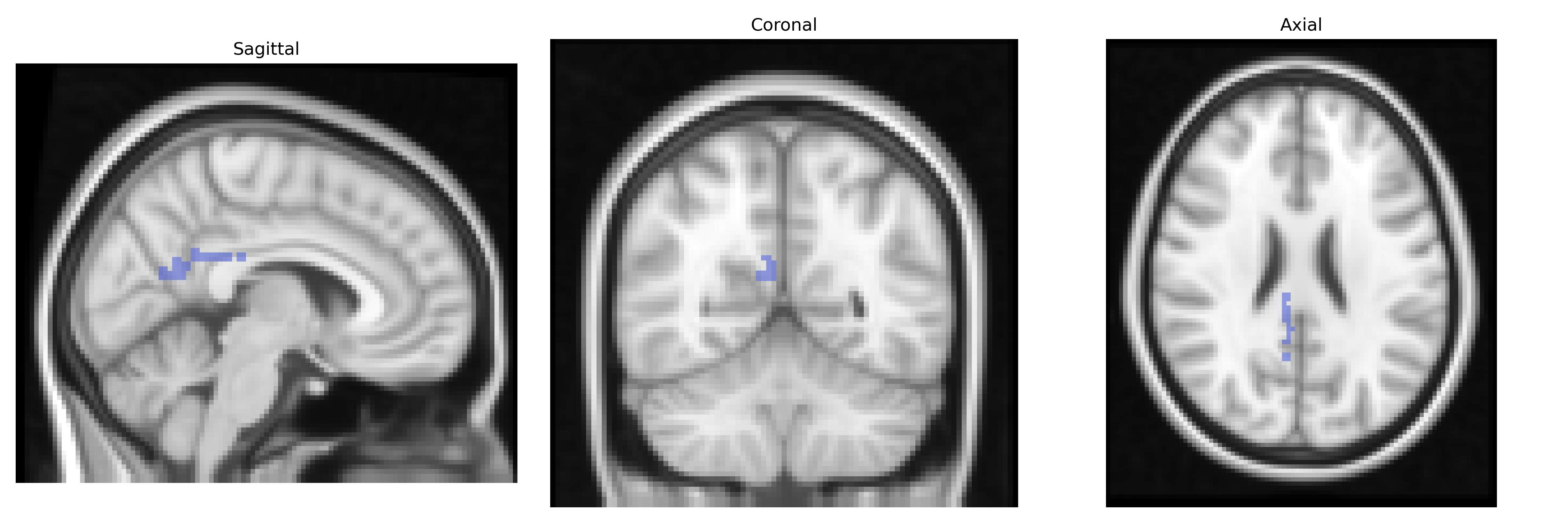
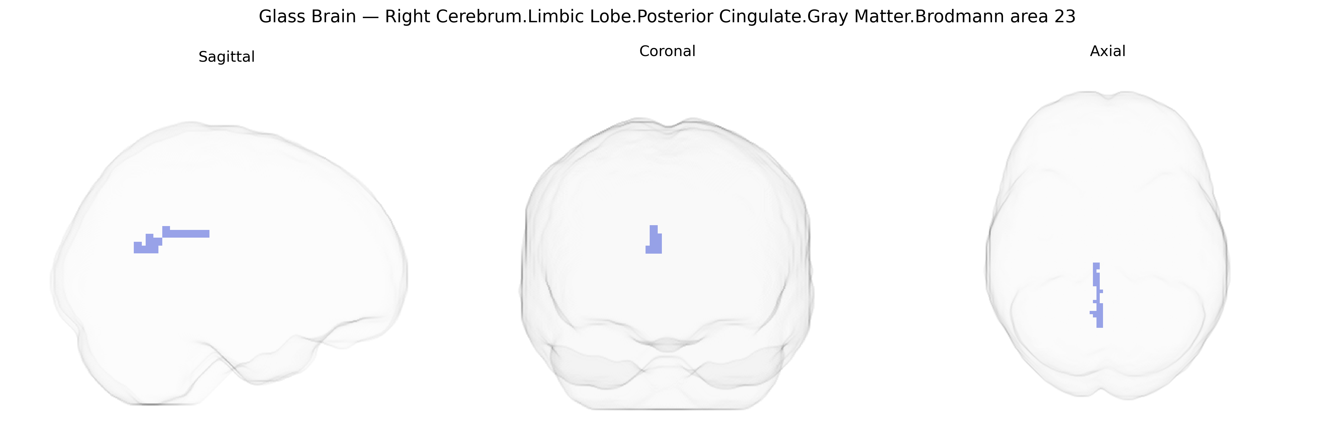
The bilateral Right Cerebrum.Limbic Lobe.Posterior Cingulate.Gray Matter.Brodmann area 23 corresponds to cortical tissue within the posterior segment of the cingulate gyrus, part of the limbic lobe situated on the medial surface of the cerebral hemisphere. Brodmann area 23 is implicated in internally directed cognition, autobiographical memory retrieval, emotional processing, and integration of visuospatial and somatosensory information with affective states, and it forms a key node in the posterior cingulate cortex within the default mode network. Cytoarchitectonically, BA23 is agranular to dysgranular association cortex, receiving multimodal inputs from medial parietal, hippocampal, and prefrontal regions and projecting to other limbic and association areas, supporting functions related to self-referential thought, evaluation of emotional significance, and regulation of attention between internal and external stimuli. There is no direct Wikipedia article for “Brodmann area 23,” but it is contained within the Posterior cingulate cortex.
Genetic associations involving the bilateral Right Cerebrum Limbic Lobe Posterior Cingulate Gray Matter (Brodmann area 23) are typically inferred from imaging genetics and GWAS of brain structure, function, and related psychiatric or cognitive traits rather than from region-specific candidate gene studies. GWAS of cortical thickness and surface area have implicated common variants in genes such as TCF4, GRIN2A, CACNA1C, and other neurodevelopmental and synaptic genes in shaping posterior cingulate morphology and connectivity within the default mode network. Imaging genetics work has linked APOE ε4 and other Alzheimer’s disease risk variants (e.g., in CLU, CR1, PICALM) to altered posterior cingulate volume, hypometabolism, and functional connectivity, consistent with this region’s vulnerability in early Alzheimer’s pathology. Polygenic risk for major depressive disorder, schizophrenia, and autism spectrum disorder has been associated with structural and functional alterations of the posterior cingulate, suggesting that pleiotropic neurodevelopmental and synaptic genes (including those in glutamatergic and calcium-channel pathways) may influence risk via effects on this hub region. Additional GWAS and large-scale consortia analyses have linked posterior cingulate features to individual differences in mind wandering, self-referential processing, and cognitive control through variants in genes involved in axon guidance, myelination, and synaptic plasticity, though specific loci and effect sizes remain modest and distributed, reflecting highly polygenic architecture.
Overview generated by GPT-4o (2026).
Region ID: 763
Hemisphere: bilateral
Atlas: Talairach labels 2mm
Full Quality Version: Download MP4
Full Quality Version: Download MP4
Wali Sidiqyar*, Gaurav Rudravaram*, Elyssa M. McMaster, Trent M. Schwartz, Adam M. Saunders, Kurt G. Schilling, Bennett A. Landman "Introducing SPINS: A Shared Public Visualization Library of Neuroanatomical Structures." Medical Imaging with Deep Learning- short paper
This resource is licensed under CC0 1.0 Universal (Public Domain).