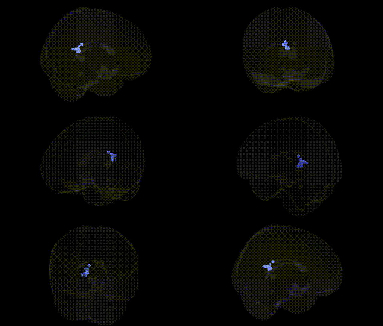
The bilateral Right Cerebrum.Limbic Lobe.Posterior Cingulate.Gray Matter.Brodmann area 29 corresponds to the retrosplenial cortex, a small agranular cortical area located in the posterior cingulate region adjacent to the splenium of the corpus callosum. Brodmann area 29 is part of the limbic network and is heavily interconnected with the hippocampal formation, parahippocampal cortex, and other cingulate and prefrontal regions. Functionally, this area is implicated in episodic memory, spatial navigation, contextual association, and integration of multimodal information relevant to autobiographical recollection and scene processing. It contributes to the broader default mode and limbic systems involved in internally directed cognition, emotional context, and memory-guided behavior. There is no direct link; see the related structure: Retrosplenial cortex.
The posterior cingulate cortex (PCC), encompassing Brodmann area 29 in the limbic lobe, has been implicated in multiple genetic and GWAS-based associations through studies of network-level and regional morphology rather than Talairach-specific labels. Large neuroimaging GWAS consortia (e.g., ENIGMA, UK Biobank) have identified common variants in genes such as APOE, CLU, BIN1, and CR1 that modulate PCC structure, connectivity, or metabolism in the context of Alzheimer’s disease risk, with the ε4 allele of APOE consistently linked to reduced PCC gray matter volume, altered functional connectivity, and hypometabolism. PCC volume and cortical thickness show polygenic influences that overlap with genetic architectures for general cognitive ability, educational attainment, and default mode network (DMN) connectivity, with loci near genes involved in synaptic function and neurodevelopment (e.g., genes in glutamatergic signaling pathways) contributing to variance in this region’s anatomy. GWAS of major depressive disorder, schizophrenia, and anxiety-related traits have reported PCC and broader limbic-DMN alterations that co-localize with risk variants, particularly in genes regulating synaptic plasticity, immune function, and myelination, although the associations are typically network-based and not exclusive to BA29. Additionally, neurodegenerative GWAS (Alzheimer’s, frontotemporal dementia) and imaging genetics studies of amyloid and tau load have highlighted the PCC as a genetically vulnerable hub, showing that polygenic risk scores for Alzheimer’s disease and cognitive decline predict PCC atrophy and connectivity disruption. Overall, genetic influences on bilateral PCC gray matter reflect highly polygenic, pleiotropic architectures, with risk and trait-associated loci affecting this region as part of broader limbic and DMN circuits rather than being specifically constrained to Talairach BA29.
Overview generated by GPT-4o (2026).
Region ID: 671
Hemisphere: bilateral
Atlas: Talairach labels 2mm
Full Quality Version: Download MP4
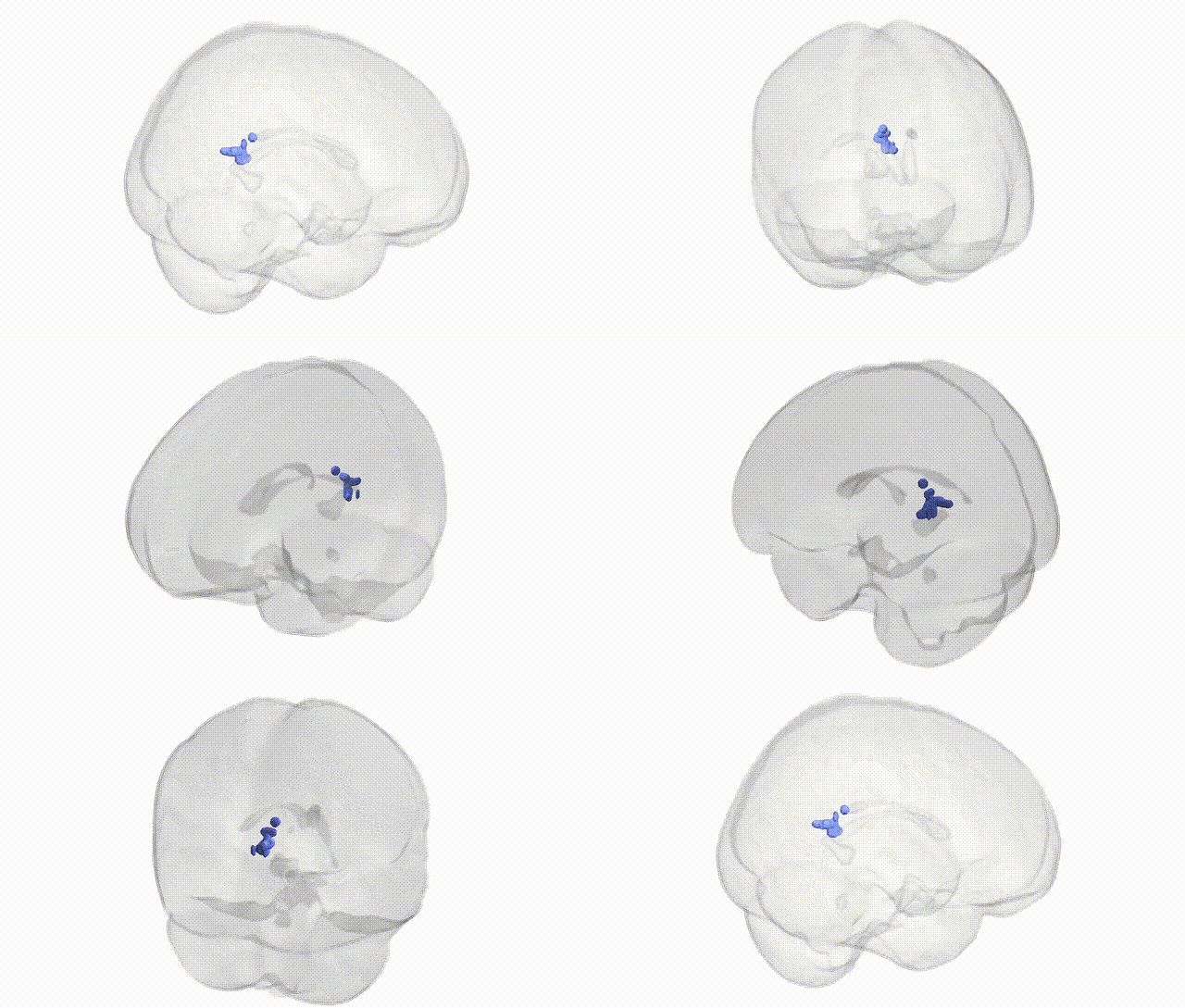
Full Quality Version: Download MP4
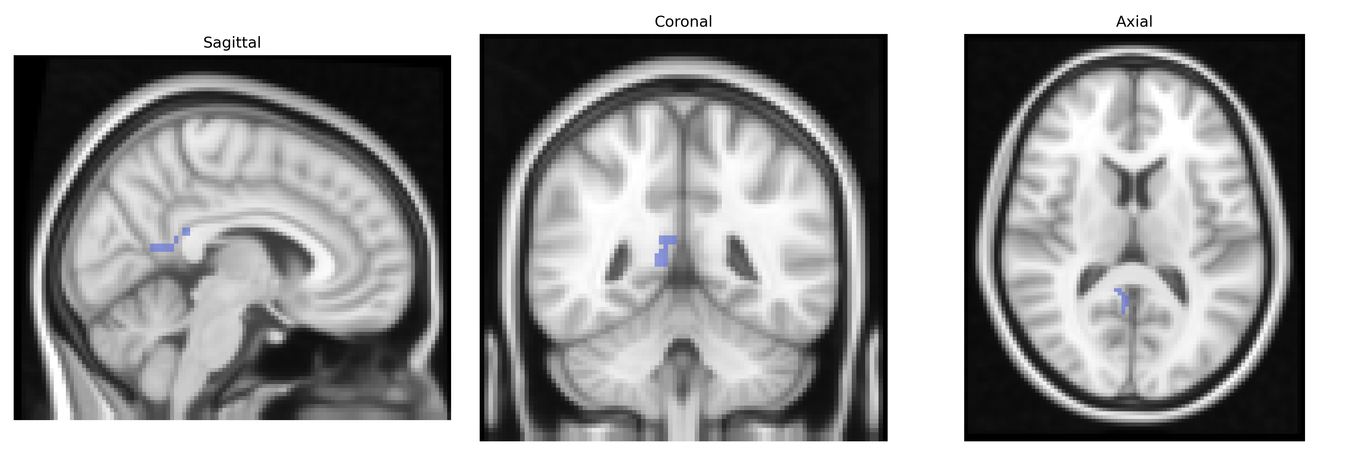
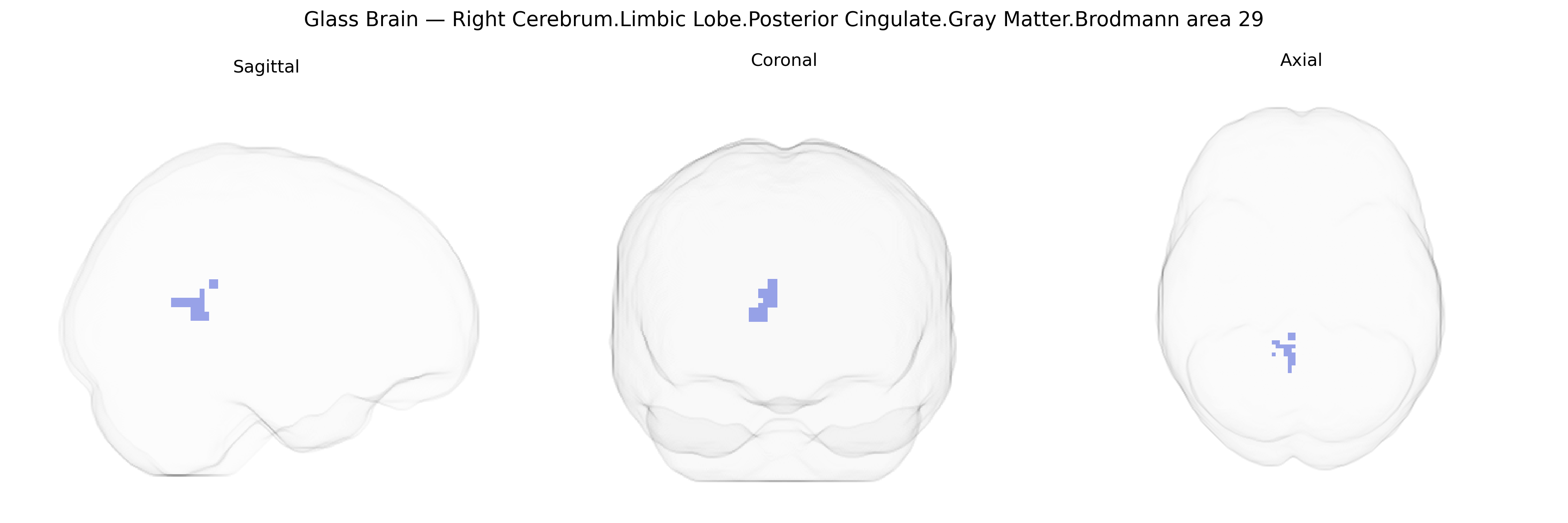
Wali Sidiqyar*, Gaurav Rudravaram*, Elyssa M. McMaster, Trent M. Schwartz, Adam M. Saunders, Kurt G. Schilling, Bennett A. Landman "Introducing SPINS: A Shared Public Visualization Library of Neuroanatomical Structures." Medical Imaging with Deep Learning- short paper
This resource is licensed under CC0 1.0 Universal (Public Domain).