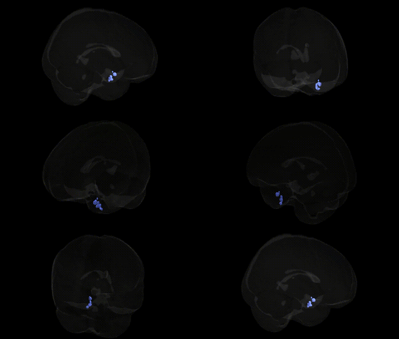
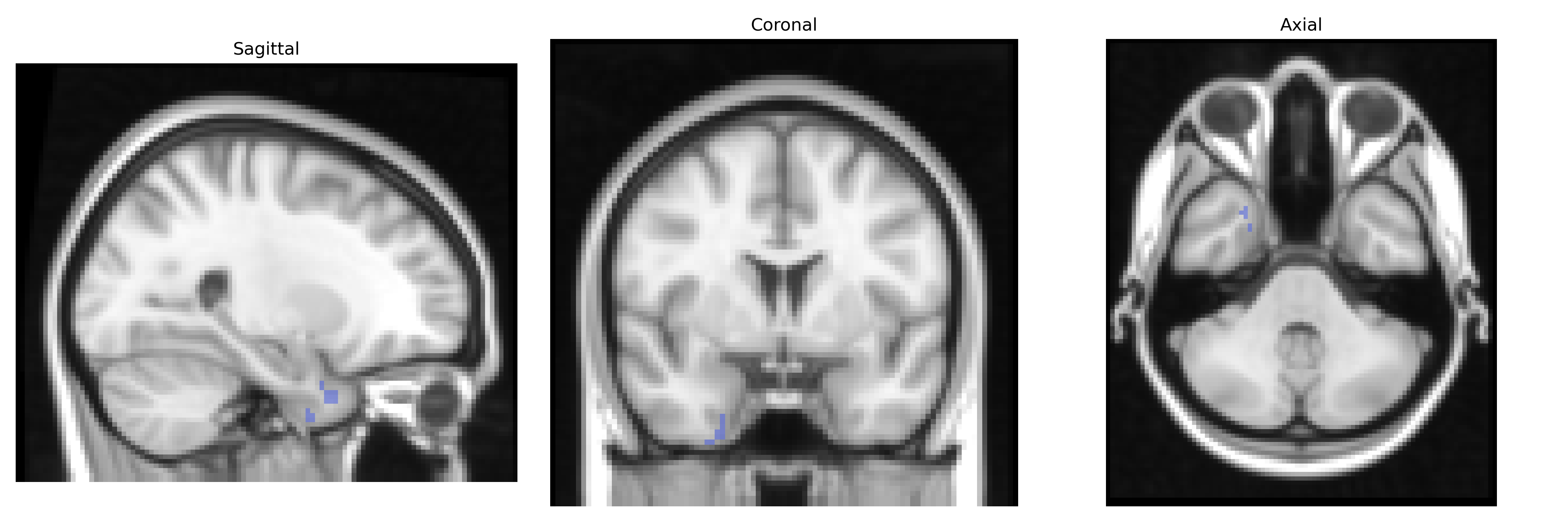
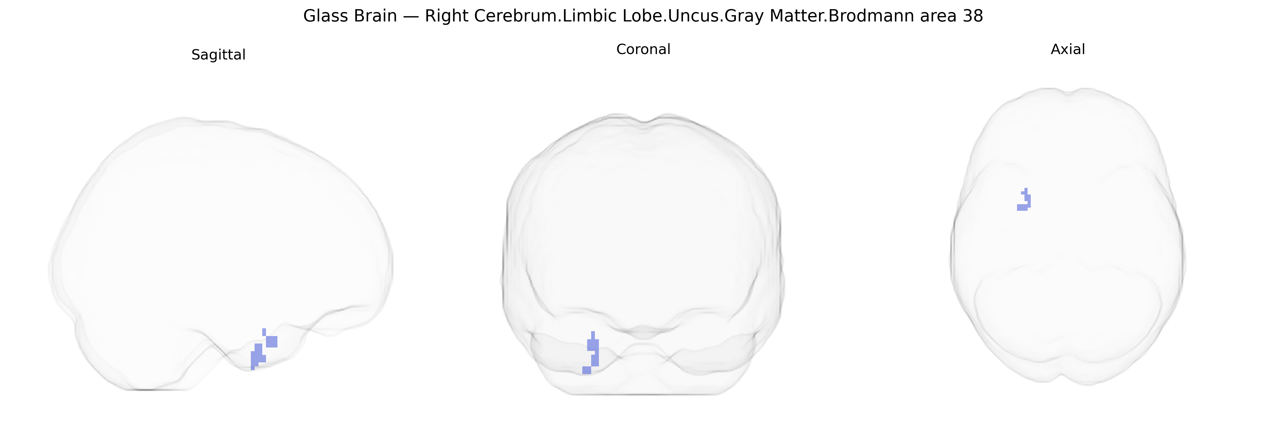
The bilateral Right Cerebrum.Limbic Lobe.Uncus.Gray Matter.Brodmann area 38 corresponds to the anterior portion of the temporal pole within the limbic-associated uncus, encompassing granular cortical gray matter involved in high-level integration of emotional, mnemonic, and complex social information. Functionally, this region participates in multimodal association processes, including semantic memory, emotional valence assignment to sensory stimuli, and aspects of social cognition, often acting as an interface between limbic structures (such as the amygdala and hippocampus) and higher-order association cortices. Brodmann area 38 is also implicated in language-related semantic processing and autobiographical memory, and lesions or dysfunction in this area have been associated with disturbances in emotion processing and certain temporal lobe epilepsies. There is no direct Wikipedia article specifically for “Brodmann area 38”; a closely related and encompassing structure is the temporal pole: Temporal pole.
The bilateral right Brodmann area 38 (temporal pole/uncus within the limbic lobe) has been implicated indirectly in genetic studies through its roles in socio-emotional processing, semantic memory, and affective regulation rather than via region-specific GWAS hits. Imaging genetics and large consortia (e.g., ENIGMA) have linked common variants in genes such as BDNF (e.g., Val66Met), APOE (particularly ε4), and MAPT, as well as loci near GRIN2B, CNTNAP2, and neuregulin-pathway genes, to temporal lobe and limbic gray matter volumes, cortical thickness, or functional connectivity patterns that include the temporal pole. GWAS of psychiatric and neurodevelopmental disorders—schizophrenia, major depressive disorder, bipolar disorder, autism spectrum disorder, and frontotemporal dementia—identify polygenic risk involving synaptic, glutamatergic, and immune-related pathways, and neuroimaging work frequently shows structural and functional abnormalities in BA38 in these conditions, suggesting that the convergent genetic architecture influencing synaptic plasticity and neurodevelopment affects this region among others. Alzheimer’s disease risk alleles (notably APOE ε4 and several GWAS-identified loci such as CLU, PICALM, and CR1) are associated with medial temporal and temporal pole atrophy and altered limbic connectivity, while temporopolar and uncus abnormalities have also been tied to genetic epilepsies (e.g., mesial temporal lobe epilepsy with hippocampal sclerosis) where heritability is high but specific BA38-focused loci remain unclear. Overall, genetic associations relevant to BA38 reflect broad, distributed neurogenetic effects on temporal and limbic networks rather than discrete, uniquely identified susceptibility loci for this specific Talairach-defined region.
Overview generated by GPT-4o (2026).
Region ID: 35
Hemisphere: bilateral
Atlas: Talairach labels 2mm
Full Quality Version: Download MP4
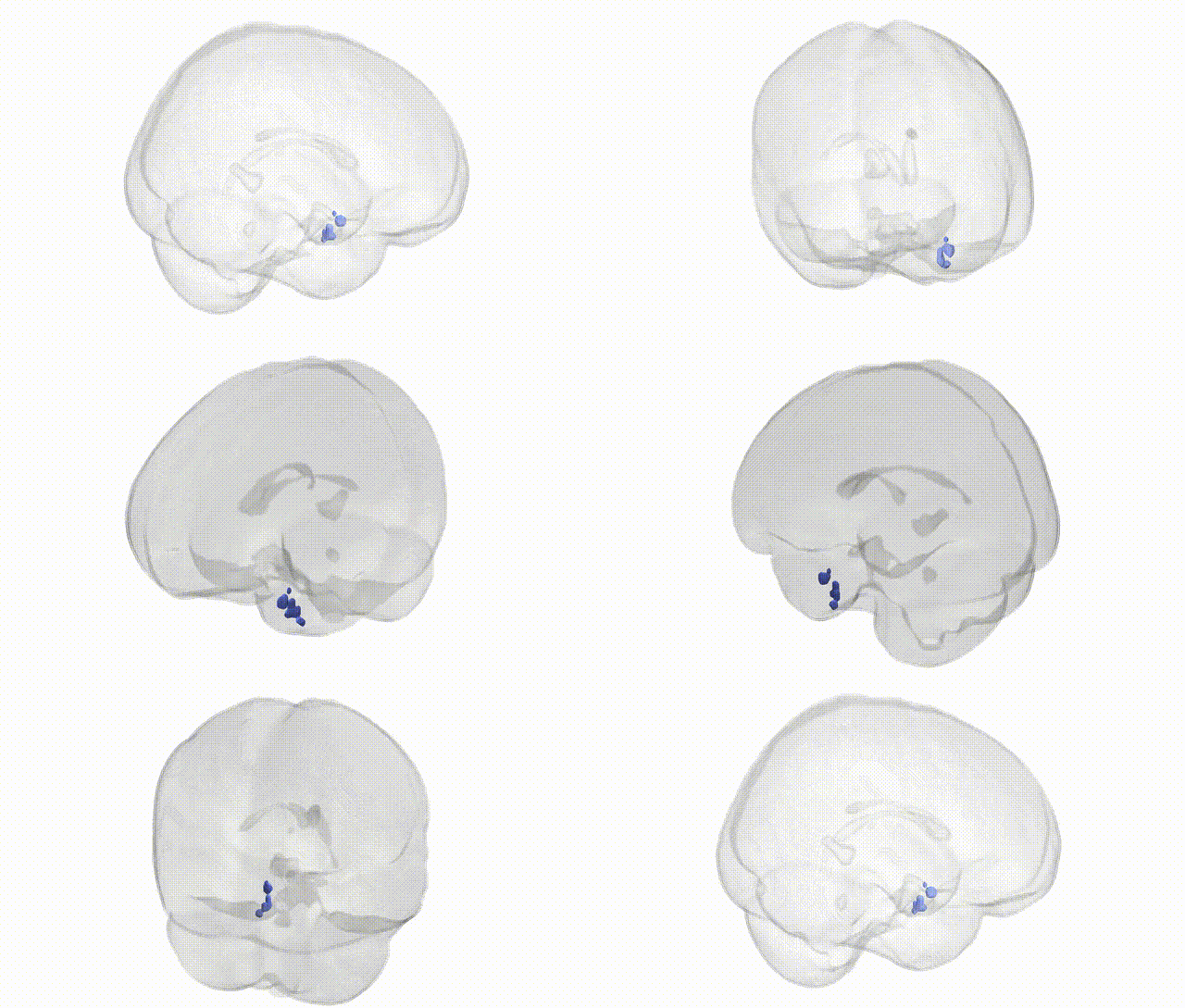
Full Quality Version: Download MP4
Wali Sidiqyar*, Gaurav Rudravaram*, Elyssa M. McMaster, Trent M. Schwartz, Adam M. Saunders, Kurt G. Schilling, Bennett A. Landman "Introducing SPINS: A Shared Public Visualization Library of Neuroanatomical Structures." Medical Imaging with Deep Learning- short paper
This resource is licensed under CC0 1.0 Universal (Public Domain).