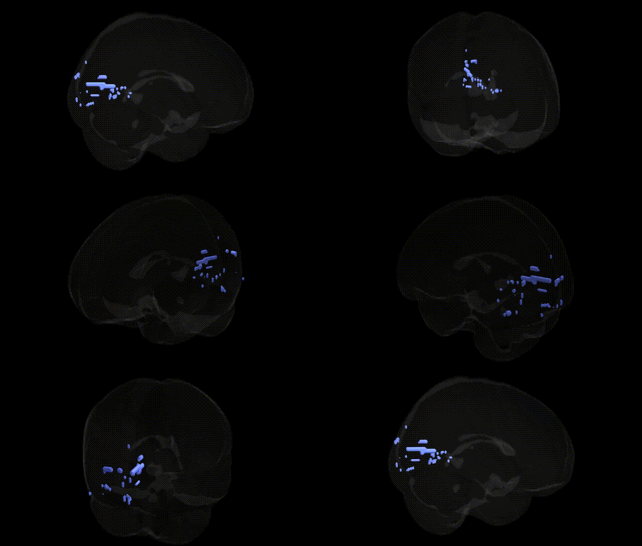
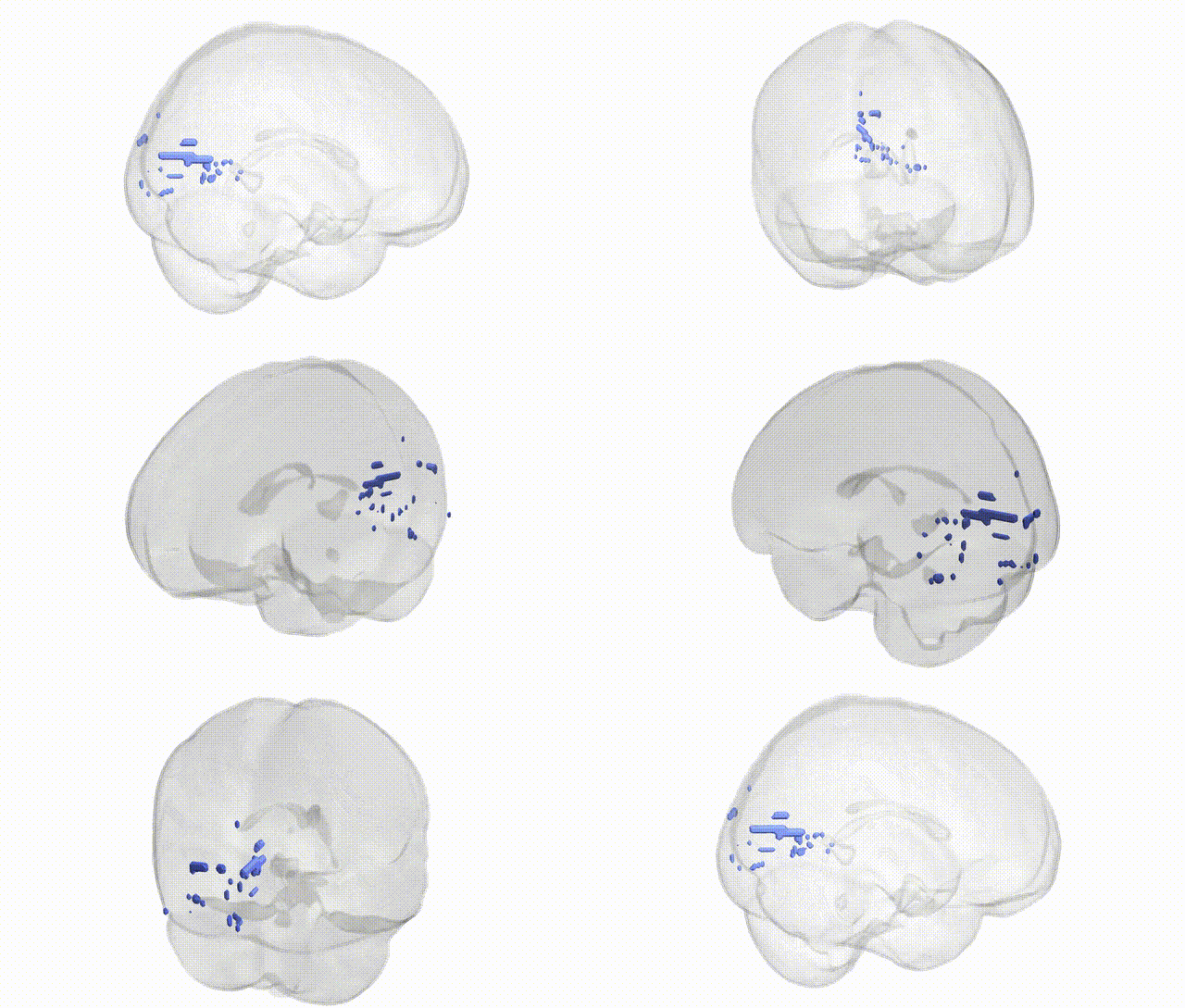
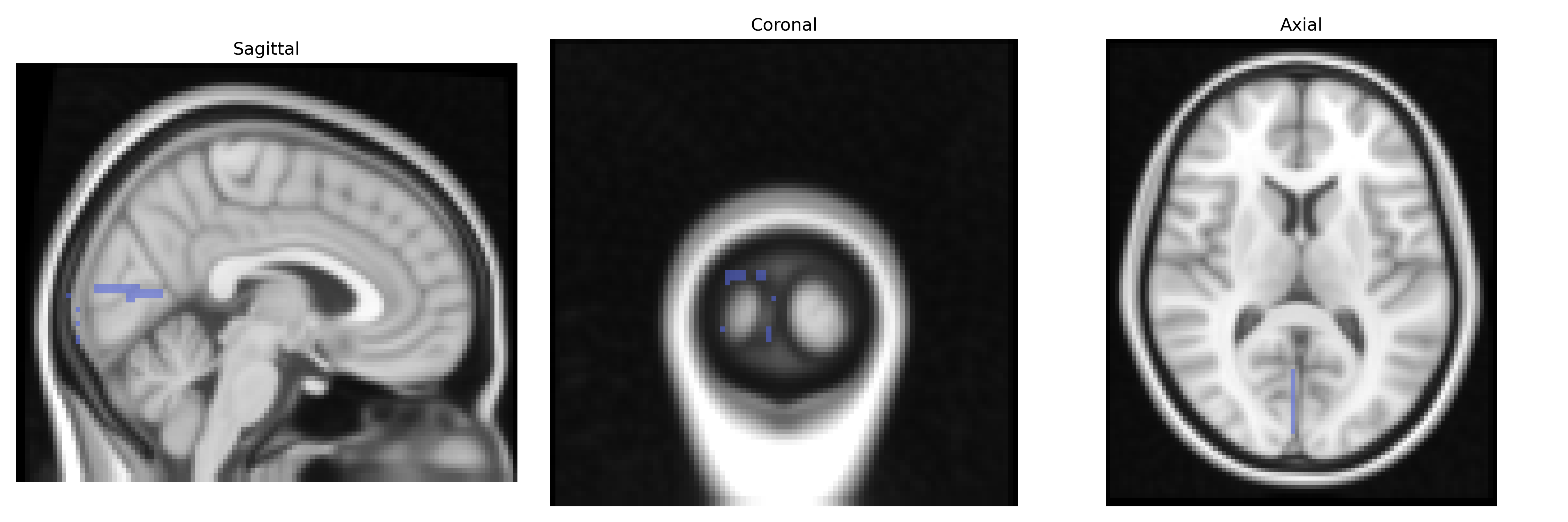
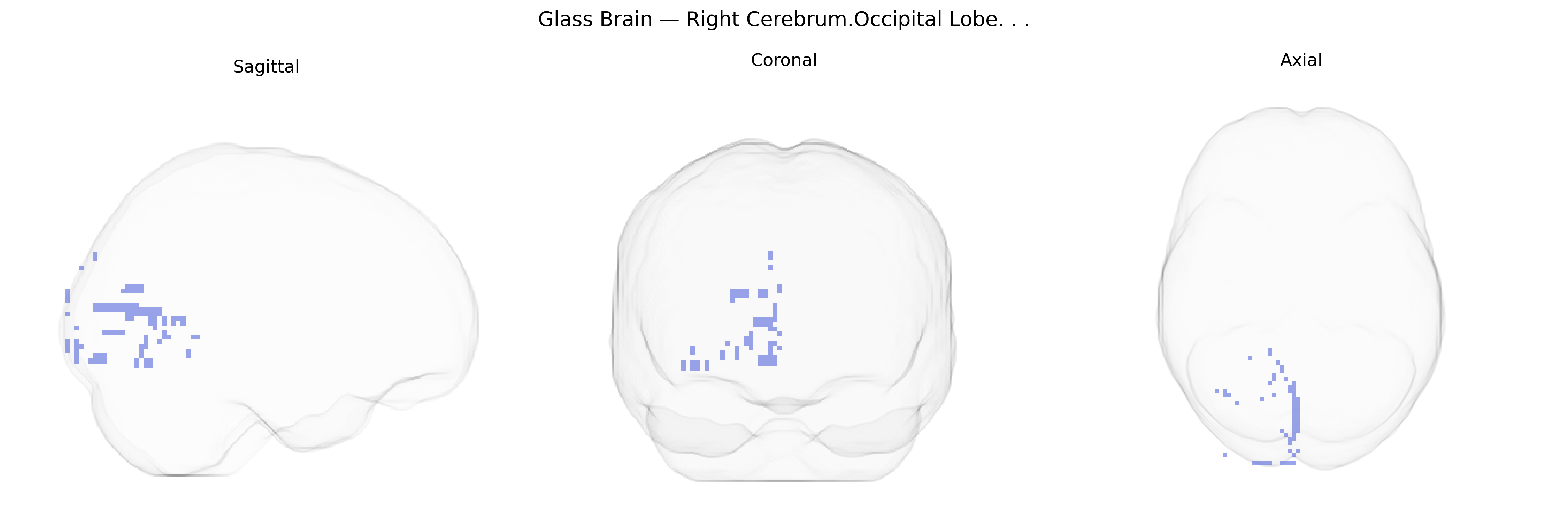
The bilateral Right Cerebrum Occipital Lobe, as defined in the Talairach 2 mm Atlas, corresponds to cortical territories in the posterior portion of the right cerebral hemisphere involved primarily in visual processing. This lobe encompasses regions such as the primary visual cortex (V1) and surrounding extrastriate areas, which receive and integrate input from the retina via the lateral geniculate nucleus of the thalamus. Neuronal populations in this lobe are organized retinotopically and support functions including basic feature detection (orientation, contrast, motion), visuospatial analysis, and higher-level visual recognition when interacting with dorsal and ventral visual pathways. Damage or dysfunction in the right occipital lobe can lead to contralateral (left visual field) deficits, including scotomas or hemianopia, and may contribute to visuoperceptual disturbances.
The bilateral right occipital lobe, encompassing primary and associative visual cortex in the Talairach 2 mm atlas, has been implicated in multiple genetic studies of brain structure, function, and disease, though most work targets occipital or visual regions more broadly rather than this specific bilateral right parcel. Large-scale neuroimaging GWAS (e.g., ENIGMA, UK Biobank) have identified common variants in genes related to neurodevelopment and synaptic function—such as HMGA2, IGF1, PAPPA, and several loci near regulatory regions—that associate with occipital cortical thickness, surface area, and overall occipital lobe volume. Visual cortex activation and connectivity phenotypes show heritability and have been linked in GWAS and candidate-gene studies to variants affecting glutamatergic and GABAergic signaling, myelination, and plasticity, including genes such as BDNF and GRIN family members, which influence visual learning and perceptual adaptation. Occipital regions, including right-sided visual cortex, emerge in imaging genetics studies of migraine (with variants in TRPM8, LRP1, and others influencing cortical excitability and visual aura susceptibility), schizophrenia and bipolar disorder (where risk loci in genes like CACNA1C, ZNF804A, and GRM3 are associated with altered occipital activation and connectivity), and autism spectrum disorder (with synaptic and neurodevelopmental genes such as CNTNAP2, SHANK3, and NRXN-related loci linked to atypical visual processing and occipital structural differences). Additionally, GWAS of reading ability, face recognition, and visuospatial skills identify variants near genes involved in cortical patterning and axonal guidance (e.g., ROBO/SLIT pathways) that modulate occipital and occipitotemporal structure–function measures, suggesting that genetic influences on visual cortex development and connectivity underpin individual differences in higher-order visual cognition and vulnerability to neuropsychiatric conditions that prominently affect occipital lobe function.
Overview generated by GPT-4o (2026).
Region ID: 248
Hemisphere: bilateral
Atlas: Talairach labels 2mm
Full Quality Version: Download MP4
Full Quality Version: Download MP4
Wali Sidiqyar*, Gaurav Rudravaram*, Elyssa M. McMaster, Trent M. Schwartz, Adam M. Saunders, Kurt G. Schilling, Bennett A. Landman "Introducing SPINS: A Shared Public Visualization Library of Neuroanatomical Structures." Medical Imaging with Deep Learning- short paper
This resource is licensed under CC0 1.0 Universal (Public Domain).