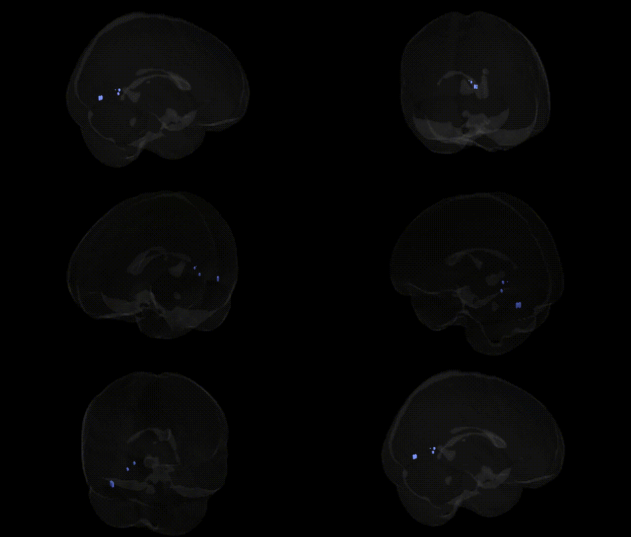
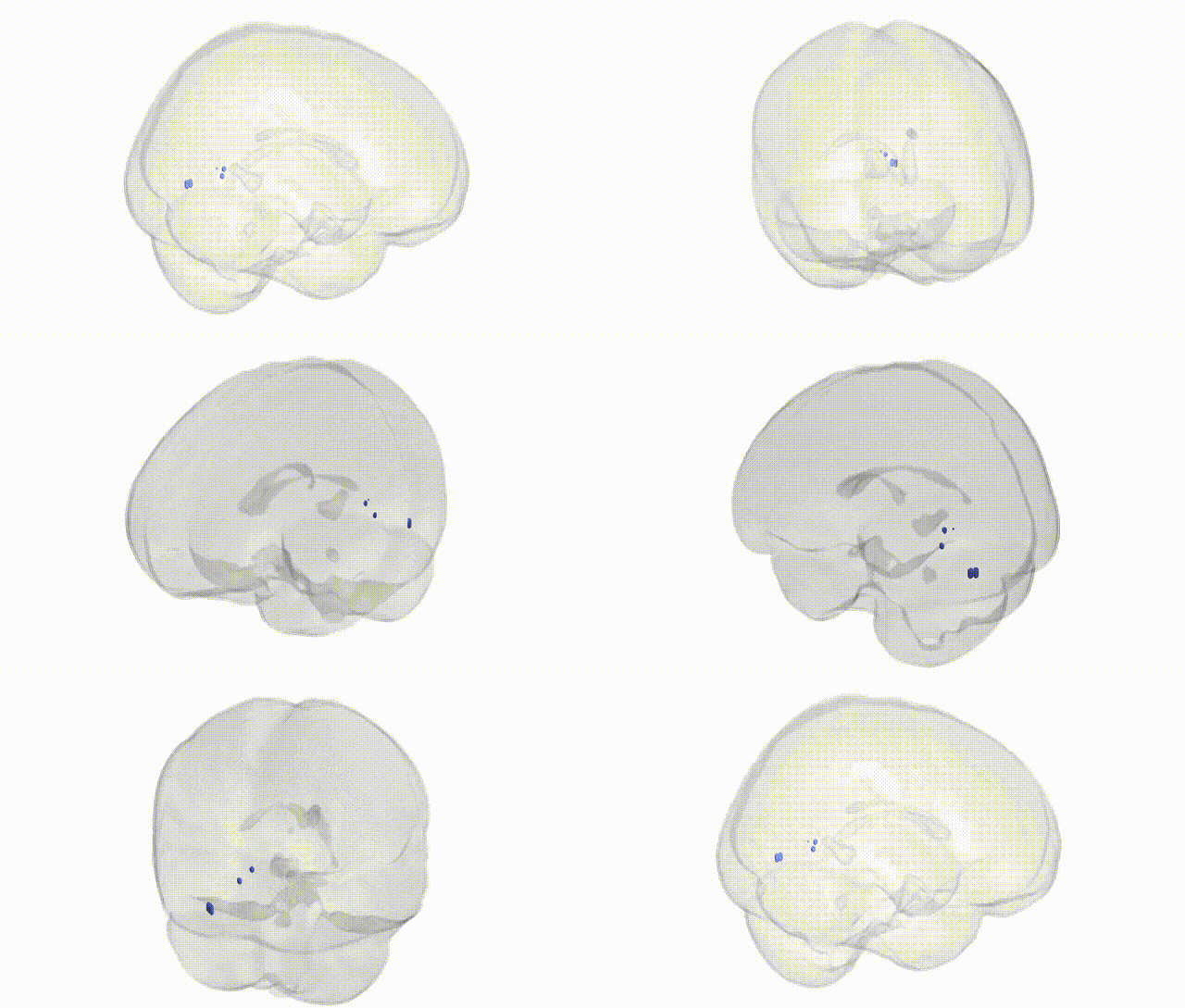
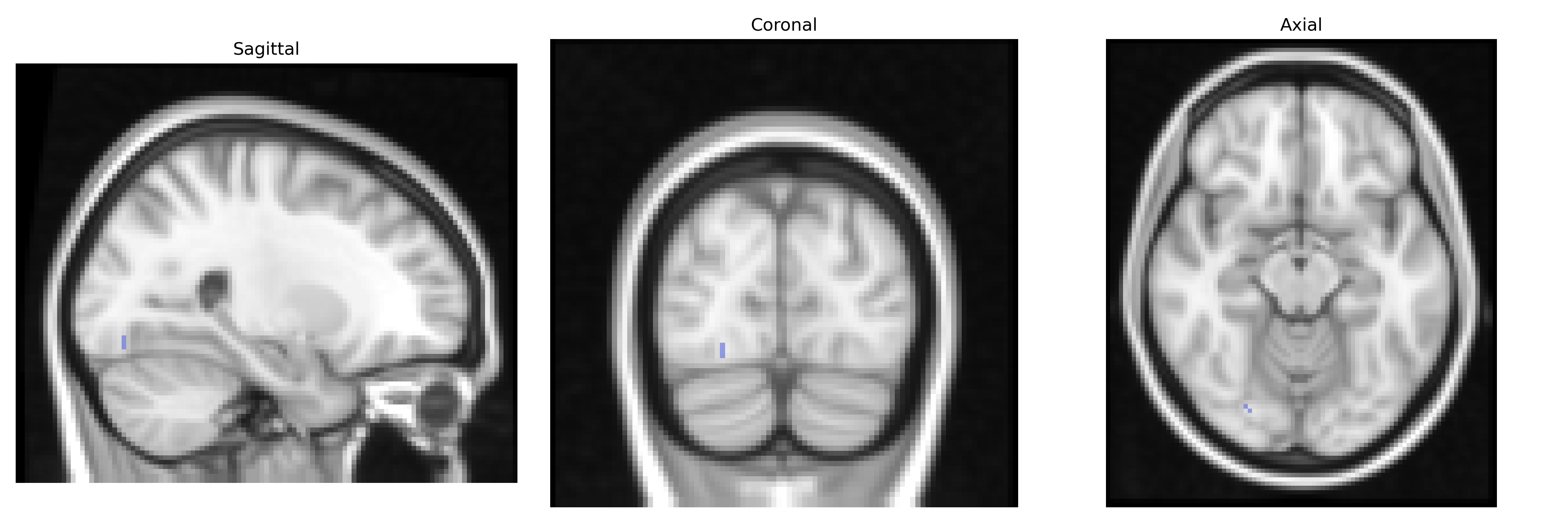
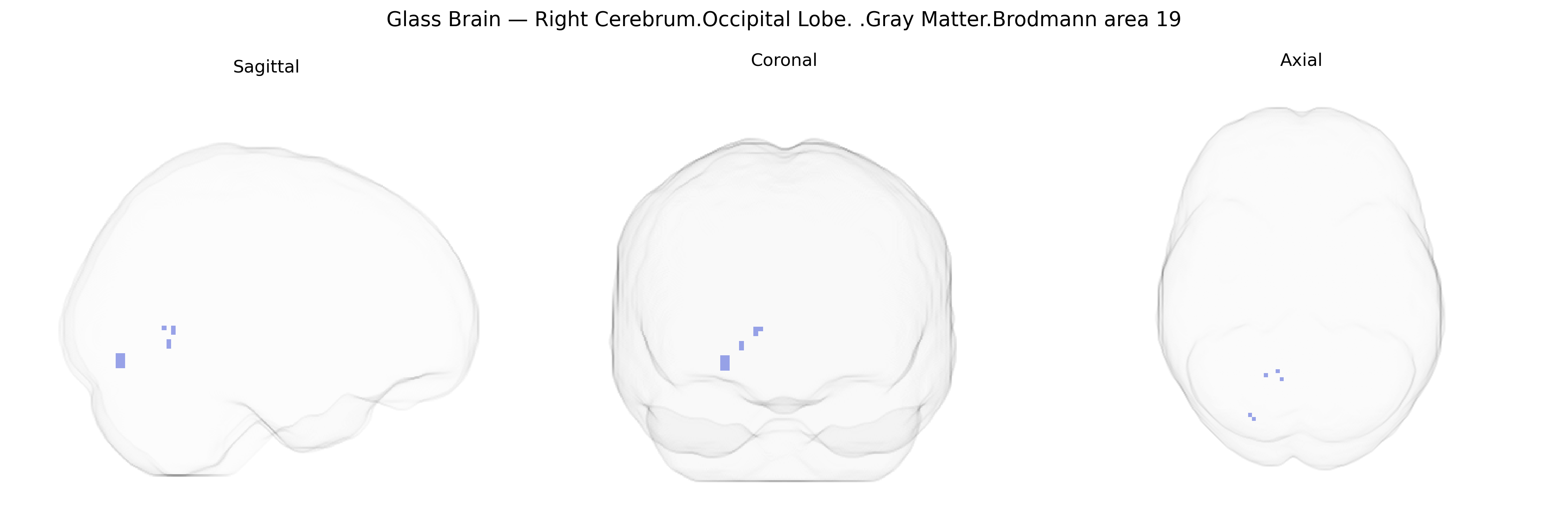
The bilateral Right Cerebrum.Occipital Lobe.Gray Matter.Brodmann area 19, as defined in the Talairach 2 mm Atlas, corresponds to a secondary visual association cortex located in the occipital lobe, surrounding and extending beyond the primary visual cortex (Brodmann area 17) and the parastriate cortex (Brodmann area 18). This region participates in higher-order visual processing, including complex feature integration, object recognition, motion and form analysis, and the integration of visual input with other sensory modalities and memory systems. It receives extensive feedforward and feedback connections from primary visual areas and projects to temporal and parietal association cortices, contributing to visuospatial processing and visual perception necessary for tasks such as scene analysis and visually guided behavior. There is no direct Wikipedia article for this precise Talairach-defined bilateral region; a closely related structure is Brodmann area 19.
Genetic associations involving bilateral Brodmann area 19 gray matter in the occipital lobe, as defined in the Talairach 2 mm atlas, largely reflect its role in higher-order visual processing and visual–cognitive integration. GWAS and imaging genetics studies implicate multiple loci that influence cortical thickness, surface area, and volume in occipital regions encompassing BA19, including variants near genes such as CENPW, TESC, PAX6, and others identified in large ENIGMA and UK Biobank cohorts, though findings are typically reported for broader occipital or visual cortex rather than BA19 specifically. Occipital and visual association cortex measures overlapping BA19 have been linked to polygenic risk for neuropsychiatric conditions (e.g., schizophrenia, major depression, and bipolar disorder), with higher genetic liability often associated with subtle alterations in occipital gray matter structure or functional activation during visual tasks. Rare monogenic and CNV disorders that affect visual perception and cortical development (for example, some forms of posterior cortical atrophy, developmental visual agnosia, and syndromes involving PAX6 or other neurodevelopmental genes) can involve structural or functional disruption in BA19. Additionally, GWAS of cognitive traits such as general intelligence, educational attainment, and reading-related skills have reported associations with occipital/visual association cortical measures, suggesting that genetically driven variation in BA19 contributes to individual differences in complex visual cognition and visuospatial abilities, although precise BA19-specific genetic effects remain incompletely resolved.
Overview generated by GPT-4o (2026).
Region ID: 263
Hemisphere: bilateral
Atlas: Talairach labels 2mm
Full Quality Version: Download MP4
Full Quality Version: Download MP4
Wali Sidiqyar*, Gaurav Rudravaram*, Elyssa M. McMaster, Trent M. Schwartz, Adam M. Saunders, Kurt G. Schilling, Bennett A. Landman "Introducing SPINS: A Shared Public Visualization Library of Neuroanatomical Structures." Medical Imaging with Deep Learning- short paper
This resource is licensed under CC0 1.0 Universal (Public Domain).