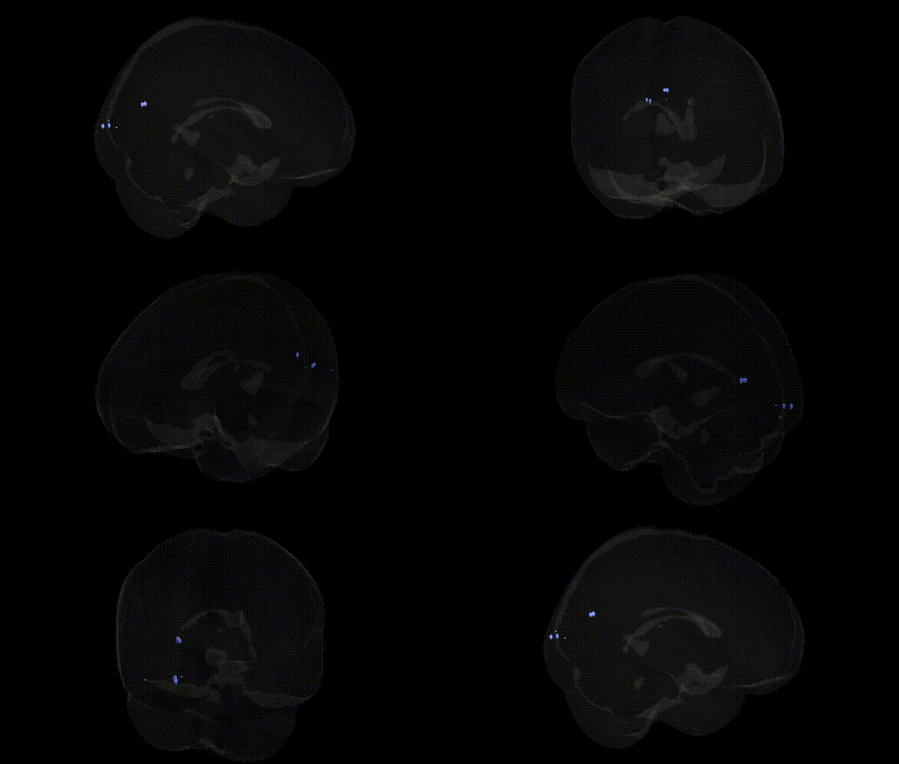
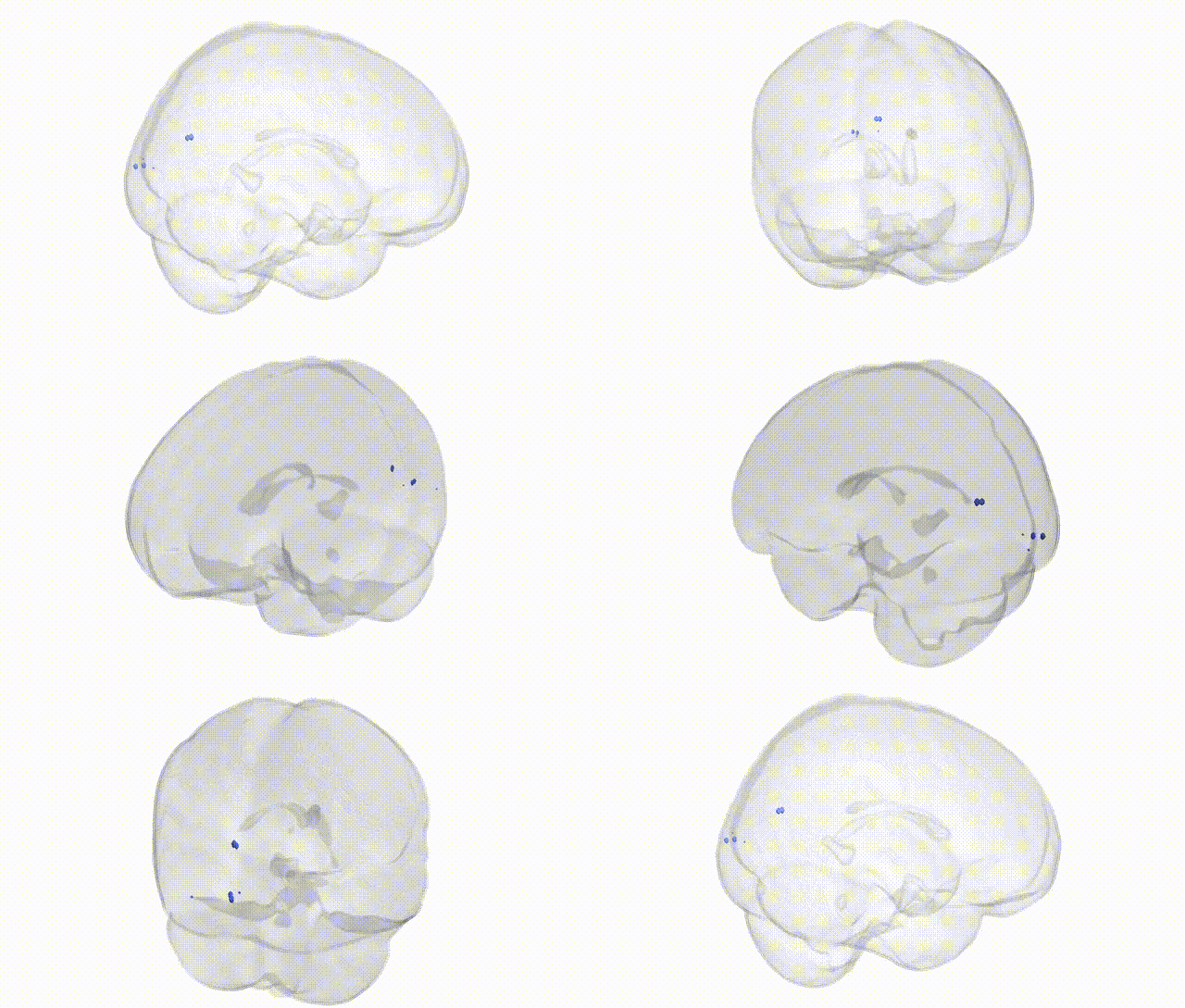
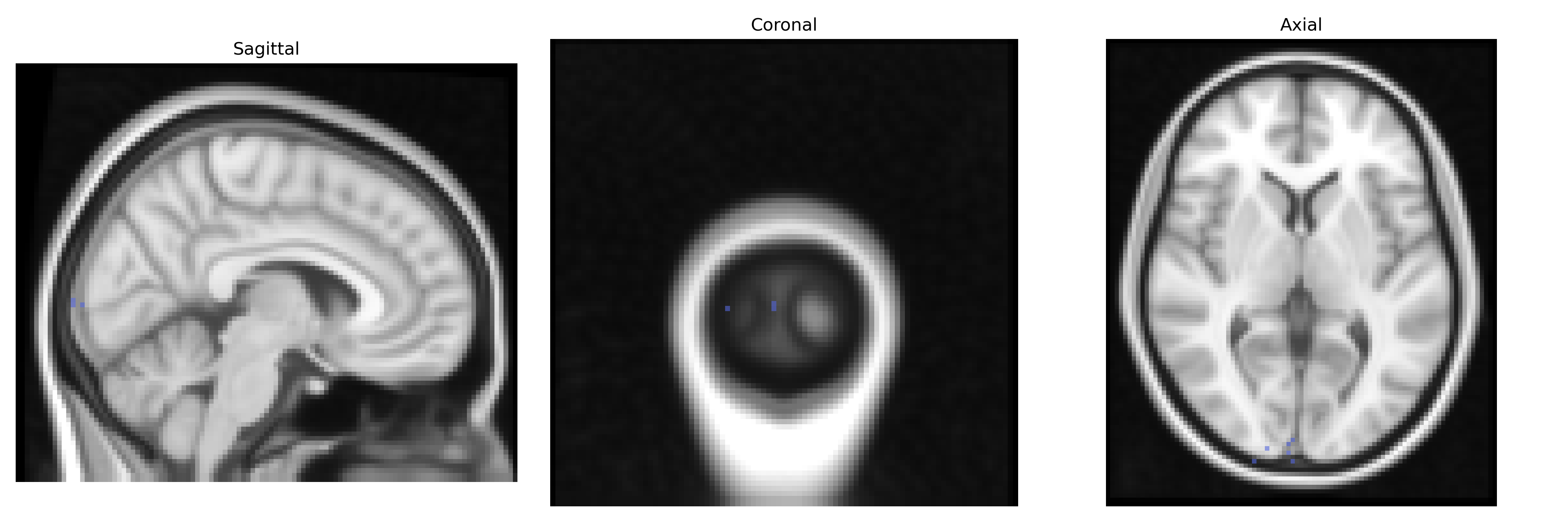
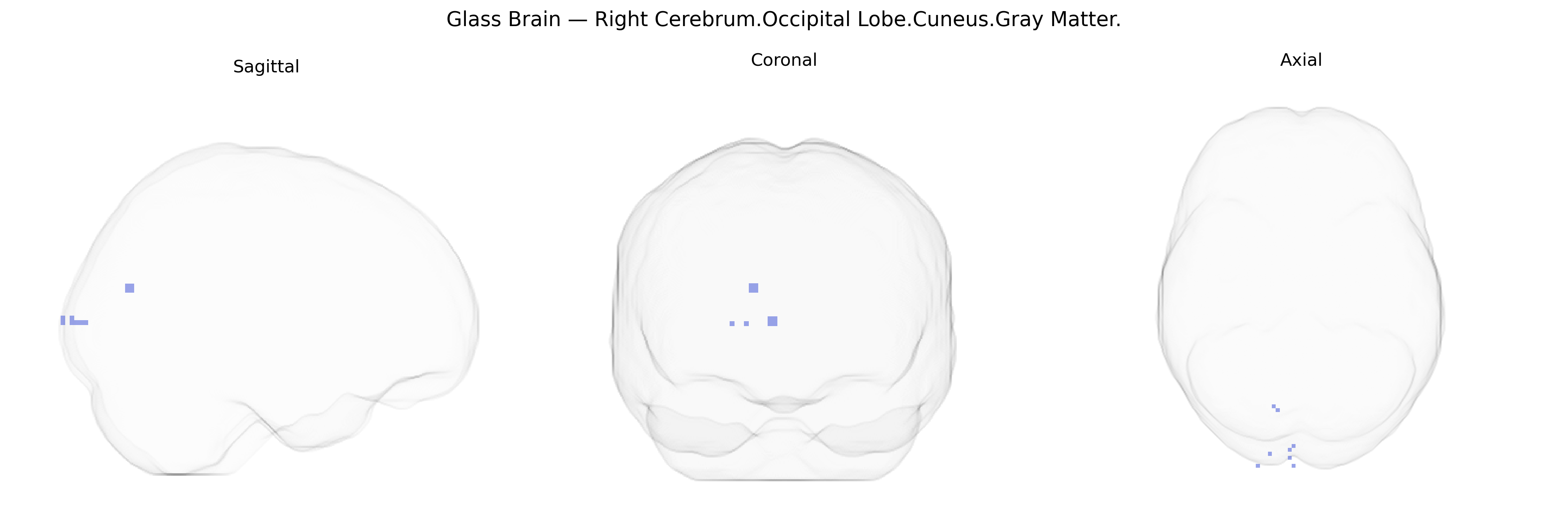
The bilateral Right Cerebrum.Occipital Lobe.Cuneus.Gray Matter corresponds to cortical gray matter within the cuneus of the occipital lobe in the right hemisphere, as defined in the Talairach 2 mm Atlas. The cuneus is a wedge-shaped portion of the medial occipital cortex located between the parieto-occipital sulcus and the calcarine sulcus, and it participates predominantly in visual processing. It is involved in basic visual functions such as visual field mapping and integration of visual input from the retina, and it contributes to higher-order functions including visual attention, visuospatial processing, and aspects of visual memory. As part of the primary and associative visual cortices, activity in this region is modulated by both bottom-up sensory input and top-down cognitive influences such as attention and expectation. There is no direct Wikipedia article specifically for “Right cerebrum occipital lobe cuneus gray matter”; a related entry describing the broader structure is Cuneus.
The bilateral right cuneus gray matter in the occipital lobe, as defined in the Talairach 2 mm atlas, has been repeatedly implicated in genetic studies of brain structure, cognition, and neuropsychiatric conditions. Numerous GWAS of cortical morphology (e.g., ENIGMA and UK Biobank–based analyses) show significant heritability of occipital and cuneus thickness and surface area, with associations involving variants in genes related to neurodevelopment, synaptic organization, and axon guidance (such as those in Wnt signaling and cell-adhesion pathways), although specific loci are often regionally broad rather than uniquely confined to the cuneus. Cuneus volume and cortical thickness have been associated with polygenic risk scores and individual variants linked to schizophrenia, major depressive disorder, and bipolar disorder, reflecting shared genetic influences on visual processing networks and higher-order integration; similar structural and functional cuneus alterations have been reported in GWAS-informed imaging studies of autism spectrum disorder and attention-deficit/hyperactivity disorder. Genetic influences on visual cognition and perceptual traits, including visual memory and processing speed, sometimes highlight occipital/cuneus regions in imaging–genetics analyses, with implicated genes involved in glutamatergic signaling and neural plasticity. In Alzheimer’s disease and other neurodegenerative GWAS, APOE and additional risk loci show downstream associations with patterns of cortical atrophy that include the cuneus, especially in longitudinal imaging–genetics frameworks. Overall, the cuneus is part of heritable occipital networks whose structure and function are modulated by polygenic architecture spanning neurodevelopmental, synaptic, and disease-related pathways, rather than by a single, region-specific genetic determinant.
Overview generated by GPT-4o (2026).
Region ID: 535
Hemisphere: bilateral
Atlas: Talairach labels 2mm
Full Quality Version: Download MP4
Full Quality Version: Download MP4
Wali Sidiqyar*, Gaurav Rudravaram*, Elyssa M. McMaster, Trent M. Schwartz, Adam M. Saunders, Kurt G. Schilling, Bennett A. Landman "Introducing SPINS: A Shared Public Visualization Library of Neuroanatomical Structures." Medical Imaging with Deep Learning- short paper
This resource is licensed under CC0 1.0 Universal (Public Domain).