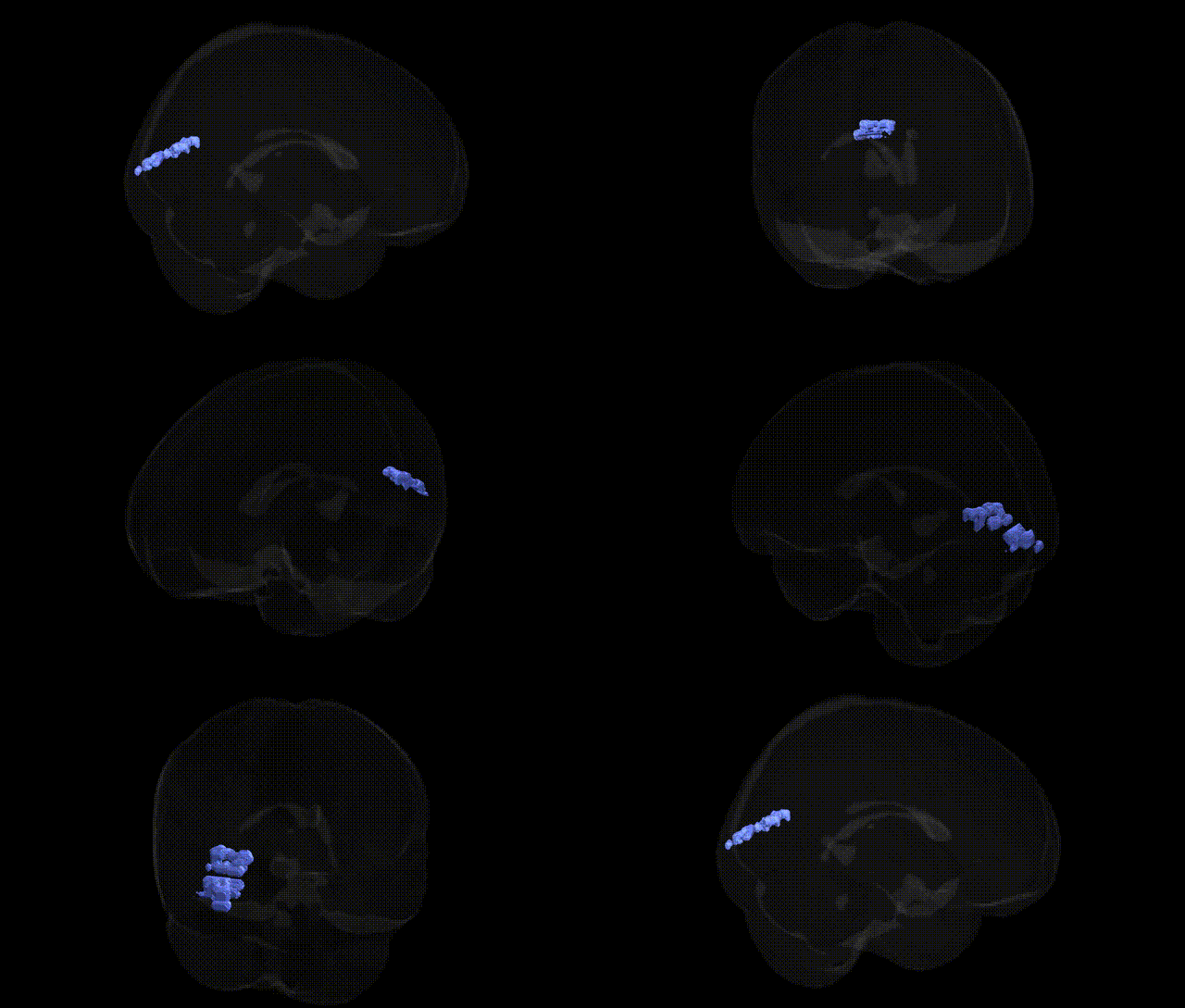
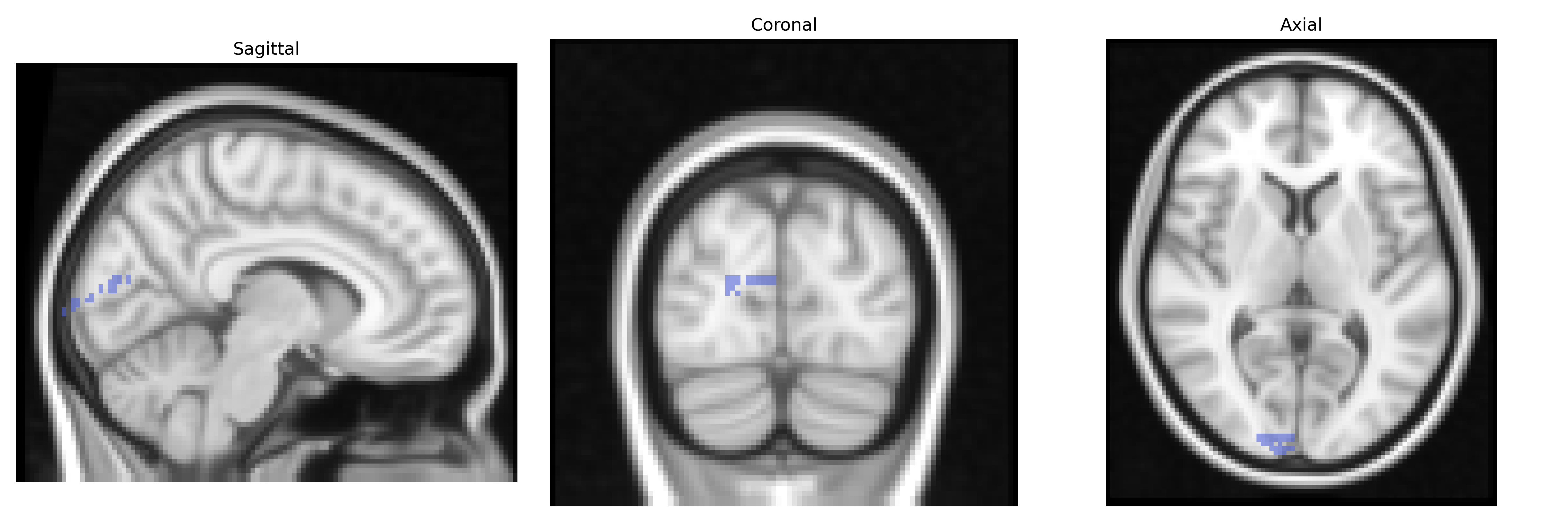
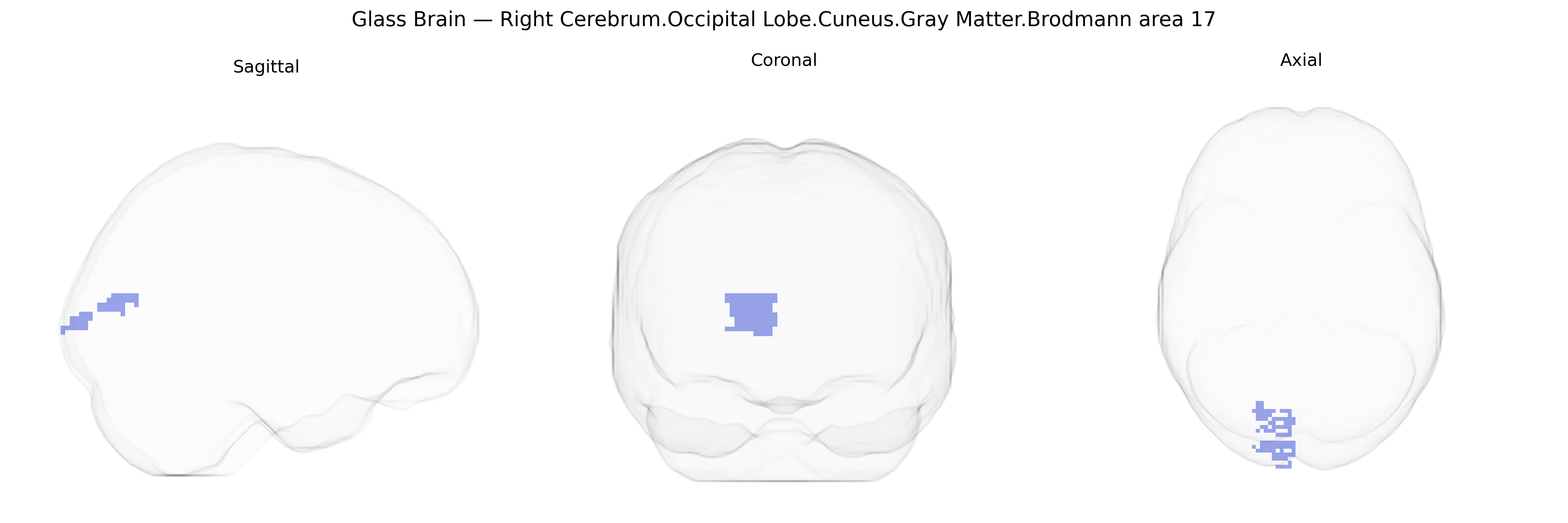
The bilateral Right Cerebrum.Occipital Lobe.Cuneus.Gray Matter.Brodmann area 17 corresponds to the primary visual cortex (V1) located in the cuneus of the occipital lobe, representing the initial cortical stage of visual processing. This region receives highly organized input from the lateral geniculate nucleus of the thalamus via the optic radiations and is characterized cytoarchitectonically by dense granular layers (notably layer IV) and a distinctive band of myelinated fibers (the line of Gennari). Functionally, Brodmann area 17 encodes basic visual features such as orientation, spatial frequency, contrast, and retinotopic position, forming a precise map of the visual field with a strong emphasis on central (foveal) vision. Activity in this area is essential for conscious visual perception, and damage can result in scotomas or cortical blindness, depending on lesion extent and laterality.
Primary visual cortex (Brodmann area 17)
The bilateral right cerebrum occipital lobe cuneus gray matter Brodmann area 17 (primary visual cortex, V1) has been implicated in several genetic and GWAS-based associations largely through imaging genetics and neuropsychiatric studies rather than direct region-specific GWAS. Variants in genes involved in synaptic development and plasticity (e.g., BDNF, NRG1, GRIN2B), neurotransmission (e.g., GABAergic and glutamatergic pathway genes), and neurodevelopmental patterning (e.g., PAX6, EMX2, visual system–related transcription factors) have been associated with structural measures such as cortical thickness or gray matter volume in or near BA17 in large MRI-GWAS cohorts (e.g., ENIGMA, UK Biobank), although results are typically reported for “occipital” or “visual cortex” regions rather than Talairach-defined BA17 specifically. GWAS of occipital/visual cortex volume and surface area have identified loci near genes such as MAPT, KIAA0586, and others related to axonal transport, ciliary function, and cortical development, some of which also overlap with risk loci for neurodevelopmental and neurodegenerative disorders. Functional and structural alterations in BA17/cuneus have been linked, often in conjunction with genetic risk, to disorders including schizophrenia, bipolar disorder, major depression, autism spectrum disorder, and migraine with aura, where risk variants in glutamatergic and synaptic genes, calcium-channel genes (e.g., CACNA1A/CACNA1C), and serotonergic genes may modulate visual cortex excitability and connectivity. In addition, genetic influences on visual acuity and refractive error (e.g., GWA-identified loci near GJD2, RASGRF1, and others) are indirectly tied to BA17 function through their impact on retinal input and visual pathway integrity, and polygenic scores for cognitive traits and educational attainment have shown associations with occipital/visual cortex morphology, suggesting shared genetic architecture between visual processing capacity and higher cognitive phenotypes. Overall, the genetic landscape related to BA17 is distributed across many neurodevelopmental, synaptic, and sensory-processing genes, with region involvement inferred from imaging genetics rather than single, region-specific “BA17 genes.”
Overview generated by GPT-4o (2026).
Region ID: 477
Hemisphere: bilateral
Atlas: Talairach labels 2mm
Full Quality Version: Download MP4
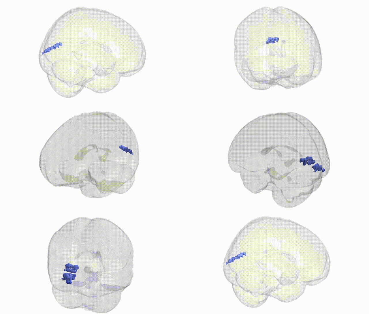
Full Quality Version: Download MP4
Wali Sidiqyar*, Gaurav Rudravaram*, Elyssa M. McMaster, Trent M. Schwartz, Adam M. Saunders, Kurt G. Schilling, Bennett A. Landman "Introducing SPINS: A Shared Public Visualization Library of Neuroanatomical Structures." Medical Imaging with Deep Learning- short paper
This resource is licensed under CC0 1.0 Universal (Public Domain).