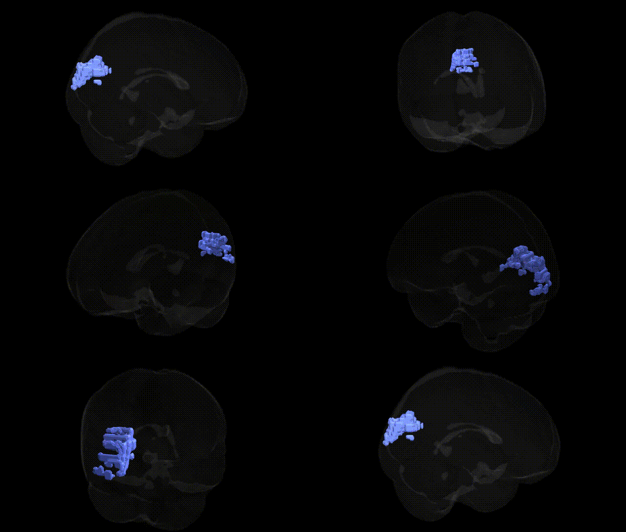
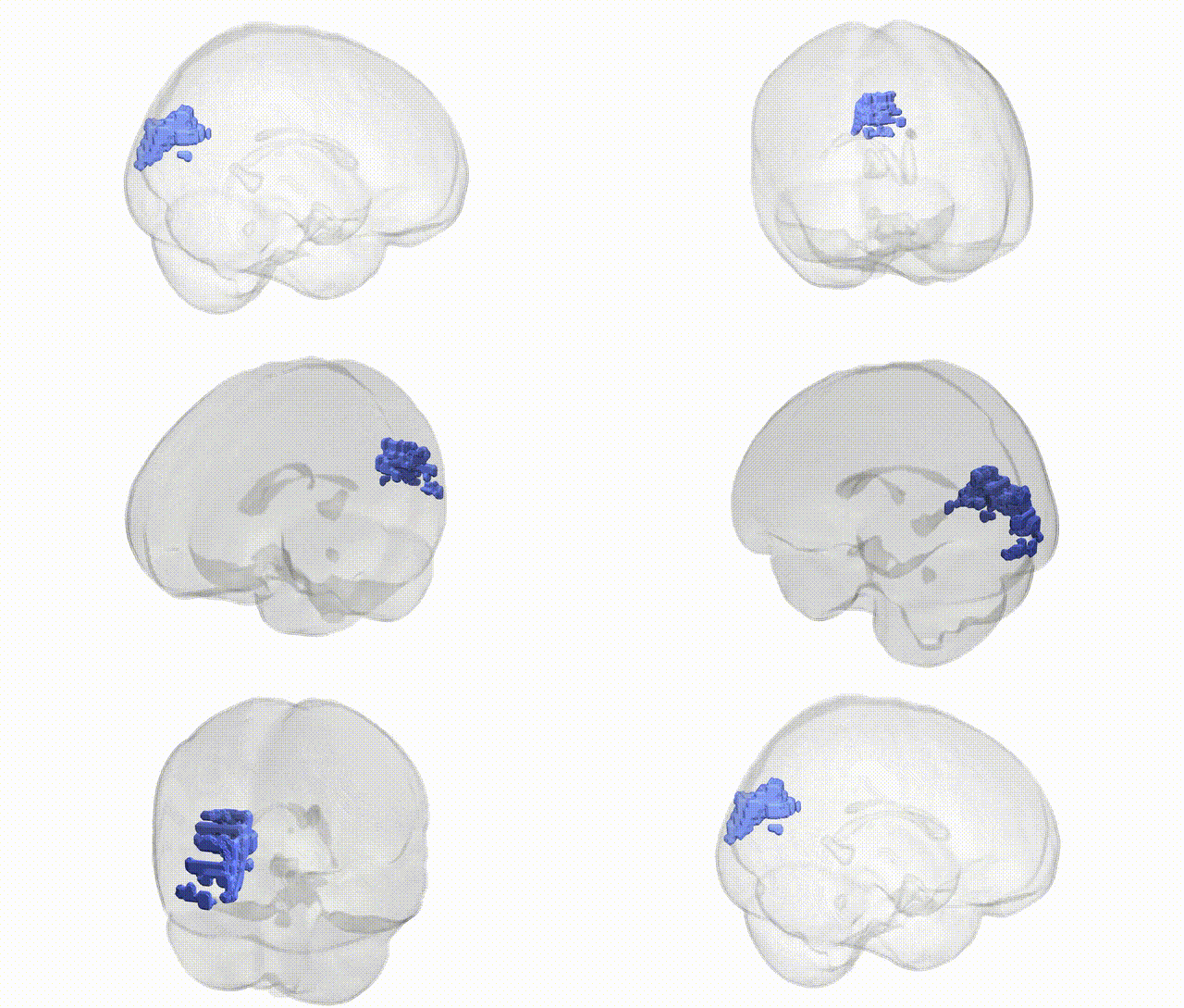
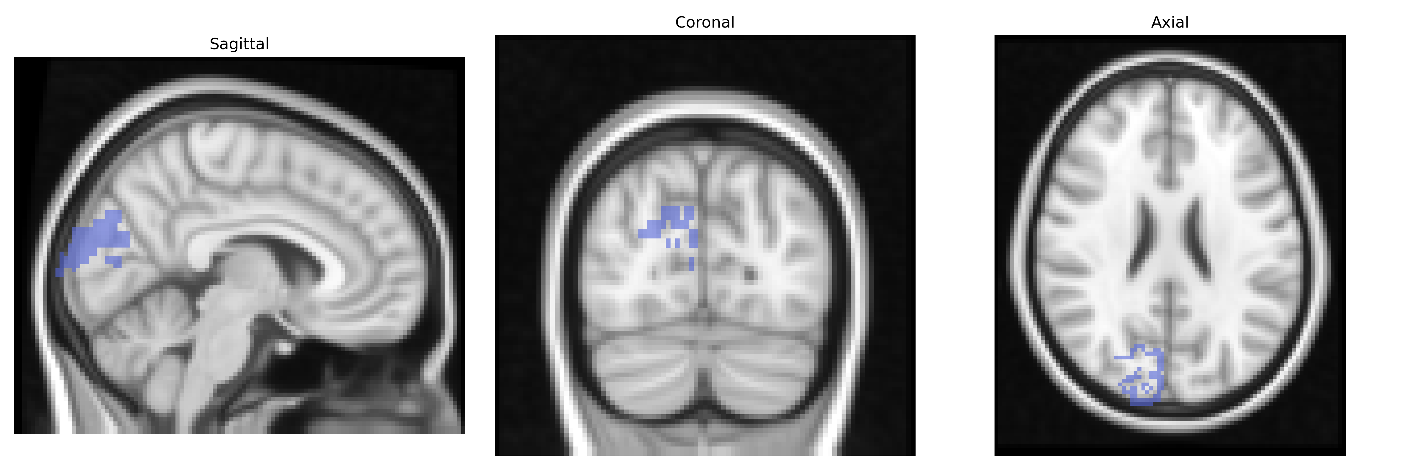
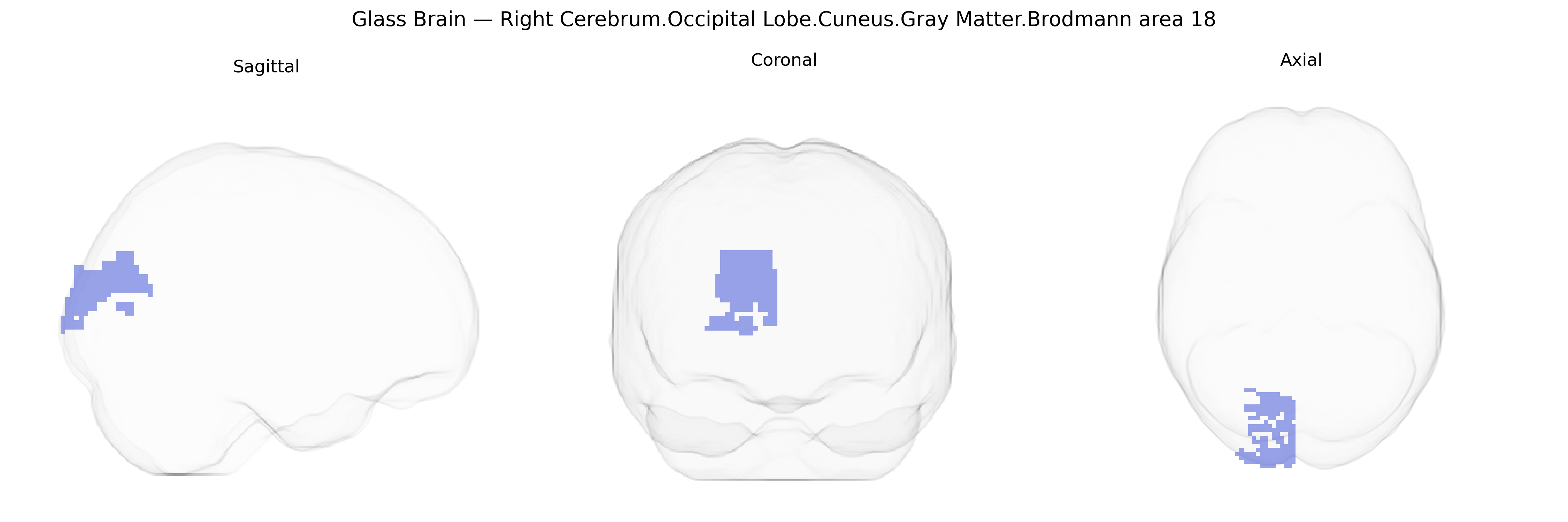
The bilateral Right Cerebrum.Occipital Lobe.Cuneus.Gray Matter.Brodmann area 18 corresponds to the secondary visual cortex (V2) located in the cuneus of the occipital lobe, adjacent to the primary visual cortex (Brodmann area 17). This region is involved in early-stage visual processing, including the integration of information about orientation, spatial frequency, color, and more complex visual patterns, acting as an intermediate processing hub between primary visual input and higher-order visual association areas. Neuronal populations within Brodmann area 18 exhibit retinotopic organization and contribute to the analysis of contours, binocular disparity, and figure–ground segregation, supporting functions such as visual perception of shapes, motion, and depth. There is no direct link for “Brodmann area 18” as a standalone page; a closely related structure and functional description is provided at Visual cortex.
The bilateral right cerebrum occipital lobe cuneus gray matter Brodmann area 18 (secondary visual cortex) is implicated in genetic studies primarily through traits and disorders involving visual processing, cognition, and neuropsychiatric phenotypes. Imaging genetics and GWAS that combine structural or functional MRI with genotypes have linked variation in BA18/cuneus volume or activation to common variants in genes related to synaptic function and neurodevelopment (for example, BDNF, COMT, and glutamatergic signaling genes) and to polygenic scores for educational attainment, general cognitive ability, and schizophrenia. Large ENIGMA and UK Biobank MRI-GWAS consortia have identified multiple loci associated with occipital and cuneus cortical thickness or surface area, including variants near genes involved in neuronal proliferation and cortical patterning (such as MEF2C, TBR1-related pathways, and other neurodevelopmental transcription factors), suggesting that genetic architecture of BA18 shares components with global cortical development. Functional activation of the cuneus/BA18 has been associated with genetic risk for major depressive disorder and anxiety in task-based fMRI studies, while altered structure or connectivity in this region appears in GWAS-informed studies of visual hallucinations in Parkinson’s disease and psychosis. Additionally, rare variant and copy-number analyses in neurodevelopmental disorders (autism spectrum conditions and intellectual disability) frequently show altered occipital and cuneus morphometry, although these findings are typically regionally distributed and not specific to BA18 alone. Overall, genetic associations for this region are indirect and primarily arise from MRI-GWAS of occipital/cuneus cortical metrics and imaging-genetic studies of cognition and psychiatric risk rather than single, well-established BA18-specific susceptibility loci.
Overview generated by GPT-4o (2026).
Region ID: 478
Hemisphere: bilateral
Atlas: Talairach labels 2mm
Full Quality Version: Download MP4
Full Quality Version: Download MP4
Wali Sidiqyar*, Gaurav Rudravaram*, Elyssa M. McMaster, Trent M. Schwartz, Adam M. Saunders, Kurt G. Schilling, Bennett A. Landman "Introducing SPINS: A Shared Public Visualization Library of Neuroanatomical Structures." Medical Imaging with Deep Learning- short paper
This resource is licensed under CC0 1.0 Universal (Public Domain).