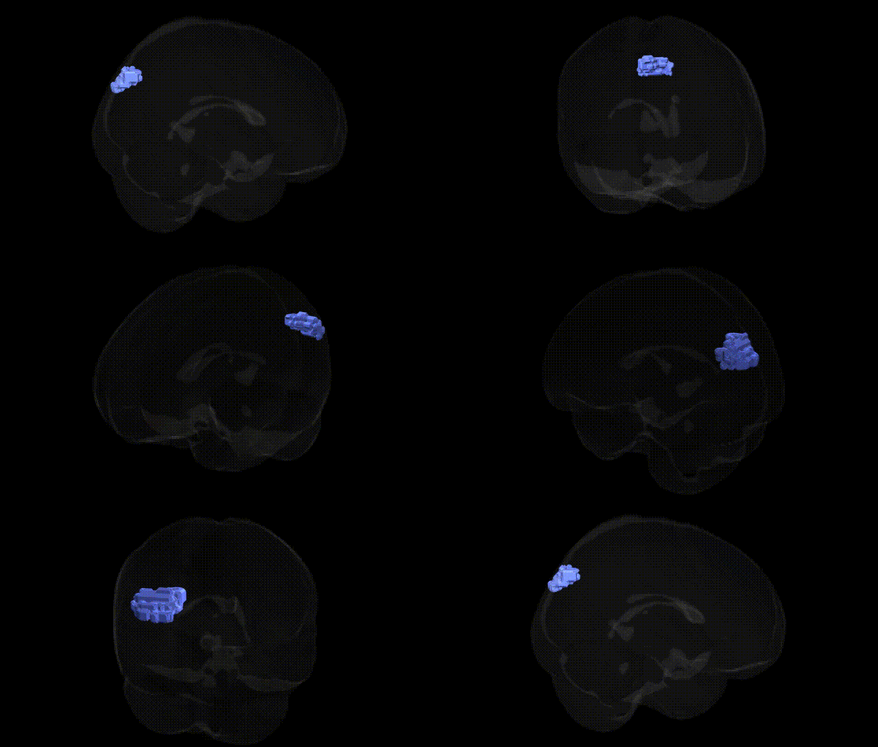
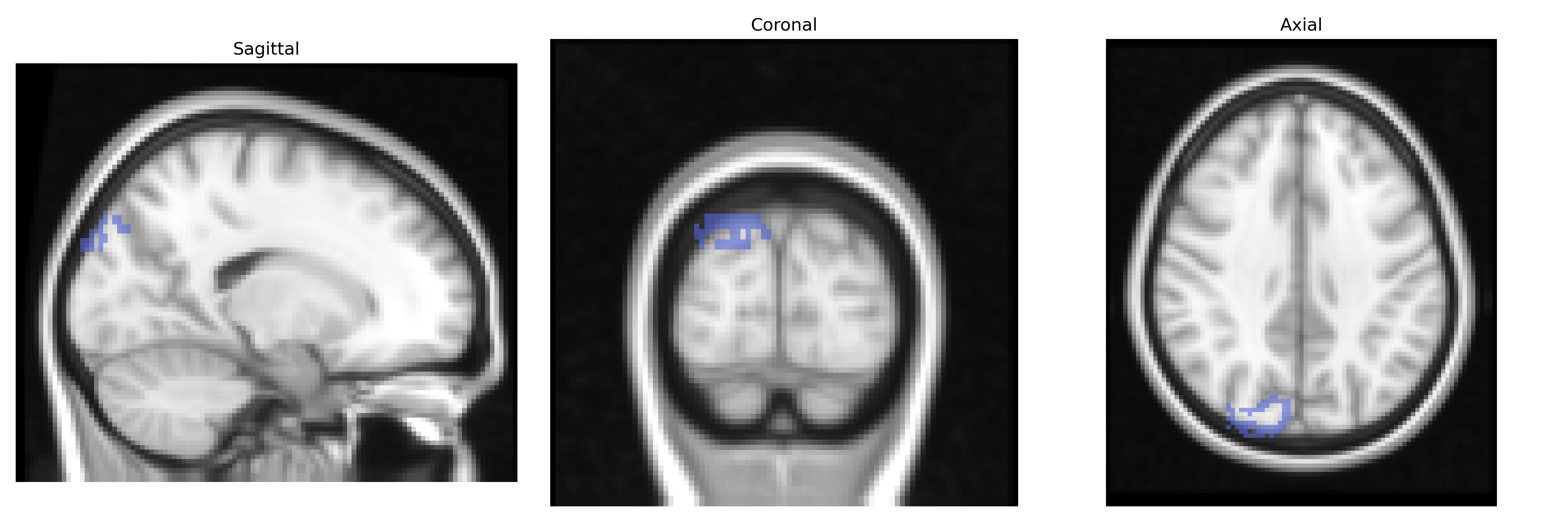
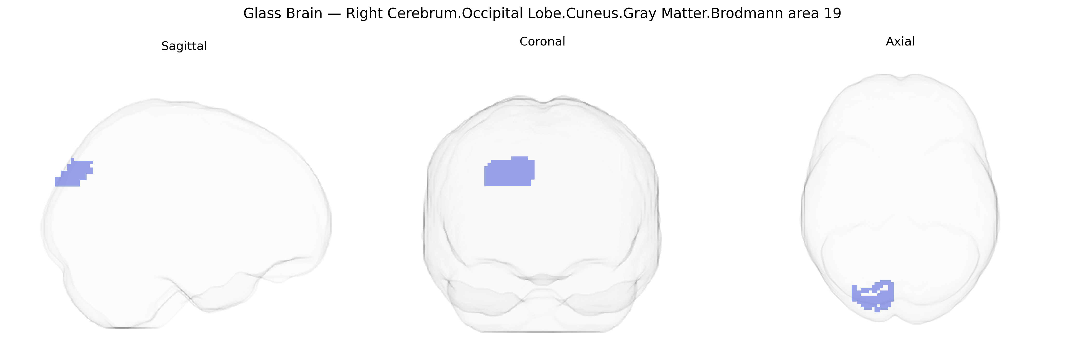
The bilateral Right Cerebrum.Occipital Lobe.Cuneus.Gray Matter.Brodmann area 19 corresponds to higher-order visual association cortex located in the dorsal portion of the occipital lobe, within the cuneus of the right hemisphere. This region receives processed visual input from primary and secondary visual areas (Brodmann areas 17 and 18) and participates in complex visual functions such as object recognition, visual form and motion integration, and aspects of visuospatial processing. Its gray matter contains neurons organized in cortical layers typical of association cortex, with extensive reciprocal connections to parietal and temporal visual areas, supporting integration of visual information with attention, memory, and spatial cognition. There is no direct link for this exact compound region; a related structure is Brodmann area 19.
The bilateral right cuneus gray matter in Brodmann area 19 (occipital association cortex) has been repeatedly implicated in genetic studies of brain structure and function, particularly through imaging genetics and GWAS of cortical thickness, surface area, and functional activation. Large-scale neuroimaging GWAS (e.g., ENIGMA and UK Biobank) have identified common variants near genes involved in neurodevelopment, synaptic function, and axonal guidance (such as variants in or near HMGA2, KIAA0586, WNT signaling components, and other loci affecting global occipital cortical morphology) that are associated with structural measures encompassing BA19, including cuneus thickness and volume. Functionally, the cuneus/BA19 region participates in higher-order visual processing, visuospatial attention, and integration of visual information, and genetic influences on these processes have been observed in studies of visual perception, reading ability, and dyslexia, where variants in genes like DCDC2, KIAA0319, and FOXP2 show associations with altered activation or connectivity in occipital and temporo-parietal visual word-form networks that include BA19. Psychiatric and neurological GWAS have linked structural or functional alterations in BA19/cuneus to schizophrenia, major depression, bipolar disorder, and Alzheimer’s disease, with risk variants in loci such as CACNA1C, GRIN2A, APOE, and complement pathway genes correlating with occipital cortical thickness, perfusion, or task-related activity. Additionally, migraine and visual aura studies have connected genetic risk factors (e.g., variants in TRPM8, PRDM16, and ion-channel genes) to excitability and cortical spreading depression in visual association cortex, including BA19. While most GWAS report regionally aggregated occipital measures rather than Talairach-defined BA19 cuneus specifically, convergent evidence indicates that this association visual cortex region is a key target of polygenic influences on visual cognition, neurodevelopmental reading disorders, affective and psychotic illness, and neurodegeneration.
Overview generated by GPT-4o (2026).
Region ID: 844
Hemisphere: bilateral
Atlas: Talairach labels 2mm
Full Quality Version: Download MP4
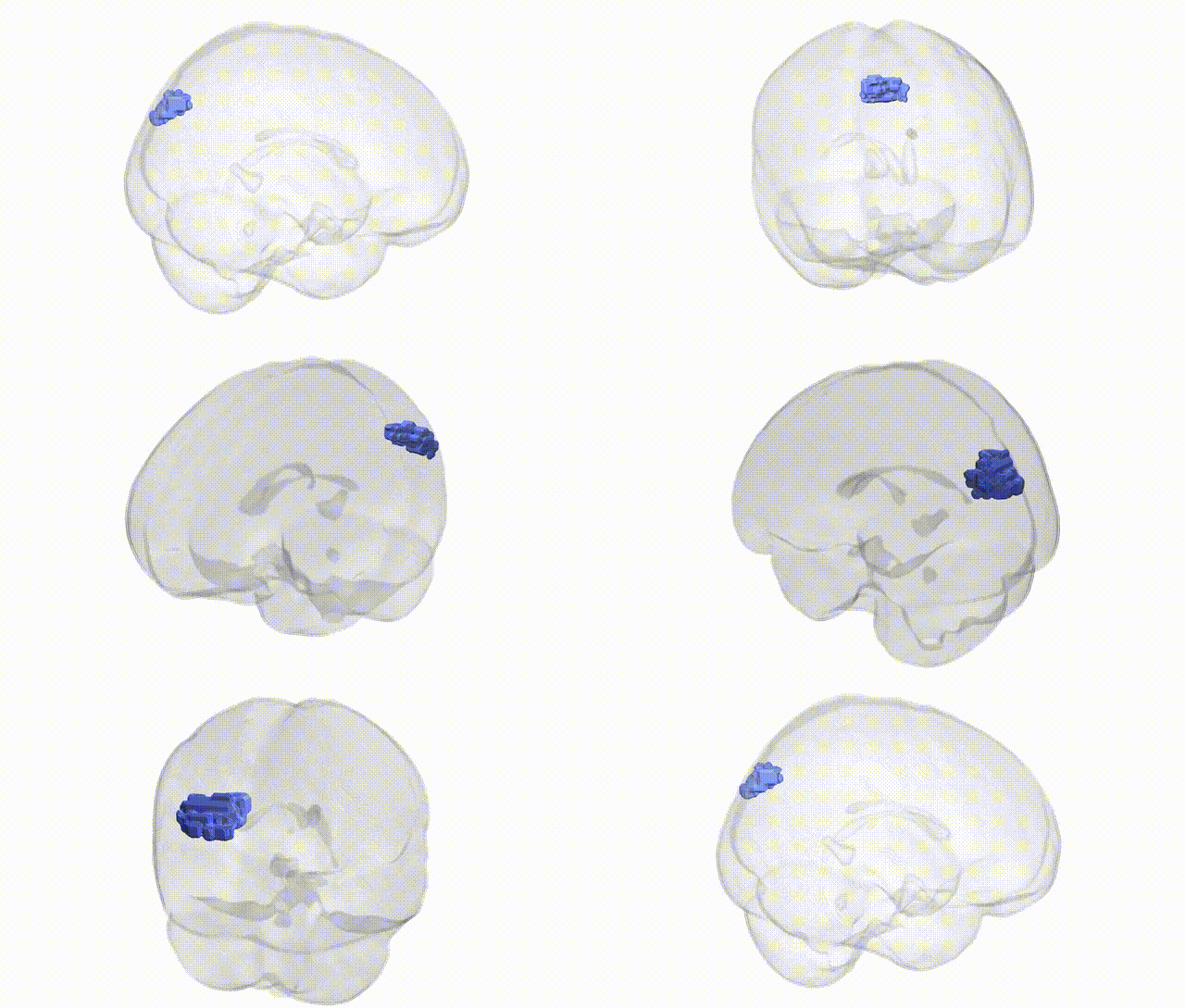
Full Quality Version: Download MP4
Wali Sidiqyar*, Gaurav Rudravaram*, Elyssa M. McMaster, Trent M. Schwartz, Adam M. Saunders, Kurt G. Schilling, Bennett A. Landman "Introducing SPINS: A Shared Public Visualization Library of Neuroanatomical Structures." Medical Imaging with Deep Learning- short paper
This resource is licensed under CC0 1.0 Universal (Public Domain).