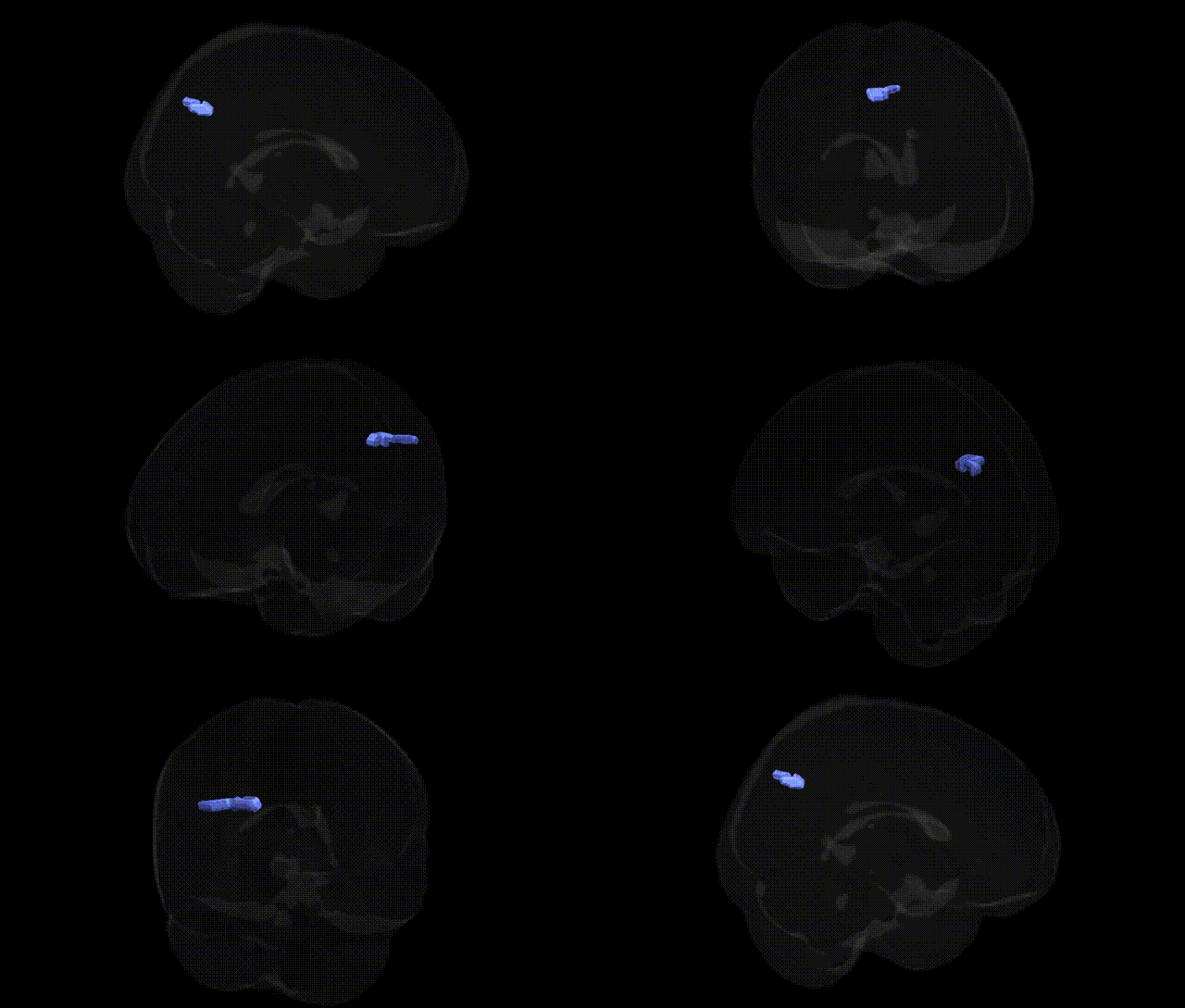
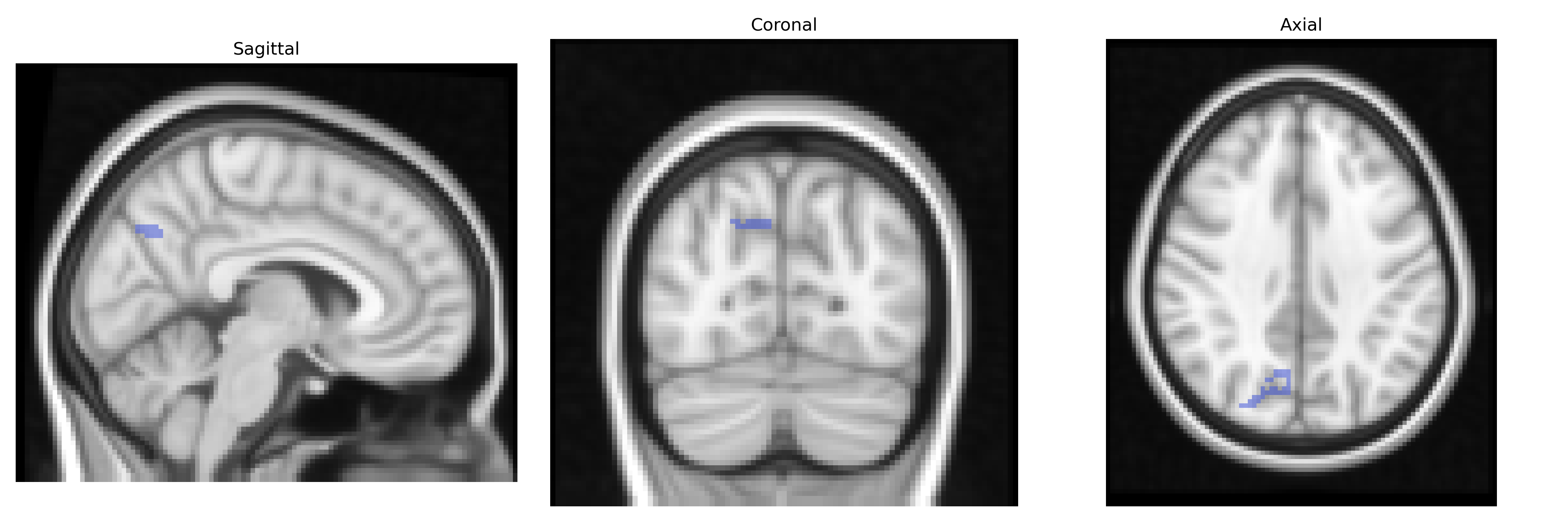
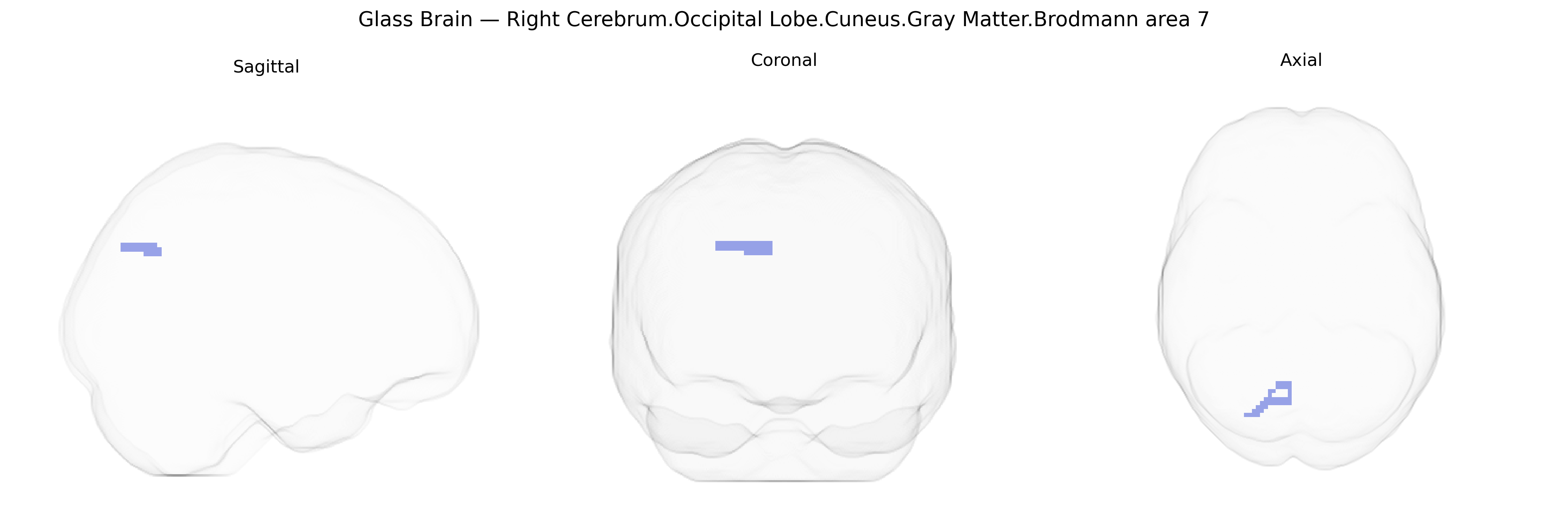
The bilateral Right Cerebrum.Occipital Lobe.Cuneus.Gray Matter.Brodmann area 7, as defined in the Talairach 2 mm Atlas, corresponds to portions of the superior occipital cortex within the cuneus that are functionally associated with the posterior parietal region designated as Brodmann area 7. Brodmann area 7 is implicated in visuospatial processing, integration of visual input with somatosensory information, and coordination of attention and eye–hand movements, supporting higher-order visual perception and spatial orientation. Although located in the occipital cuneus in this atlas labeling, its functional profile aligns with the broader parietal association cortex network involved in visual–motor integration and spatial cognition. There is no direct link for this exact composite label; a closely related structure is Brodmann area 7.
The bilateral right cerebral occipital lobe cuneus gray matter corresponding to Brodmann area 7 (per the Talairach 2 mm atlas) lies at the parieto–occipital junction and is frequently implicated in genetic studies of visuospatial processing, attention, and higher-order visual integration. GWAS and imaging‑genetics analyses using MRI‑derived cortical thickness and surface area have identified multiple common variants—particularly near genes involved in synaptic plasticity, neuronal migration, and neurodevelopment (e.g., MAPT, BDNF, and loci in the MHC region)—that modulate structure and function in superior parietal/cuneus territories overlapping BA7. Polygenic risk for schizophrenia, bipolar disorder, major depression, and ADHD has been associated with altered gray matter volume or cortical thickness in the cuneus/BA7 region, and specific risk alleles in CACNA1C, ZNF804A, and GRM3 have been linked to functional activation changes during attention and working‑memory tasks engaging BA7. Autism spectrum disorder and dyslexia cohorts show genetically influenced differences in parietal–occipital networks including BA7, with variants in genes such as CNTNAP2 and DCDC2 contributing to atypical connectivity and visuospatial or reading‑related performance. Large brain‑wide GWAS (e.g., ENIGMA and UK Biobank studies) report heritability and significant SNP associations for cuneus thickness and surface area, with implicated genes enriched for neurodevelopmental and synaptic pathways, suggesting that BA7 structural variability is a genetically regulated intermediate phenotype connecting common variants to cognitive traits and neuropsychiatric disorders.
Overview generated by GPT-4o (2026).
Region ID: 953
Hemisphere: bilateral
Atlas: Talairach labels 2mm
Full Quality Version: Download MP4
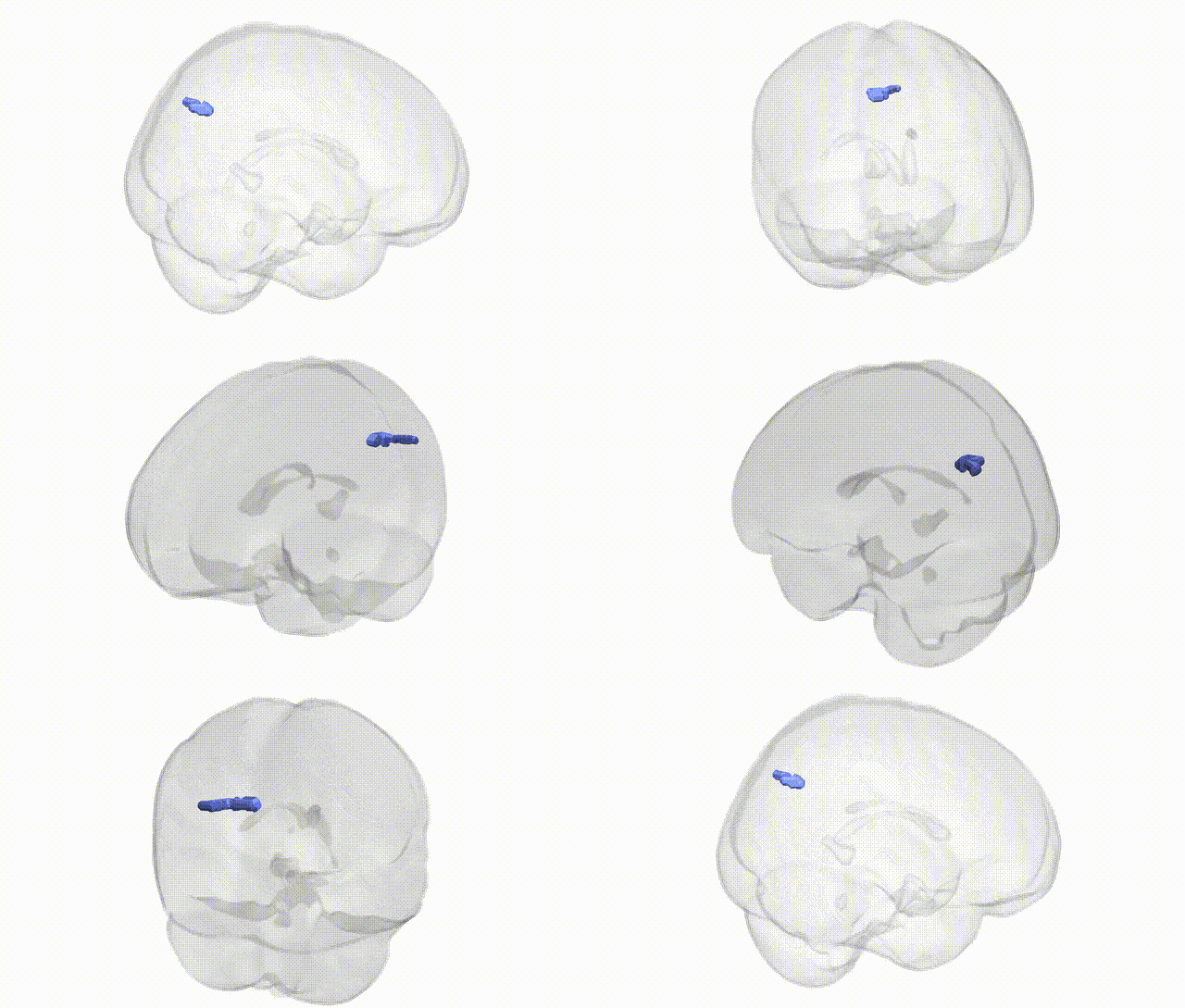
Full Quality Version: Download MP4
Wali Sidiqyar*, Gaurav Rudravaram*, Elyssa M. McMaster, Trent M. Schwartz, Adam M. Saunders, Kurt G. Schilling, Bennett A. Landman "Introducing SPINS: A Shared Public Visualization Library of Neuroanatomical Structures." Medical Imaging with Deep Learning- short paper
This resource is licensed under CC0 1.0 Universal (Public Domain).