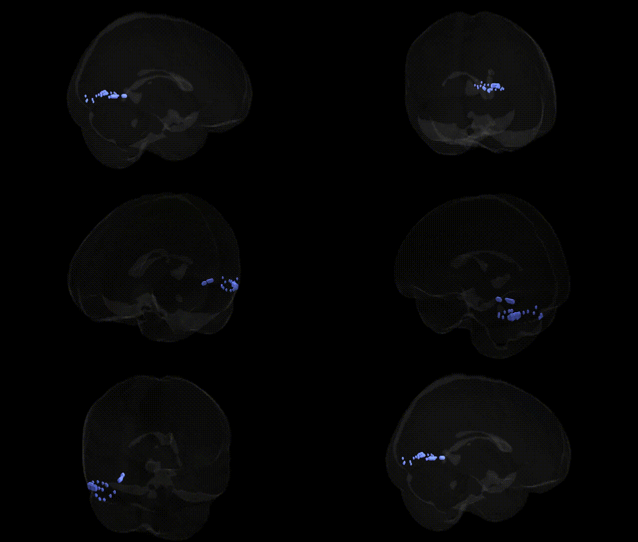
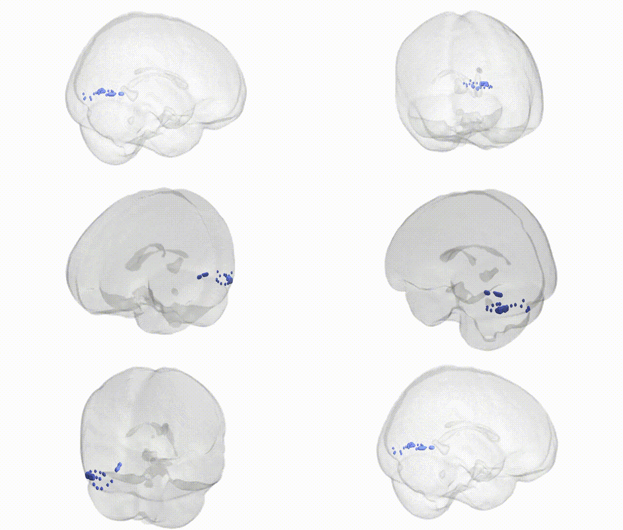
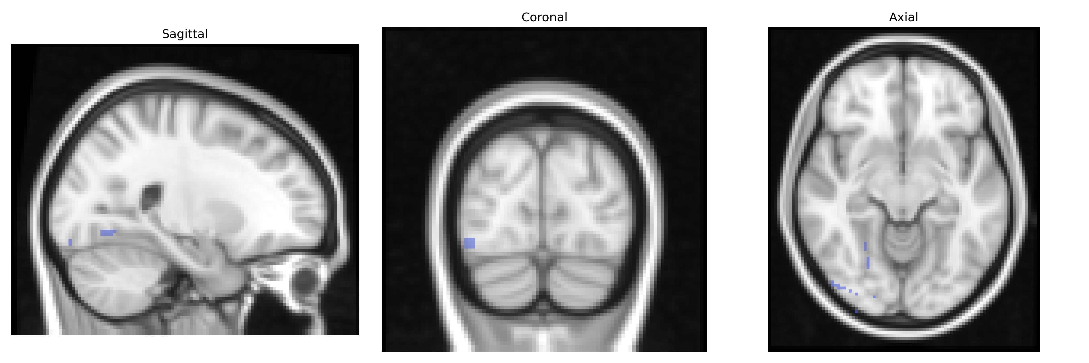
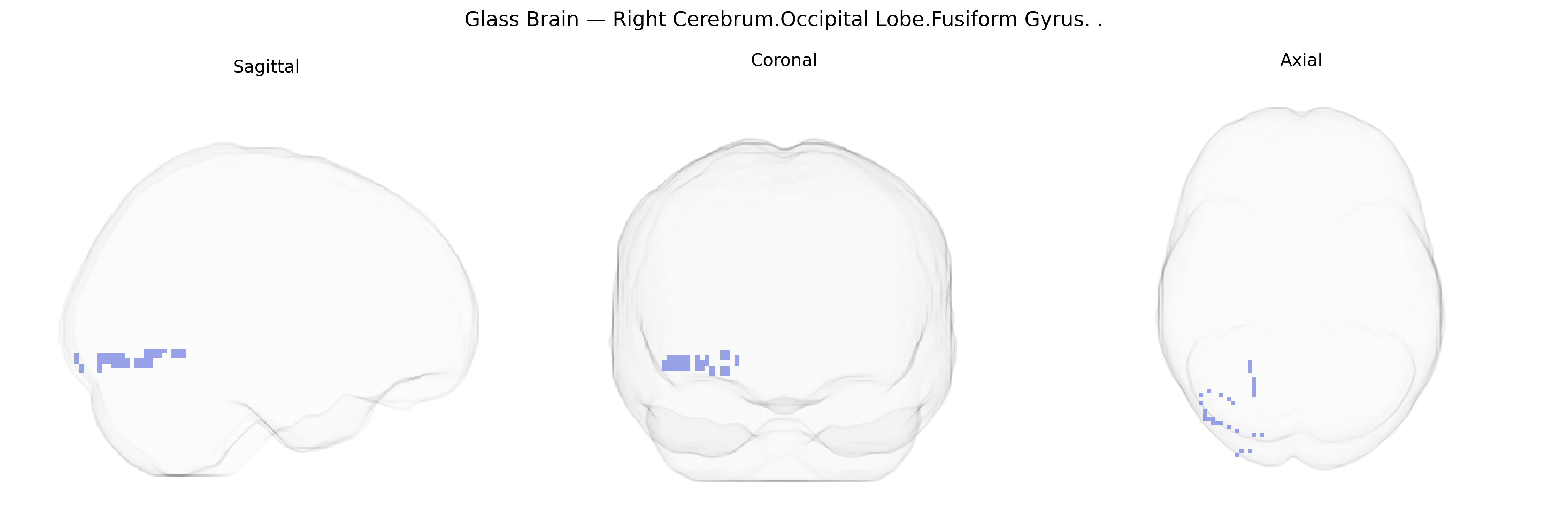
The bilateral Right Cerebrum.Occipital Lobe.Fusiform Gyrus corresponds to portions of the fusiform gyrus located in the occipital lobe of the right cerebral hemisphere, encompassing ventral occipito-temporal cortex involved in high-level visual processing. This region contributes to the perception and integration of complex visual stimuli, including object features, shapes, and patterns, and forms part of the ventral “what” pathway that supports visual recognition and categorization. It is closely associated with neighboring areas in the temporal lobe that subserve face, word, and category-selective processing, and participates in distributed networks for visual memory and semantic association. There is no direct Wikipedia article for this specific Talairach-defined region; see the related structure Fusiform gyrus.
The bilateral right occipital lobe fusiform gyrus (Talairach 2 mm atlas region) has been implicated in several genetic and GWAS-based associations through its roles in high-level visual processing, face recognition, and word-form processing. Twin and heritability studies show substantial genetic influence on fusiform gyrus volume and functional activation, particularly in face-selective regions (e.g., the fusiform face area), with variants in genes related to cortical development (such as those in neurogenesis and synaptic plasticity pathways) contributing to structural variability. Large-scale imaging genetics consortia (e.g., ENIGMA) have identified heritable variation in occipito-temporal cortical thickness and surface area, with genome-wide significant loci near genes involved in neuronal migration and axon guidance (including KIAA0586, PAX6, and other developmental regulators), although findings are generally reported for broader occipital or temporal parcels rather than the specific Talairach-defined fusiform region. GWAS of reading and dyslexia have linked polymorphisms in genes such as DCDC2, KIAA0319, and ROBO1 to altered activation and connectivity in left and right fusiform regions that subserve orthographic processing, while face-recognition abilities show genetic correlation with fusiform structure and activity, with rare variants and familial aggregation reported in developmental prosopagnosia affecting right fusiform function. Psychiatric GWAS and imaging-genetics studies in schizophrenia, autism spectrum disorder, and major depression have associated risk polygenic scores with reduced fusiform gray matter volume or atypical activation, implicating synaptic and immune-related genes (e.g., complement pathway loci like C4 and synaptic scaffolding genes) in occipito-temporal circuitry disruption. In Alzheimer’s disease and frontotemporal dementia, APOE ε4 and other risk variants have been linked to atrophy patterns that include fusiform and adjacent occipital regions, while GWAS of visual hallucinations in Parkinson’s disease and Lewy body dementia suggest polygenic contributions to occipital and fusiform vulnerability. Overall, genetic associations involving this right occipital fusiform region arise mainly from imaging-genetics work on fusiform structure and function and from disorder-related GWAS that affect occipito-temporal networks, rather than from studies targeting this specific Talairach-defined subregion.
Overview generated by GPT-4o (2026).
Region ID: 204
Hemisphere: bilateral
Atlas: Talairach labels 2mm
Full Quality Version: Download MP4
Full Quality Version: Download MP4
Wali Sidiqyar*, Gaurav Rudravaram*, Elyssa M. McMaster, Trent M. Schwartz, Adam M. Saunders, Kurt G. Schilling, Bennett A. Landman "Introducing SPINS: A Shared Public Visualization Library of Neuroanatomical Structures." Medical Imaging with Deep Learning- short paper
This resource is licensed under CC0 1.0 Universal (Public Domain).