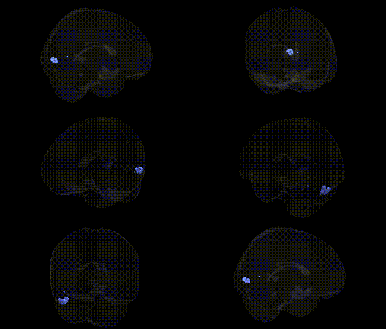
The bilateral Right Cerebrum.Occipital Lobe.Fusiform Gyrus.Gray Matter.Brodmann area 18 corresponds to secondary visual cortex (V2) located in the occipital lobe, extending onto the fusiform gyrus where it participates in intermediate-level visual processing. As part of Brodmann area 18, this region receives strong input from primary visual cortex (V1, BA17) and contributes to the analysis of complex visual features such as contours, textures, and binocular disparity, as well as early stages of form and object processing that feed into higher-order ventral visual stream areas. Neurons in BA18 show more complex receptive field properties than those in V1 and are involved in transforming retinotopic visual information into more abstract representations used for recognition. There is no direct link for “Brodmann area 18” on the fusiform gyrus; a closely related structure is Visual cortex.
The bilateral Right Cerebrum Occipital Lobe Fusiform Gyrus Gray Matter Brodmann area 18 region, a visual association area bordering primary visual cortex, has been implicated in several genetically influenced traits and disorders through imaging genetics and GWAS of brain structure and function. Large-scale neuroimaging GWAS (e.g., ENIGMA, UK Biobank) have identified common variants in and near genes involved in neurodevelopment, synaptic signaling, and axonal guidance (including KIAA0586, DLG2, TCF4, PAX6, and others) that are associated with occipital and fusiform cortical thickness, surface area, and volume, though findings are typically reported at lobar or regional visual cortex levels rather than a Talairach-defined BA18 subregion. Variants in genes such as GRIN2B, NRXN1, CNTNAP2, and SHANK3, which influence synaptic function and cortical circuitry, have been repeatedly linked to autism spectrum disorder, where altered fusiform and occipital activation during face and social perception tasks is common, suggesting a genetically modulated vulnerability of this circuitry. Schizophrenia and bipolar disorder GWAS (implicating loci near CACNA1C, ZNF804A, and others) have also been connected to structural and functional abnormalities in occipital and fusiform regions, particularly in visual processing and hallucination-related circuits. Additionally, GWAS of brain functional connectivity and visual processing traits (such as visual acuity endophenotypes, reading/word recognition, and face recognition) have reported associations involving occipital and fusiform networks, with candidate genes related to myelination and cortical patterning (e.g., FOXP2, ROBO1, CNTN4) contributing to individual differences in these skills. However, current genetic evidence is largely region-level and network-level rather than specific to the Talairach 2 mm Right BA18 label, and most associations reflect polygenic influences that modulate development and function of the broader visual association cortex rather than a single, sharply localized genetic effect.
Overview generated by GPT-4o (2026).
Region ID: 205
Hemisphere: bilateral
Atlas: Talairach labels 2mm
Full Quality Version: Download MP4
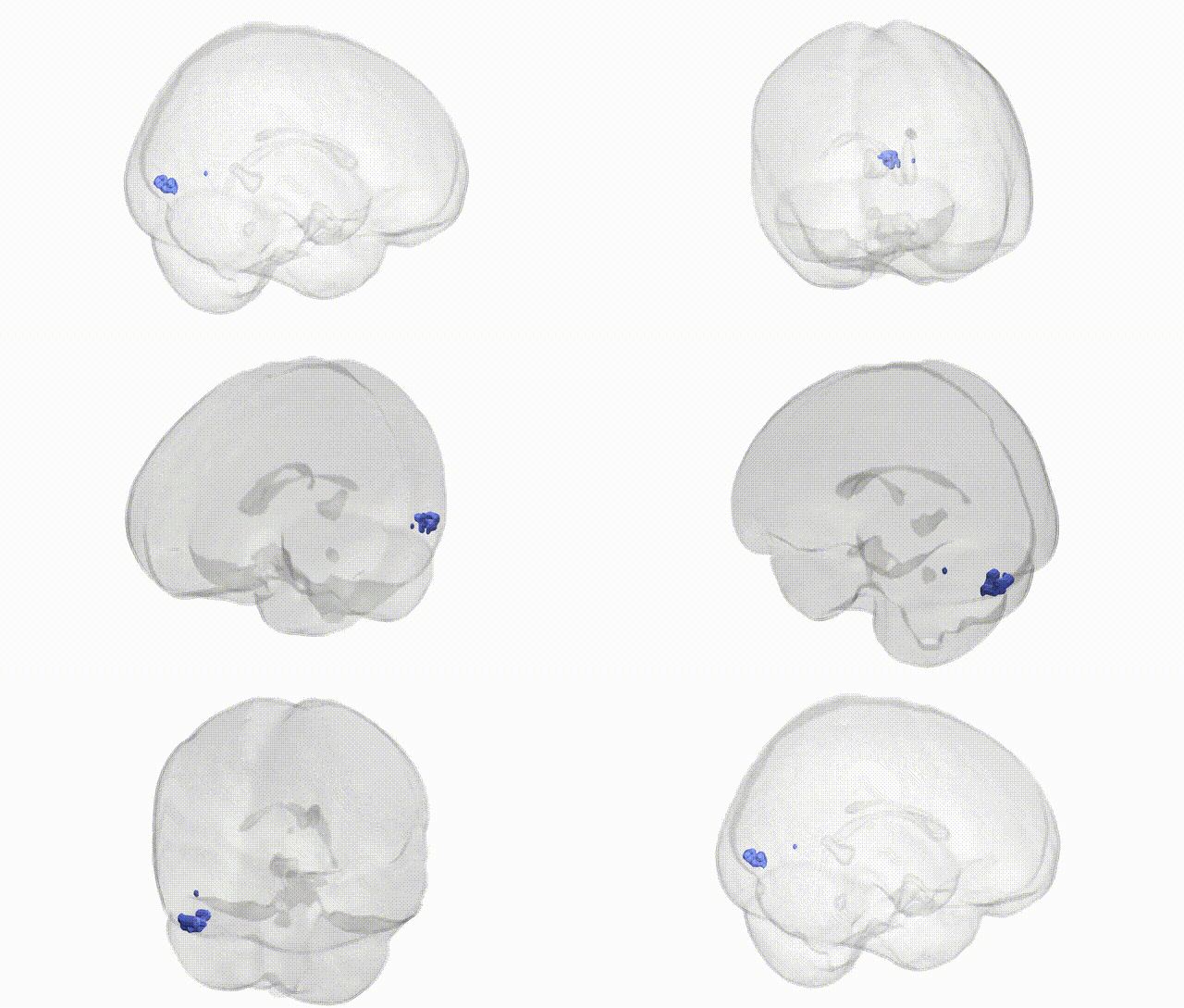
Full Quality Version: Download MP4
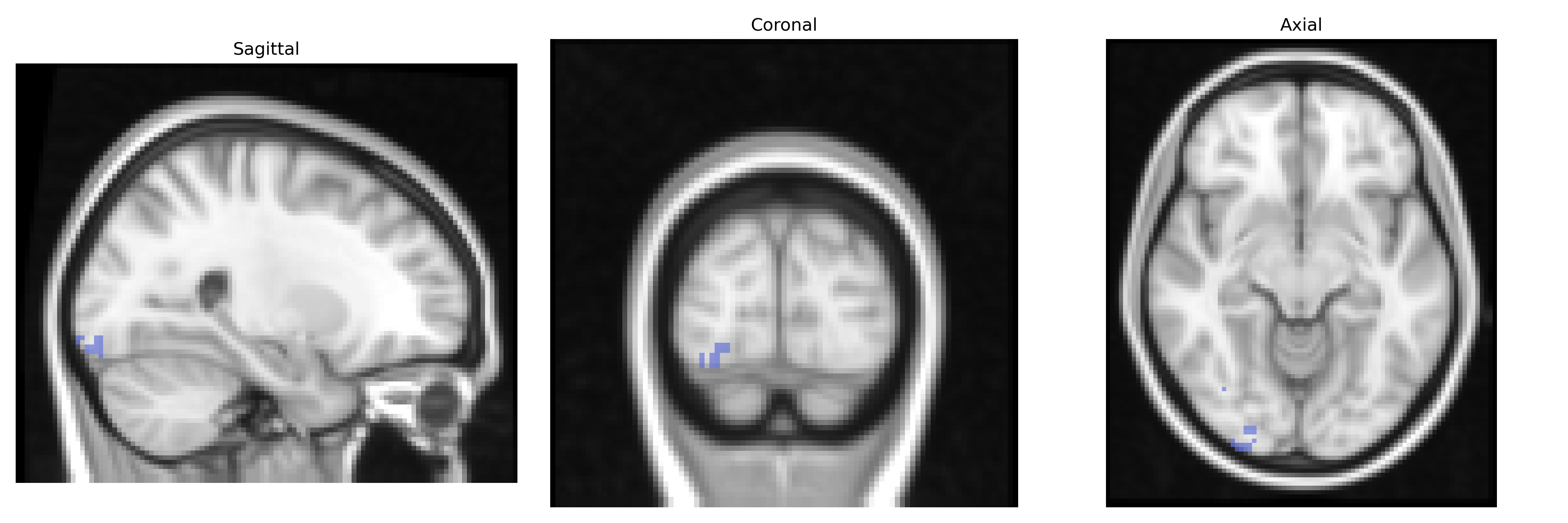
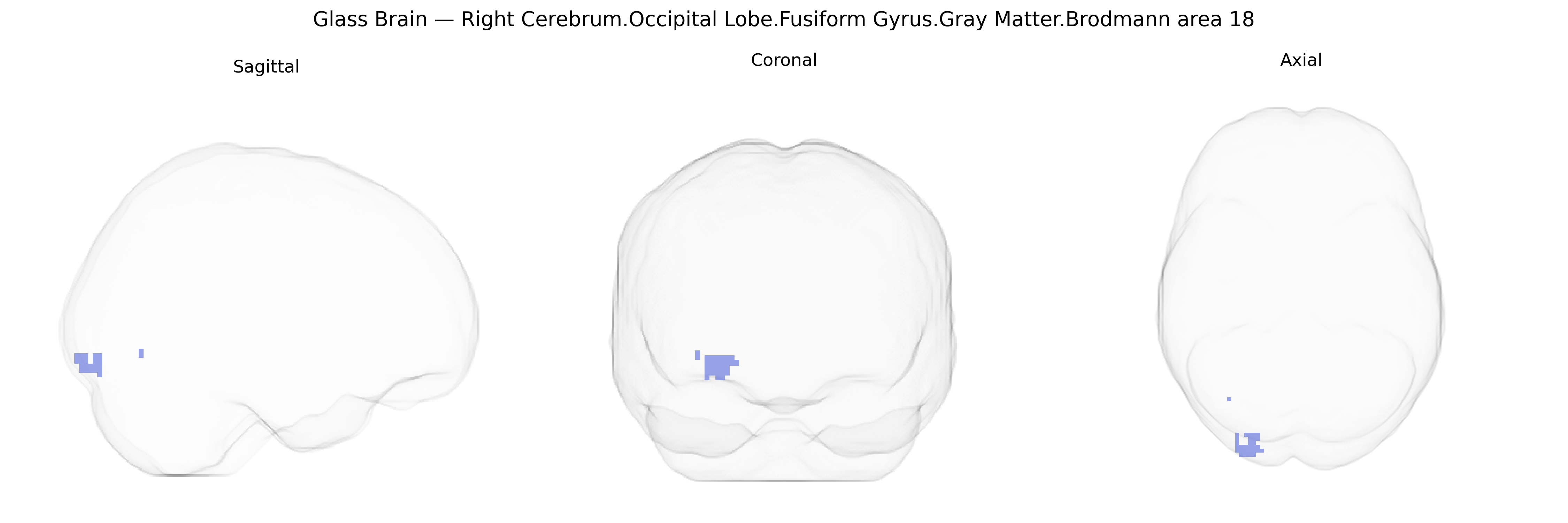
Wali Sidiqyar*, Gaurav Rudravaram*, Elyssa M. McMaster, Trent M. Schwartz, Adam M. Saunders, Kurt G. Schilling, Bennett A. Landman "Introducing SPINS: A Shared Public Visualization Library of Neuroanatomical Structures." Medical Imaging with Deep Learning- short paper
This resource is licensed under CC0 1.0 Universal (Public Domain).